. 2023 Jun 25;44(24):2244-2253.
doi: 10.1093/eurheartj/ehad274.

Takotsubo syndrome is a coronary microvascular disease: experimental evidence

Affiliations

Takotsubo syndrome is a coronary microvascular disease: experimental evidence

Feng Dong et al. Eur Heart J. .

Abstract

Background and aims: Takotsubo syndrome (TTS) is a conundrum without consensus about the cause. In a murine model of coronary microvascular dysfunction (CMD), abnormalities in myocardial perfusion played a key role in the development of TTS.

Methods and results: Vascular Kv1.5 channels connect coronary blood flow to myocardial metabolism and their deletion mimics the phenotype of CMD. To determine if TTS is related to CMD, wild-type (WT), Kv1.5-/-, and TgKv1.5-/- (Kv1.5-/- with smooth muscle-specific expression Kv1.5 channels) mice were studied following transaortic constriction (TAC). Measurements of left ventricular (LV) fractional shortening (FS) in base and apex, and myocardial blood flow (MBF) were completed with standard and contrast echocardiography. Ribonucleic Acid deep sequencing was performed on LV apex and base from WT and Kv1.5-/- (control and TAC). Changes in gene expression were confirmed by real-time-polymerase chain reaction. MBF was increased with chromonar or by smooth muscle expression of Kv1.5 channels in the TgKv1.5-/-. TAC-induced systolic apical ballooning in Kv1.5-/-, shown as negative FS (P < 0.05 vs. base), which was not observed in WT, Kv1.5-/- with chromonar, or TgKv1.5-/-. Following TAC in Kv1.5-/-, MBF was lower in LV apex than in base. Increasing MBF with either chromonar or in TgKv1.5-/- normalized perfusion and function between LV apex and base (P = NS). Some genetic changes during TTS were reversed by chromonar, suggesting these were independent of TAC and more related to TTS.

Conclusion: Abnormalities in flow regulation between the LV apex and base cause TTS. When perfusion is normalized between the two regions, normal ventricular function is restored.

Keywords: Broken heart syndrome; Coronary circulation; Myocardial hibernation; Stress-induced cardiomyopathy.


Conflict of interest statement

Conflict of interest Drs. Chilian, Ohanyan, and Yin are co-founders of KromTherapeutics, and have filed a patent for the use of chromonar in the treatment of Takotsubo Syndrome. The remaining authors have nothing to disclose.

Figures

Structured Graphical Abstract
This pictorial summary of our study represents the induction of Takotsubo Syndrome by stress resulting in tissue hypoxia in the apex compared to the base of the left ventricle as shown by the blue color. The cause of this difference in tissue hypoxia is related to the lower myocardial blood flow (MBF) in the apex compared to the base. These changes in perfusion and the resultant hypoxia in the apex led to a drastic change in gene expression. The array shown reveals the dramatic changes between control and Takotsubo hearts, in which the expression of several genes appears to be ‘flipped’ between the two conditions with red–brown color indicating higher expression (Control vs. Takotsubo) and the blue the reverse. The summary also shows the effects of chromonar, a coronary-specific vasodilator, in term of restoring myocardial blood flow to the apex and restoration of normal function.
Figure 1
Figure 1
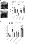
(A) B-Mode ultrasound end-systolic images of the left ventricular of Kv1.5−/− mice under control conditions, following transaortic constriction [2 weeks (2W)], and debanding (removal of the transaortic constriction) for 2 weeks. (B) Fractional shortening (%FS) of the left ventricular apex and base of wild-type and Kv1.5−/− mice under baseline conditions, 2 weeks after transaortic constriction (2W transaortic constriction), 2 weeks after debanding (2W transaortic constriction + 2W deband, 4 weeks after debanding (2W transaortic constriction + 4W deband). Sample sizes: wild-type-control, base n = 18; wild-type -control, apex n = 11; wild-type-transaortic constriction, n = 16; wild-type-2W transaortic constriction + 2W deband, n = 12; 2W transaortic constriction + 4W deband base, n = 12; 2W transaortic constriction + 4W deband apex, n = 11; Kv1.5−/−-control base, n = 6; Kv1.5−/−-control apex, n = 11; Kv1.5−/− −2W transaortic constriction + 2W deband, n = 12; Kv1.5−/− −2W transaortic constriction + 4W deband, n = 12. If base and apex are not mentioned, the sample sizes are equal for the two regions in the group. The data sets were analyzed by a one-way ANOVA followed by Šídák’s multiple comparison test. ANOVA, analysis of variance; CI, confidence interval.
Figure 2
Figure 2
Myocardial blood flow in the left ventricular base and apex of wild-type and Kv1.5−/− mice (presenting with Takotsubo syndrome) after transaortic constriction surgery. MBF was measured under baseline conditions and during a norepinephrine stress test. Sample sizes: wild-type -control base, n = 6; wild-type -control apex, n = 3; wild-type -NE, n = 6, Kv1.5−/−-Control, n = 10; Kv1.5−/−-NE, n = 8. The data sets were analyzed by a one-way ANOVA followed by Šídák’s multiple comparison test. ANOVA, analysis of variance.
Figure 3
Figure 3
(A). Left: B-mode ultrasound end-systolic images of the left ventricle of Kv1.5−/− mice following transaortic constriction or transaortic constriction + chromonar (chrom). Note, despite the transaortic constriction, chromonar eliminated the Takotsubo phenotype. Right: fractional shortening (%FS) of the left ventricular apex and base of Kv1.5−/− mice under baseline conditions (n = 7), 2 weeks after transaortic constriction [2W transaortic constriction (n = 11 base; n = 10 apex)], 2 weeks of chromonar treatment after transaortic constriction [2W transaortic constriction + 2W chrom (n = 8)] and 4 weeks of chromonar treatment after transaortic constriction [2W transaortic constriction + 4W Chrom (n = 11 base; n = 9 apex)]. (B) Myocardial blood flow in the left ventricular apex and base of Kv1.5−/− mice following 2 weeks of transaortic constriction (n = 9), 2 weeks of transaortic constriction + Norepinephrine [NE (n = 9)], 2 weeks of chromonar treatment after transaortic constriction [2W transaortic constriction + 2W chrom (n = 9)] and 2 weeks of chromonar treatment after transaortic constriction + NE [2W transaortic constriction + 2W Chrom + NE (n = 9)]. The data sets were analyzed by a one-way ANOVA followed by Šídák’s multiple comparison test. ANOVA, analysis of variance.
Figure 4
Figure 4
Heatmap with significantly different genes between apex and base of kv1.5−/− mice under control conditions and after transaortic constriction to produce Takotsubo syndrome. Rows are centered and unit variance scaling is applied to each gene. Rows and columns are clustered using correlation distance and average linkage. n = 2 per group.
Figure 5
Figure 5
Enrichment plots of gene expression associated with pathways of fatty acid metabolism (left), oxidative phosphorylation (middle), and hypoxia (right). Comparisons were made between control Kv1.5−/− and those subjected to transaortic constriction exhibiting Takotsubo syndrome. Genes associated with the pathways of fatty acid metabolism and oxidative phosphorylation were downregulated in Takotsubo syndrome (vs. control), but the pathway associated with hypoxia was upregulated in Takotsubo syndrome. n = 2 per group.
Figure 6
Figure 6
Real-time-polymerase chain reaction results of genes associated with metabolism (Ucp3, Acaa2, Pfkfb1) signaling, mitochondrial-localized kinases (Sbk3), cardiac function (Mir133a-2), apoptosis (C920009B18Rik), hypoxia (lox), and interstitial remodeling (postn). Chromonar restored or partially restored the expression of C920009B18Rik, Postn, Lox, Ucp3, and Sbk3 but did not alter the expression of Mir133a-2, Acaa2, or Pfkb1. Data are mean ± 95% CI (n = 4–5 per group). *P < 0.05 vs. corresponding Kv1.5−/−-control group; # P < 0.05 vs. corresponding Kv1.5−/−-transaortic constriction (Takotsubo syndrome) group. The data were analyzed by a one-way ANOVA followed by Šídák’s multiple comparison test. ANOVA, analysis of variance; CI, confidence interval.
Figure 7
Figure 7
Real-time- polymerase chain reaction data for apex vs. Base in hearts from Kv1.5−/−- transaortic constriction and Kv1.5−/−- transaortic constriction plus chromonar (chrom) groups. Data are mean ± 95% CI (n = 4 to 5 per group). *P < 0.05 vs. apex. The data sets were analyzed by a one-way ANOVA followed by Šídák’s multiple comparison test. ANOVA, analysis of variance; CI, confidence interval.

Comment in

References

    1. Ghadri JR, Wittstein IS, Prasad A, Sharkey S, Dote K, Akashi YJ, et al. . International expert consensus document on takotsubo syndrome (part I): clinical characteristics, diagnostic criteria, and pathophysiology. Eur Heart J 2018;39:2032–2046. 10.1093/eurheartj/ehy076 - DOI - PMC - PubMed
    1. Pelliccia F, Kaski JC, Crea F, Camici PG. Pathophysiology of takotsubo syndrome. Circulation 2017;135:2426–2441. 10.1161/CIRCULATIONAHA.116.027121 - DOI - PubMed
    1. El-Battrawy I, Santoro F, Stiermaier T, Moller C, Guastafierro F, Novo G, et al. . Incidence and clinical impact of right ventricular involvement (biventricular ballooning) in takotsubo syndrome: results from the GEIST registry. Chest 2021;160:1433–1441. 10.1016/j.chest.2021.04.072 - DOI - PubMed
    1. de Chazal HM, Del Buono MG, Keyser-Marcus L, Ma L, Moeller FG, Berrocal D, et al. . Stress cardiomyopathy diagnosis and treatment: JACC state-of-the-art review. J Am Coll Cardiol 2018;72:1955–1971. 10.1016/j.jacc.2018.07.072 - DOI - PMC - PubMed
    1. Scantlebury DC, Prasad A. Diagnosis of takotsubo cardiomyopathy. Circ J 2014;78:2129–2139. 10.1253/circj.cj-14-0859 - DOI - PubMed

Publication types