

The use of single-timepoint images to link administered radioiodine activity (MBq) to a prescribed lesion radiation-absorbed dose (cGy): a regression-based prediction interval tool for the management of well-differentiated thyroid cancer patients
- PMID: 37171634
- PMCID: PMC10382352
- DOI: 10.1007/s00259-023-06240-1
The use of single-timepoint images to link administered radioiodine activity (MBq) to a prescribed lesion radiation-absorbed dose (cGy): a regression-based prediction interval tool for the management of well-differentiated thyroid cancer patients
Abstract
Purpose: To introduce a biomarker-based dosimetry method for the rational selection of a treatment activity for patients undergoing radioactive iodine 131I therapy (RAI) for metastatic differentiated thyroid cancer (mDTC) based on single-timepoint imaging of individual lesion uptake by 124I PET.
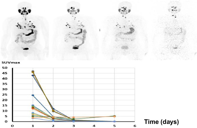
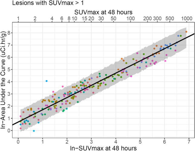
Methods: Patients referred for RAI therapy of mDTC were enrolled in institutionally approved protocols. A total of 208 mDTC lesions (in 21 patients) with SUVmax > 1 underwent quantitative PET scans at 24, 48, 72, and 120 h post-administration of 222 MBq of theranostic NaI-124I to determine the individual lesion radiation-absorbed dose. Using a general estimating equation, a prediction curve for biomarker development was generated in the form of a best-fit regression line and 95% prediction interval, correlating individual predicted lesion radiation dose metrics, with candidate biomarkers ("predictors") such as SUVmax and activity in microcurie per gram, from a single imaging timepoint.
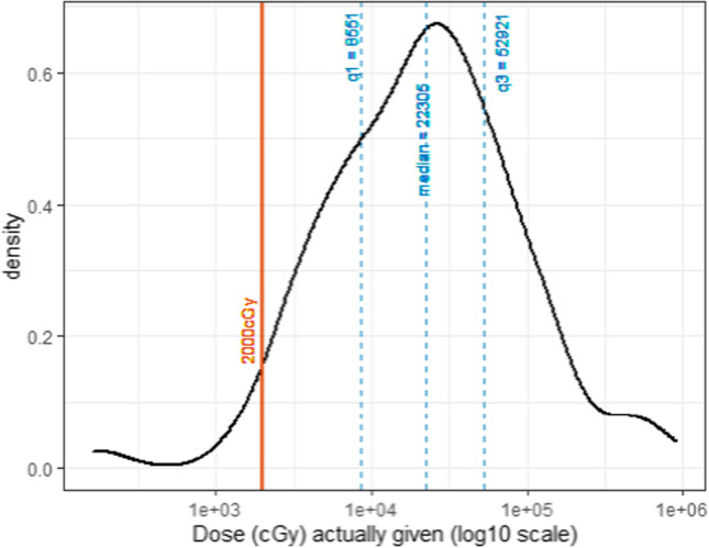
Results: In the 169 lesions (in 15 patients) that received 131I therapy, individual lesion cGy varied over 3 logs with a median of 22,000 cGy, confirming wide heterogeneity of lesion radiation dose. Initial findings from the prediction curve on all 208 lesions confirmed that a 48-h SUVmax was the best predictor of lesion radiation dose and permitted calculation of the 131I activity required to achieve a lesional threshold radiation dose (2000 cGy) within defined confidence intervals.
Conclusions: Based on MIRD lesion-absorbed dose estimates and regression statistics, we report on the feasibility of a new single-timepoint 124I-PET-based dosimetry biomarker for RAI in patients with mDTC. The approach provides clinicians with a tool to select personalized (precision) therapeutic administration of radioactivity (MBq) to achieve a desired target lesion-absorbed dose (cGy) for selected index lesions based on a single 48-h measurement 124I-PET image, provided the selected activity does not exceed the maximum tolerated activity (MTA) of < 2 Gy to blood, as is standard of care at Memorial Sloan Kettering Cancer Center.
Trial registration: NCT04462471, Registered July 8, 2020. NCT03647358, Registered Aug 27, 2018.
Keywords: Differentiated thyroid cancer; Dosimetry; Iodine-124; PET/CT; Radioactive iodine therapy.
© 2023. The Author(s).
Conflict of interest statement
Steven M. Larson, Audrey Mauguen, Alan Ho, Ravinder Grewal, and John Humm are co-inventors of provisional patent for Soothsayer: Number 63/193,700 filed on 5/27/21; conversion deadline: 5/27/22 “Soothsayer,” filed by Office of Technology Development, MSK. SM Larson reports receiving commercial research grants from Y-mAbs Therapeutics, Inc.; Genentech, Inc.; WILEX AG; Telix Pharmaceuticals Limited; and Regeneron Pharmaceuticals, Inc.; holding ownership interest/equity in Elucida Oncology, Inc., and holding stock in ImaginAb, Inc., and Y-mAbs Therapeutics. SML is the inventor of issued patents both currently unlicensed and licensed by MSK to Samus Therapeutics, Inc.; Elucida Oncology, Inc.; and Y-mAbs Therapeutics, Inc. SML serves or has served as a consultant both compensated and uncompensated to Cynvec, LLC; Eli Lilly & Co.; Prescient Therapeutics Limited; Advanced Innovative Partners, LLC; Gerson Lehrman Group; Progenics Pharmaceuticals, Inc.; Exini, Inc.; and Janssen Pharmaceuticals, Inc. See
Figures


Comment on
-
Selumetinib-enhanced radioiodine uptake in advanced thyroid cancer.N Engl J Med. 2013 Feb 14;368(7):623-32. doi: 10.1056/NEJMoa1209288. N Engl J Med. 2013. PMID: 23406027 Free PMC article. Clinical Trial.
References
Publication types
MeSH terms
Substances
Associated data
Grants and funding
LinkOut - more resources
Full Text Sources
Medical
