. 2023 Aug;53(9):1911-1918.
doi: 10.1007/s00247-023-05682-x. Epub 2023 May 12.

Ultrafast cadmium-zinc-telluride-based renal single-photon emission computed tomography: clinical validation

Affiliations

Ultrafast cadmium-zinc-telluride-based renal single-photon emission computed tomography: clinical validation

Matthieu Dietz et al. Pediatr Radiol. 2023 Aug.

Abstract

Background: One of the main limitations of 99mtechnetium-dimercaptosuccinic acid (DMSA) scan is the long acquisition time.

Objective: To evaluate the feasibility of short DMSA scan acquisition times using a cadmium-zinc-telluride-based single-photon emission computed tomography (SPECT) system in children.

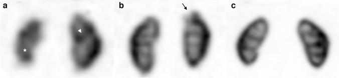
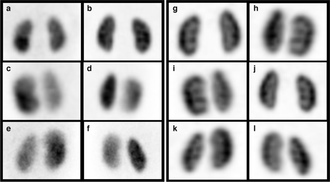
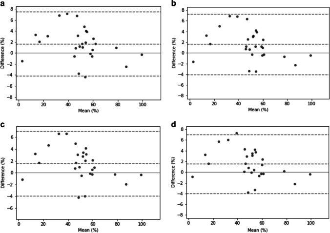
Materials and methods: The data of 27 children (median age: 4 years; 16 girls) who underwent DMSA SPECT were retrospectively analyzed. Both planar and SPECT DMSA were performed. SPECT images were analyzed using coronal-simulated planar two-dimensional images. A reduction in SPECT acquisition time was simulated to provide 4 series (SPECT-15 min, SPECT-10 min, SPECT-5 min and SPECT-2.5 min). A direct comparison of the planar and SPECT series was performed, including semi-quantification reproducibility, image quality (mean quality score on a scale of 0 to 2) and inter- and intra-observer reproducibility of the scintigraphic patterns.

Results: The overall image quality score (± standard deviation) was 1.3 (± 0.6) for the planar data set, 1.6 (± 0.5) for the SPECT-15 min data set, 1.4 (± 0.5) for the SPECT-10 min data set, 1.0 (± 0.5) for the SPECT-5 min data set and 0.6 (± 0.6) for the SPECT-2.5 min data set. Median Kappa coefficients for inter-observer agreement between planar and SPECT images were greater than 0.83 for all series and all readers except one reader for the SPECT-2.5 min series (median Kappa coefficient = 0.77).

Conclusion: Shortening SPECT acquisitions to 5 min is feasible with minimal impact on images in terms of quality and reproducibility.

Keywords: Children; Diagnostic imaging; Kidney disease; Radionuclide imaging.


Conflict of interest statement

None

Figures

Fig. 1
Fig. 1
Anterior 99mtechnetium-dimercaptosuccinic acid (DMSA) images of the kidneys illustrating image quality scores. a A score of 0 in an 11-month-old girl with bilateral duplex kidneys and recurrent febrile urinary tract infections (UTIs). On the DMSA images, there is evidence of a left duplex kidney (arrowhead depicting the cortical bar) and diffuse volume loss of the right lower moiety (asterisk) that is probably developmental (reflux nephropathy). b A score of 1 in a 7-year-old girl with recurrent UTIs. A small scar is seen in the upper pole of the left kidney (arrow). c A score of 2 in a 4-year-old boy with recurrent UTIs, who had a normal DMSA scan on presentation
Fig. 2
Fig. 2
Comparison of planar and single-photon emission computed tomography (SPECT) images in a 1-year-old boy who was referred for a 99mtechnetium-dimercaptosuccinicacid (DMSA) examination to exclude renal parenchymal scarring following an acute urinary tract infection. al Conventional planar images (af) and 5-min scan two-dimensional coronal projection SPECT images (gl), showing homogeneous and diffuse cortical renal absorption. Renal function was estimated to be 52% and 48% for the right and left kidneys, respectively. a, g Anterior images. b, h Posterior images. c, i Right anterior oblique images. d, j Left posterior oblique images. e, k Left anterior oblique images. f, i Right posterior oblique images
Fig. 3
Fig. 3
Comparison of planar and single-photon emission computed tomography (SPECT) images in a 3-year-old girl referred for renal scarring 5 months after urinary tract infection with Escherichia coli. Posterior planar image (a) and posterior two-dimensional coronal projection SPECT images at 15 min (b), 10 min (c), 5 min (d) and 2.5 min (e) showed a scar in the upper pole of the right kidney
Fig. 4
Fig. 4
Bland–Altman plots of split renal function comparing planar images to single-photon emission computed tomography images at 2.5 min (a), 5 min (b), 10 min (c) and 15 min (d) duration

References

    1. Do J, Ruddy TD, Wells RG. Reduced acquisition times for measurement of myocardial blood flow with 99mTc-tetrofosmin and solid-state detector SPECT. J Nucl Cardiol. 2021;28:2518–2529. doi: 10.1007/s12350-020-02048-w. - DOI - PubMed
    1. Surti S, Viswanath V, Daube-Witherspoon ME, et al. Benefit of improved performance with state-of-the art digital PET/CT for lesion detection in oncology. J Nucl Med. 2020;61:1684–1690. doi: 10.2967/jnumed.120.242305. - DOI - PMC - PubMed
    1. Dietz M, Kamani CH, Allenbach G, et al. Comparison of the prognostic value of impaired stress myocardial blood flow, myocardial flow reserve, and myocardial flow capacity on low-dose Rubidium-82 SiPM PET/CT. J Nucl Cardiol. 2022 doi: 10.1007/s12350-022-03155-6. - DOI - PMC - PubMed
    1. Vali R, Armstrong IS, Bar-Sever Z, et al. SNMMI procedure standard/EANM practice guideline on pediatric [99mTc]Tc-DMSA renal cortical scintigraphy: an update. Clin Transl Imaging. 2022;10:173–184. doi: 10.1007/s40336-022-00484-x. - DOI
    1. Reichkendler MH, Berg RMG, de Nijs R, et al. Planar scan vs. SPECT/low-dose CT for estimating split renal function by 99mTc-DMSA scintigraphy in children. Eur J Nucl Med Mol Imaging. 2020;47:729–733. doi: 10.1007/s00259-019-04575-2. - DOI - PubMed

Substances

LinkOut - more resources