Review
. 2023 Oct 1;103(4):2623-2677.
doi: 10.1152/physrev.00032.2022. Epub 2023 May 12.

Making a good egg: human oocyte health, aging, and in vitro development

Affiliations
Review

Making a good egg: human oocyte health, aging, and in vitro development

Evelyn E Telfer et al. Physiol Rev. .

Abstract

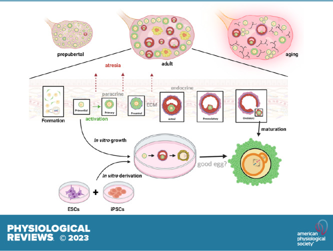
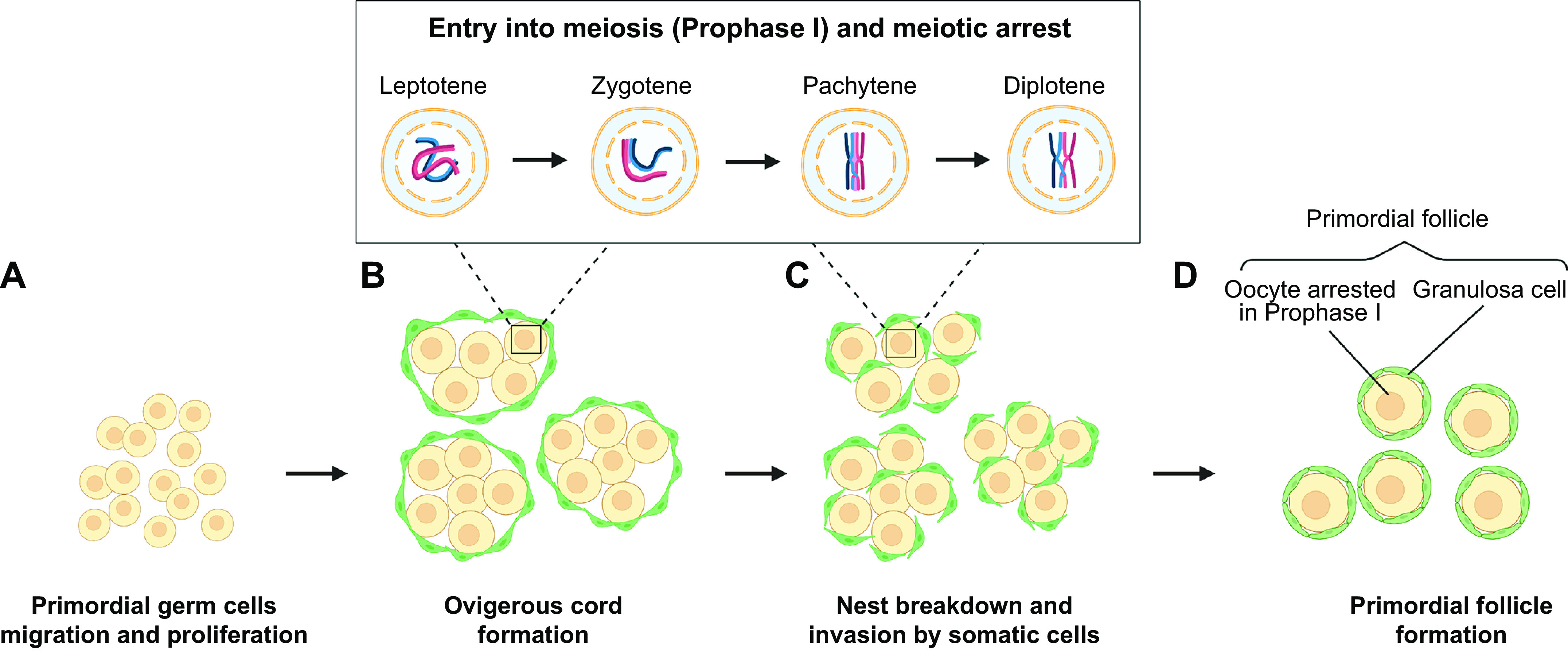
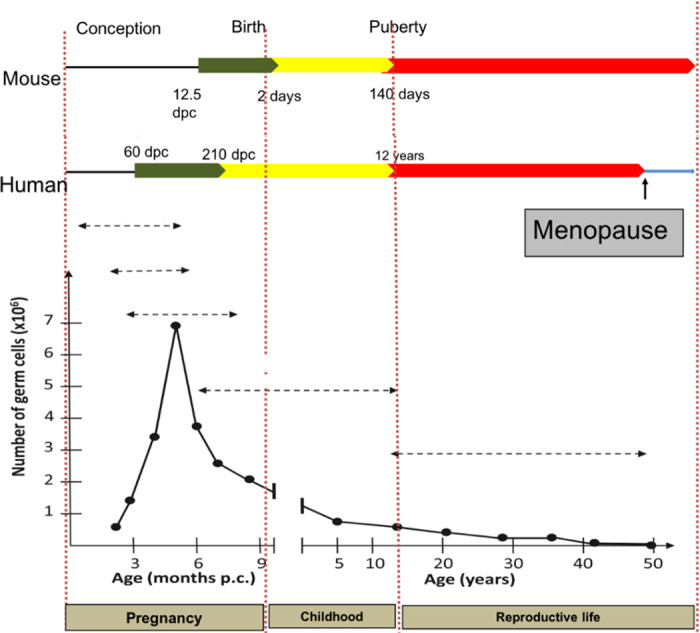
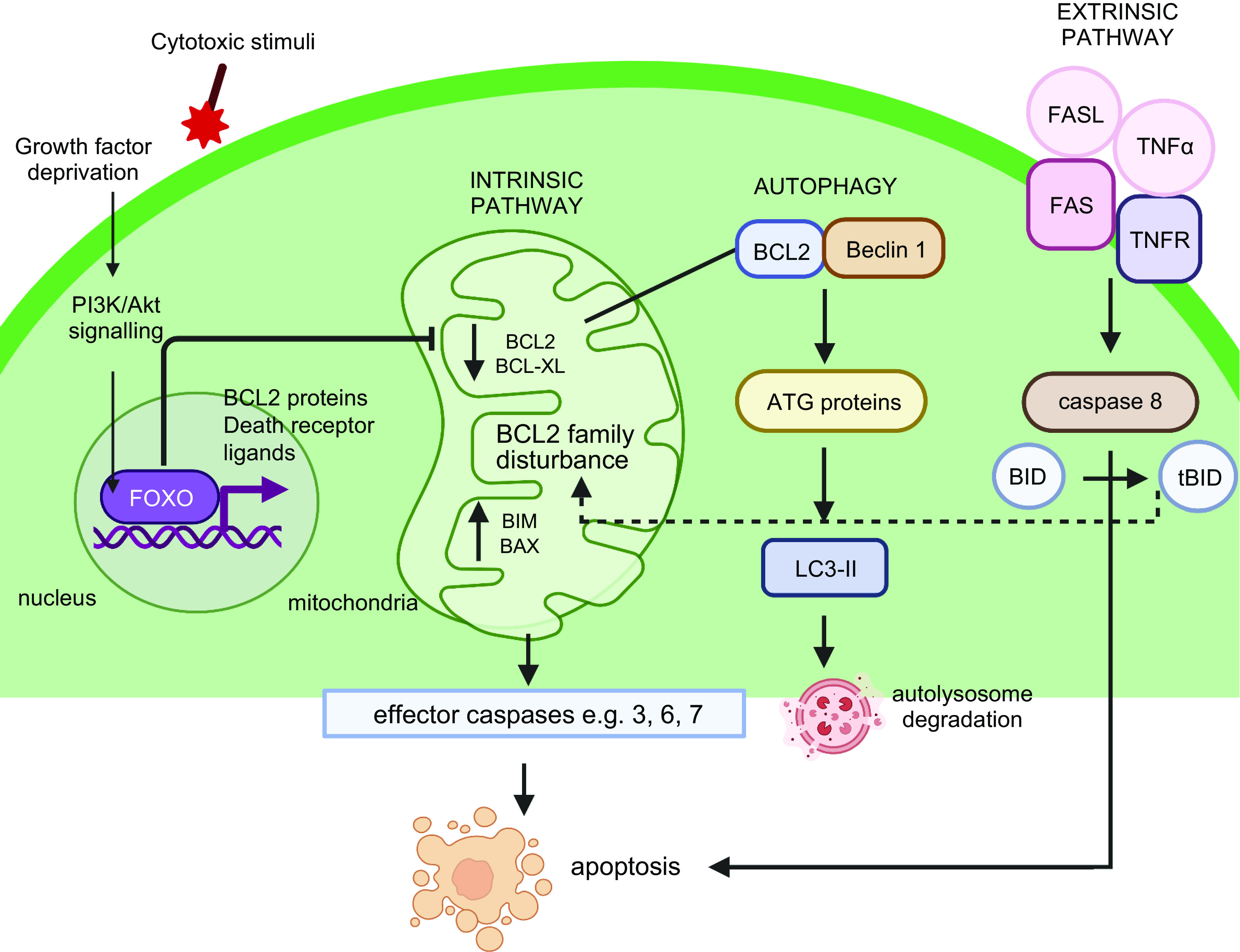
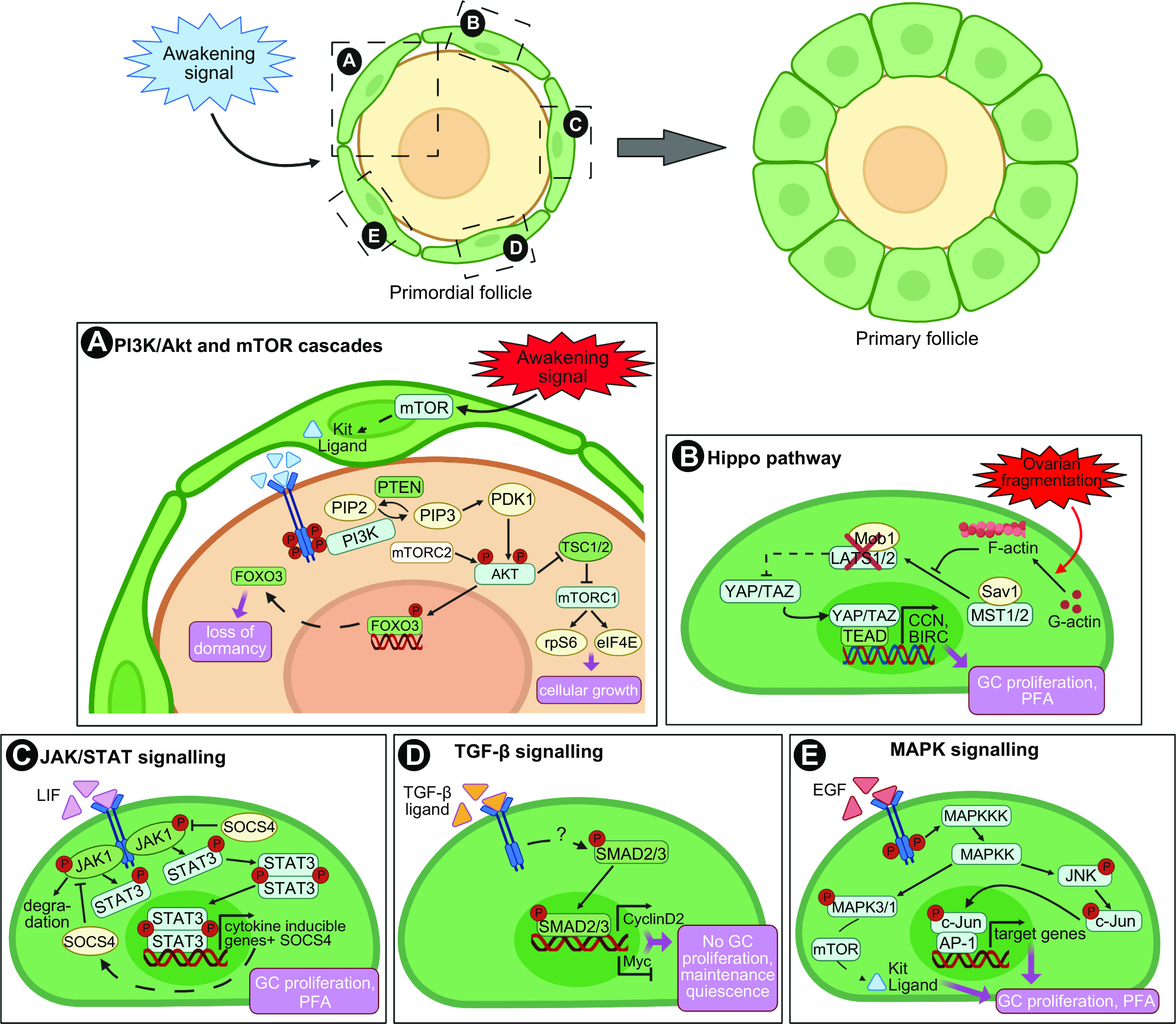
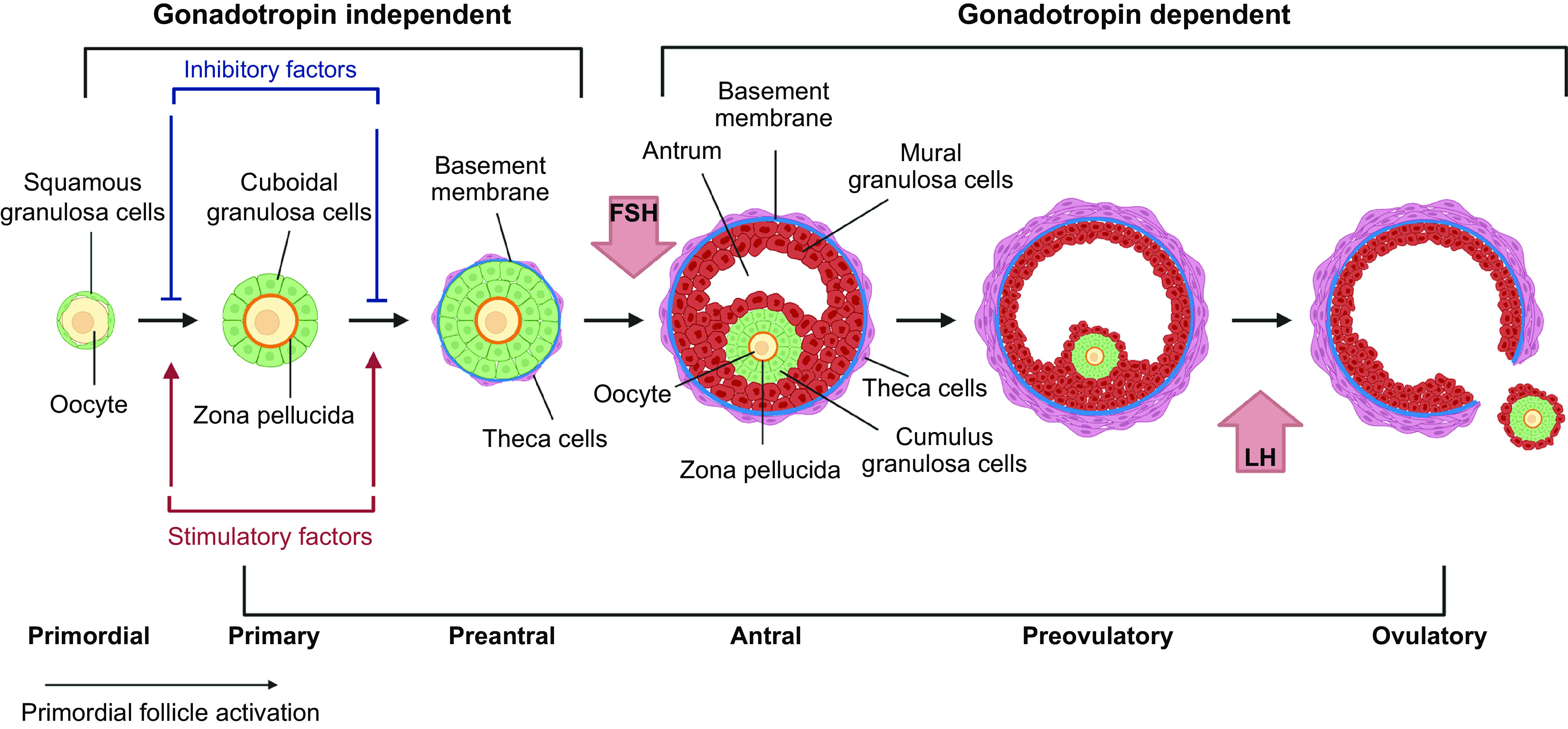
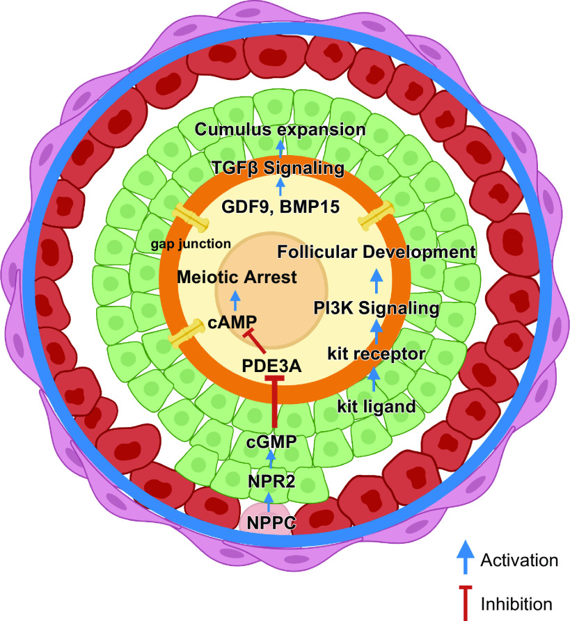
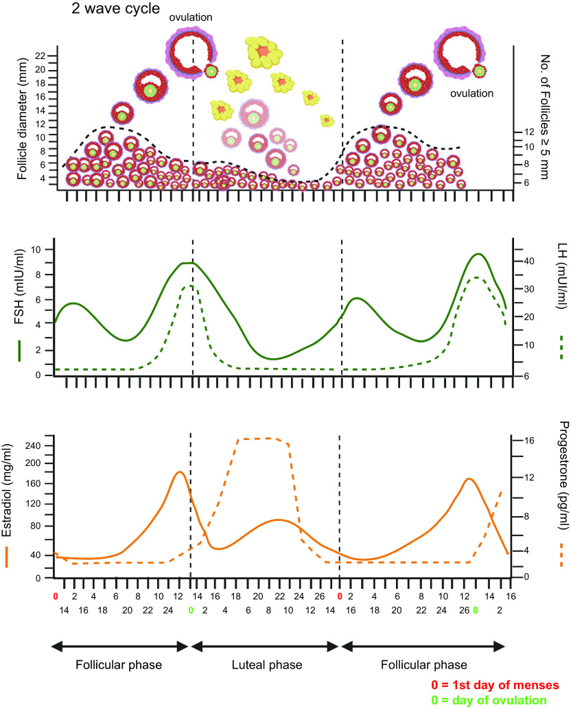
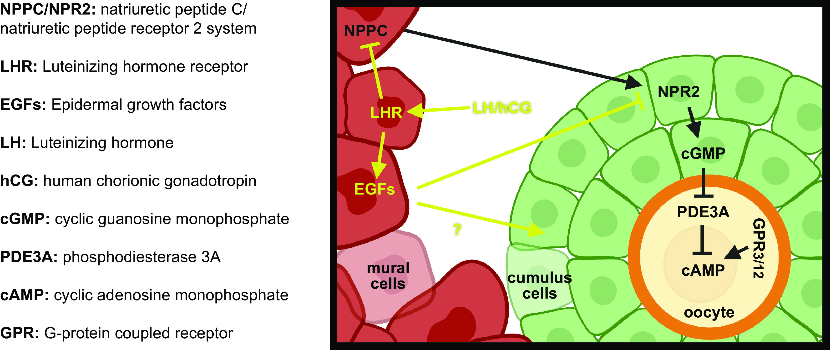
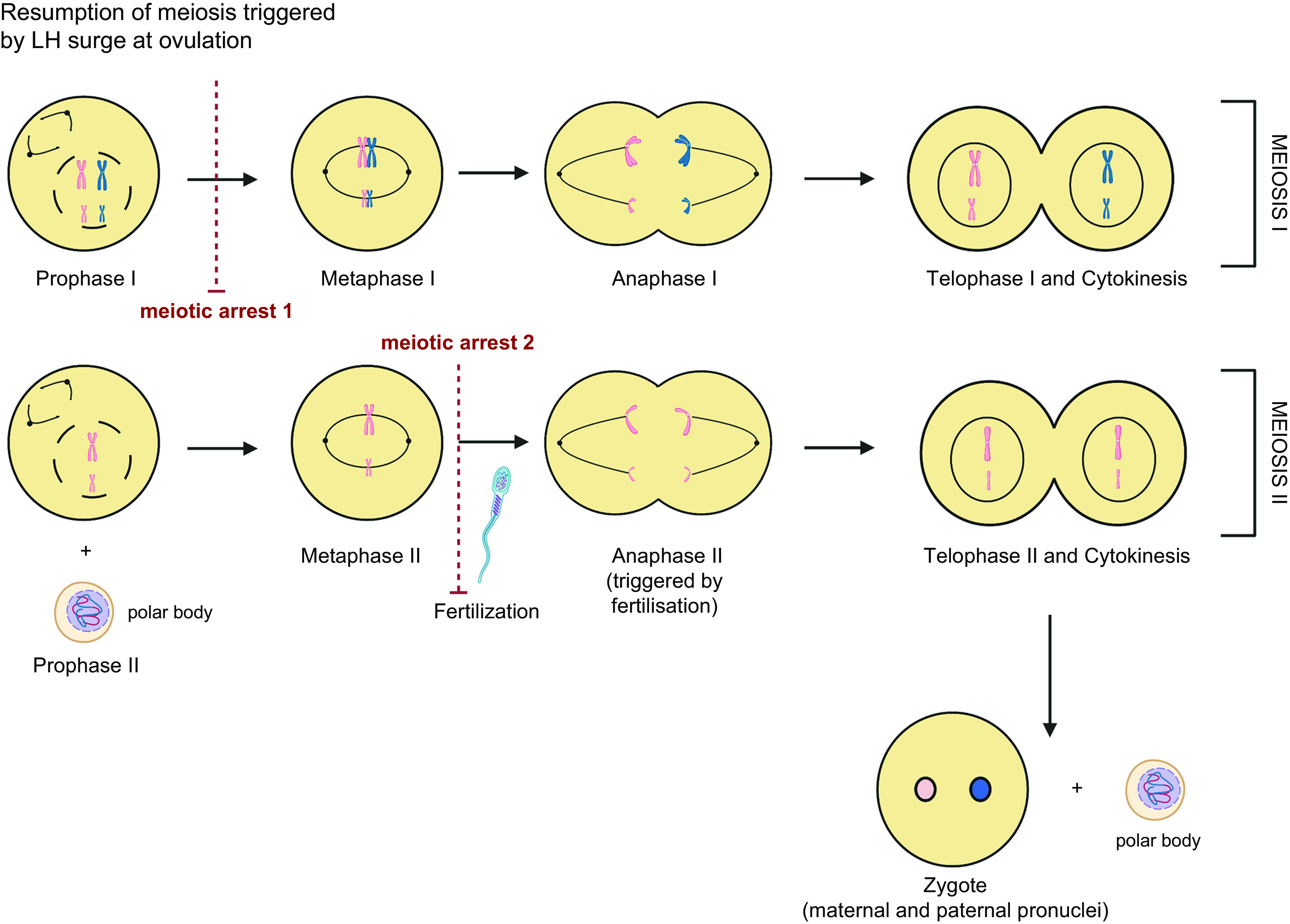
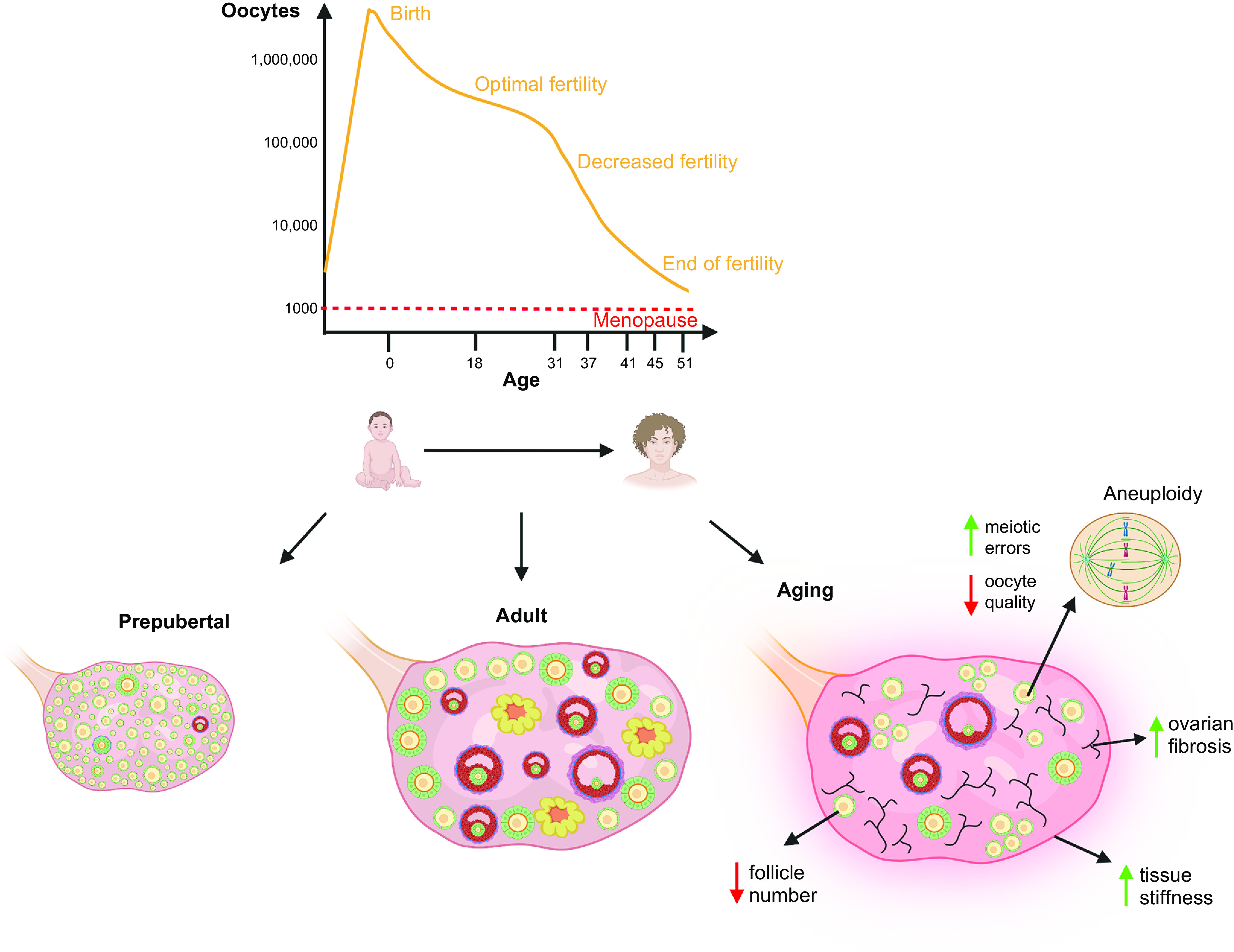
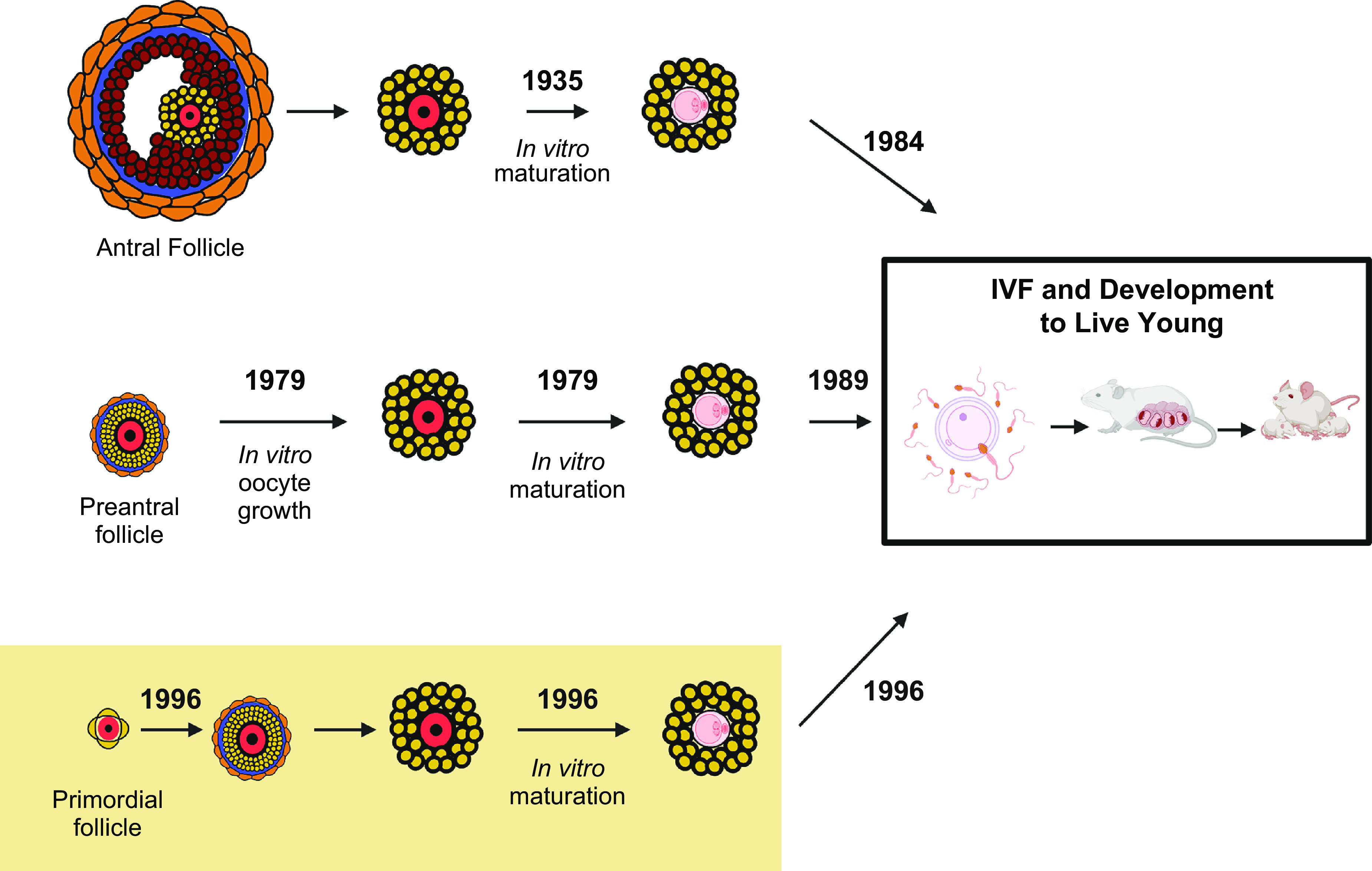
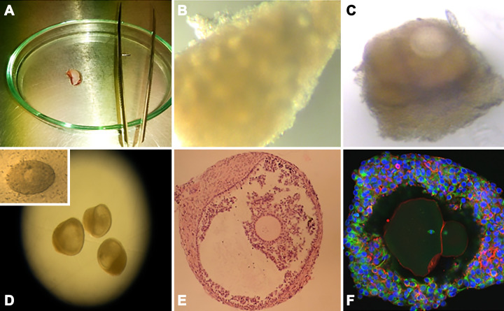
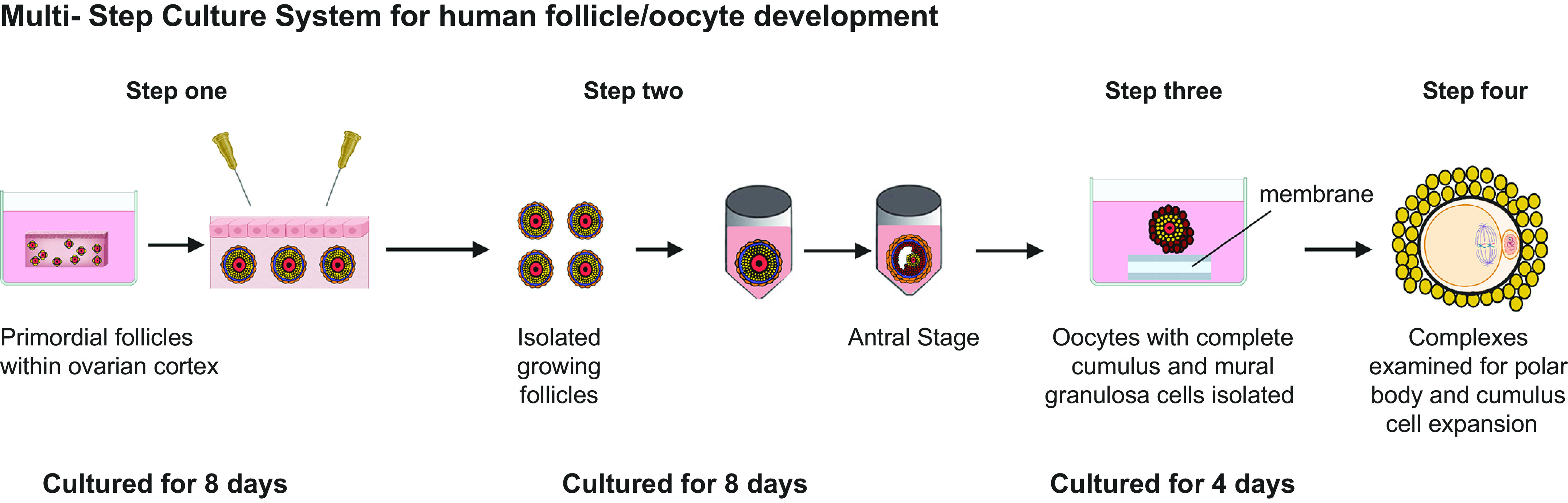
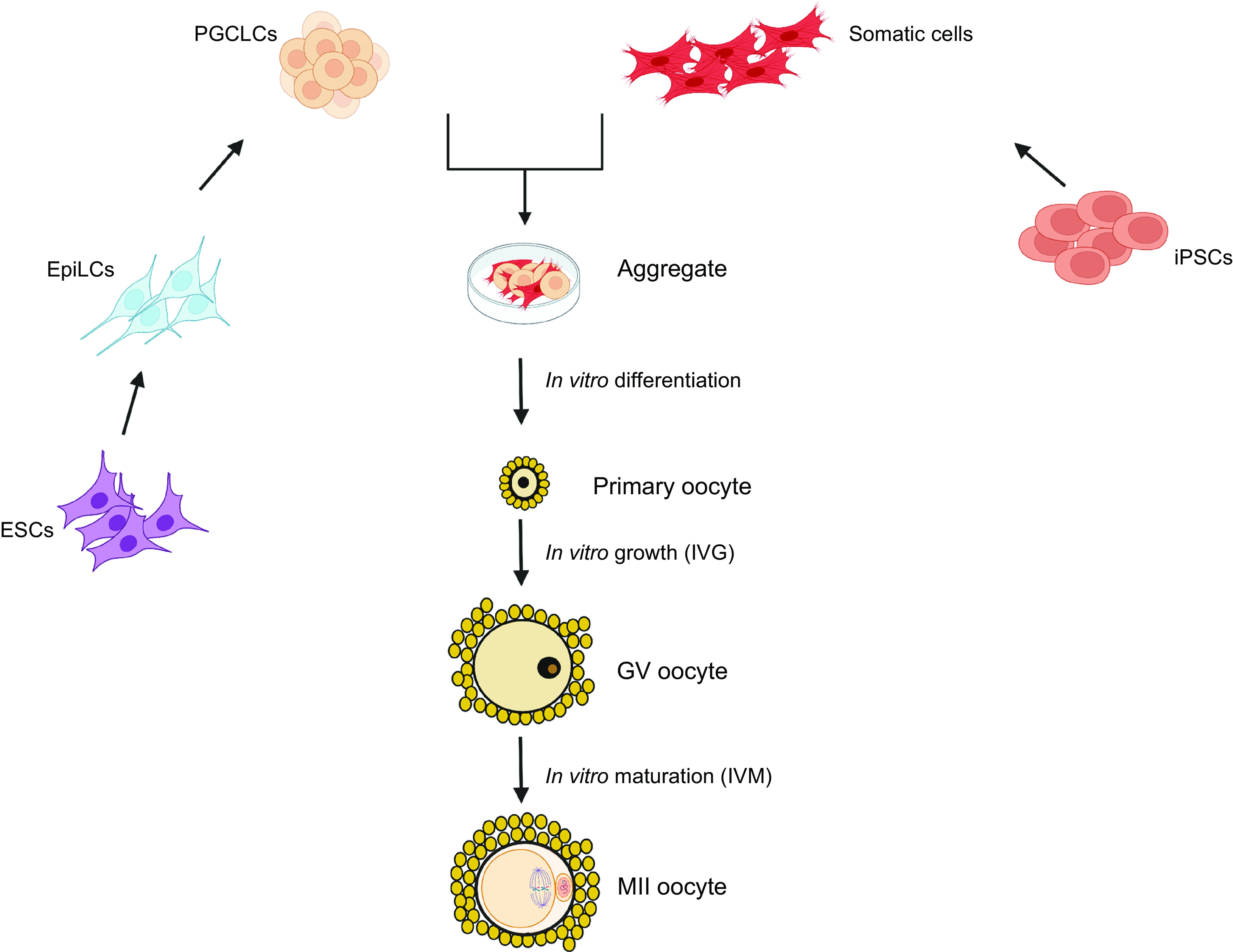
Mammalian eggs (oocytes) are formed during fetal life and establish associations with somatic cells to form primordial follicles that create a store of germ cells (the primordial pool). The size of this pool is influenced by key events during the formation of germ cells and by factors that influence the subsequent activation of follicle growth. These regulatory pathways must ensure that the reserve of oocytes within primordial follicles in humans lasts for up to 50 years, yet only approximately 0.1% will ever be ovulated with the rest undergoing degeneration. This review outlines the mechanisms and regulatory pathways that govern the processes of oocyte and follicle formation and later growth, within the ovarian stroma, through to ovulation with particular reference to human oocytes/follicles. In addition, the effects of aging on female reproductive capacity through changes in oocyte number and quality are emphasized, with both the cellular mechanisms and clinical implications discussed. Finally, the details of current developments in culture systems that support all stages of follicle growth to generate mature oocytes in vitro and emerging prospects for making new oocytes from stem cells are outlined.

Keywords: follicle culture; meiosis; oocyte maturation; ovary; reproductive aging; stem cells.


Conflict of interest statement

No conflicts of interest, financial or otherwise, are declared by the authors.

Figures

None
Graphical abstract
FIGURE 1.
FIGURE 1.
Formation of primordial follicles. Primordial germ cells (PGCs) migrate to the gonadal ridge (A) and form nests of oogonia surrounded by somatic cells (B) within the presumptive ovary. Oogonia undergo a defined period of proliferation prior to entering meiosis and forming oocytes (C). Meiosis progresses to the diplotene stage of prophase I to form oocytes that are found at 16 weeks of gestation in the human fetal ovary. Oocytes establish connections with somatic cells (granulosa cells) to form primordial follicles (D). Image created with BioRender.com, with permission.
FIGURE 2.
FIGURE 2.
Numbers and timings of oocyte formation and loss. Outline of timings (dpc, days postconception) of follicle formation in mouse and human (green) with graph depicting numbers of germ cells and rate of loss prepubertally (yellow) and until menopause (red). Only 0.1% of follicles will ever be ovulated with the rest degenerating at different stages. Image created with BioRender.com, with permission.
FIGURE 3.
FIGURE 3.
Cell death pathways utilized by oocytes and granulosa cells. The intrinsic apoptotic pathway is elicited through growth factor deprivation or cytotoxic stimuli. Signals are transduced via the phosphoinositide-3-kinase (PI3K) pathway converging on the transcription factor forkhead box O3 (FOXO3) whose targets include BCL2 family proteins and death receptor ligands, causing an imbalance in BCL2 family proteins, activating effector caspases, and leading to apoptosis. Effector caspases are also activated via the extrinsic apoptotic pathway when death receptor ligands binding their corresponding receptors on the cell membrane. Death receptor signaling can also result in BH3 interacting-domain death agonist (BID) cleavage by caspase-8, leading to the generation of active tBID, and cross talk with the intrinsic apoptosis pathway. Finally, BCL2 can interact with Beclin 1 to regulate autophagy, and downstream ATG proteins and LC3-II act to control autophagosome formation. Image created with BioRender.com, with permission.
FIGURE 4.
FIGURE 4.
Intrafollicular signaling pathways regulating primordial follicle quiescence and entry into growth. A: granulosa cell (GC) induction of mammalian target of rapamycin (mTOR) leads to the secretion of kit ligand that binds its c-KIT receptor on oocytes, triggering the phosphoinositide-3-kinase (PI3K) cascade. Phosphorylation of AKT triggers nuclear export and suppression of forkhead box O3 (FOXO3) transcription factor activity to promote follicle activation, and induces the activation of the downstream mTOR pathway components to direct cell growth. B: Hippo dysregulation by ovarian fragmentation triggers a switch in the G-actin/F-actin ratio, resulting in the inhibition of LATS1/2 activity and YAP1 dephosphorylation and translocation into the nucleus. YAP/TAZ interaction with TEAD transcription factors promotes the expression of target genes involved in granulosa cell proliferation and primordial follicle activation (PFA). C: activation of the JAK/STAT pathway leads to STAT3 phosphorylation and formation of dimers that translocate to the nucleus, bind to DNA, and regulate transcription of genes involved in GC proliferation and primordial follicle activation. The JAK/STAT activity is negatively regulated by SOCS4. D: GCs from quiescent follicles express the transcription factor SMAD3, which promotes expression of cyclin D2 and represses Myc. Cyclin D2 is bound by the inhibitory factor P27 preventing cell cycle progression while repression of Myc maintains growth arrest. E: activation of the MAPK signaling triggers the phosphorylation of MAPK3/1, which participates in mTOR pathway activation, and JNK, which controls the activity of the proto-oncogene c-Jun and downstream transcription factor AP-1, both promoting GC proliferation and follicle entry into growth. Granulosa cells in green, oocyte in yellow. Image created with BioRender.com, with permission.
FIGURE 5.
FIGURE 5.
Stages of follicle growth (primordial to preovulatory). Primordial follicles are activated grow to the primary stage which is characterized by the oocyte being surrounded by a complete layer of cuboidal granulosa cells. Under the regulation of paracrine factors, granulosa cells proliferate to form multilaminar structures (preantral), which have differentiated thecal cells organized out with the basement membrane. Follicles then form a fluid filled cavity (antral) with mural granulosa cells lining the wall of the follicle and cumulus granulosa cells surrounding the oocyte. Antral follicles undergo rapid growth to reach preovulatory stages with the oocyte-cumulus complex being released at ovulation in response to luteinizing hormone (LH) signaling. Early stages grow independently of the gonadotropin follicle-stimulating hormone (FSH), but multilaminar stages are acutely dependent on FSH for further growth. Image created with BioRender.com, with permission.
FIGURE 6.
FIGURE 6.
Bidirectional communication within the follicle. Communication between all cell types (oocyte, cumulus and mural granulosa cells and theca) within the growing follicle is facilitated through, gap junctions, transzonal projections (TZPs), and paracrine factors. This communication network is key to maintaining meiotic arrest through maintaining elevated levels of cAMP, facilitating movement of paracrine factors from the granulosa cells to the oocyte (e.g., kit ligand) and oocyte secreted factors (e.g. GDF-9, BMP-15) that affect follicle development (see text). Image created with BioRender.com, with permission.
FIGURE 7.
FIGURE 7.
Follicle waves and ovulation. Adapted from Ref. , with permission from Oxford University Press. Top: illustration of the emergence of a wave of antral follicles and selection of a dominant follicle that survives decreasing levels of follicle-stimulating hormone (FSH) and can respond to the surge of luteinizing hormone (LH) leading to ovulation. Image created with BioRender.com, with permission.
FIGURE 8.
FIGURE 8.
Meiotic activation. Resumption of meiosis and ovulation is triggered by luteinizing hormone (LH). LH signaling downregulates the NPPC/NPR2 system causing reduction in cAMP and cGMP levels within the oocyte leading to the phosphorylation of PDE3A and the degradation of cAMP triggering the resumption of meiosis and driving the formation of the first meiotic spindle. Image created with BioRender.com, with permission.
FIGURE 9.
FIGURE 9.
Stages of meiosis. As oocytes are formed during fetal life they enter meiosis, reach the dictyate stage of prophase 1, and are then held in meiotic arrest. Meiosis resumes in response to the luteinizing hormone (LH) surge at the time of ovulation and progresses through the first meiotic division with emission of the first polar body and the formation of the metaphase II spindle and arrested for a second time. Final resumption and completion of meiosis is triggered by fertilization. Image created with BioRender.com, with permission.
FIGURE 10.
FIGURE 10.
Representation of the key changes in the ovary with age. Prepubertally the ovary has a maximum endowment of follicles, with follicle growth present to early antral stages. After puberty, all stages of follicle development, ovulation, and corpora lutea are present. In later reproductive life, there is depletion of the primordial and growing follicle pool but ovulation continues until the menopausal transition. There is also an increasingly uneven distribution of primordial follicles with some clustering, and increasing fibrosis of the stroma affecting its mechanical properties. Image created with BioRender.com, with permission.
FIGURE 11.
FIGURE 11.
Development of mouse follicle/oocyte culture systems. Culture systems to support different stages of mouse oocytes through meiotic maturation, fertilization to the production of live young have been developed, initially starting with in vitro maturation of oocytes from antral stages (531), leading to successful fertilization and live young (532). Progressively moving to in vitro growth (IVG) of preantral follicles (533, 534) and now IVG of primordial follicles (535). IVF, in vitro fertilization. Image created with BioRender.com, with permission.
FIGURE 12.
FIGURE 12.
In vitro growth (IVG) of human primordial follicles. A: human ovarian cortical tissue piece prior to being prepared into microcortex for step one of culture. B: microcortex following step 1 of culture, showing evidence of follicle activation. C: growing follicle with surrounding theca cells dissected from microcortex following 8 days of culture and selected for individual culture in step 2 of multistep culture system. D: antral follicle formed following 8 days in step 1 and a further 8 days in step 2 of culture. Inset: removal of oocyte-granulosa cell complex for culture in step 3. E: histological section of in vitro grown antral follicle. F: confocal image of IVG and matured oocyte, characterized by the emission of a first polar body and meiotic spindle (543).
FIGURE 13.
FIGURE 13.
Multistep culture system for human oocyte development from primordial to maturity (376, 543). Step 1: pieces of ovarian tissue containing primordial/unilaminar follicles are prepared for culture. Once follicles have reached multilaminar stages, they can be mechanically isolated using needles. Step 2: isolated follicles are cultured individually from preantral to antral stages. Step 3: cumulus-oocyte complexes (COCs) are retrieved from the antral follicles and further cultured until oocyte diameter is >100 µm. Step 4: COCs are placed within medium for in vitro maturation (IVM) and then examined for cumulus cell expansion (yellow), Metaphase II spindle formation and the presence of a polar body (543). Image created with BioRender.com, with permission.
FIGURE 14.
FIGURE 14.
In vitro derivation of mouse oocytes from stem cells. Complete in vitro formation of ovarian follicles from embryonic stem cells (ESCs) or induced pluripotent stem cells (iPSCs) (613, 614) used embryonic tissue to obtain somatic cells to support germ cell development, whereas (614, 615) derived somatic support cells from iPSCS demonstrated the complete recapitulation of germ cell development in vitro forming competent oocytes capable of being fertilized and forming embryos. MII, metaphase II; PGCLCs, primordial germ cell-like cells; EpiLCs, epiblast-like cells. Image created with BioRender.com, with permission.

Similar articles

Cited by

References

    1. Harvey W. Exercitationes de generatione animalium. In: Du Gardanis. London: Du-Gardianis, 1651.
    1. Von Baer KE. De Ovi Mammalium et Hominis Genesi Epistolam ad Academiam Imperialem Scientiarum Petropolitanam dedit Carolus Ernestus a Baer. Leipzig, Germany: Leopold Voss, 1827.
    1. Rock J, Menkin MF. In vitro fertilization and cleavage of human ovarian eggs. Science 100: 105–107, 1944. doi:10.1126/science.100.2588.105. - DOI - PubMed
    1. Steptoe PC, Edwards RG. Birth after the reimplantation of a human embryo. Lancet 2: 366, 1978. doi:10.1016/s0140-6736(78)92957-4. - DOI - PubMed
    1. Johnson MH. A short history of in vitro fertilization (IVF). Int J Dev Biol 63: 83–92, 2019. doi:10.1387/ijdb.180364mj. - DOI - PubMed

Publication types

LinkOut - more resources