. 2023 May 12;19(5):e1010767.
doi: 10.1371/journal.pgen.1010767. eCollection 2023 May.

The molecular mechanism for carbon catabolite repression of the chitin response in Vibrio cholerae

Affiliations

The molecular mechanism for carbon catabolite repression of the chitin response in Vibrio cholerae

Virginia E Green et al. PLoS Genet. .

Abstract

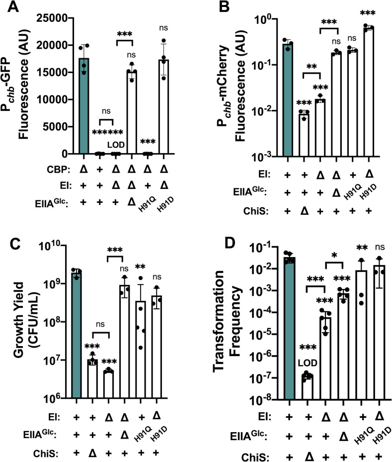
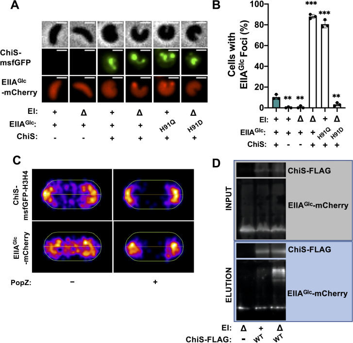
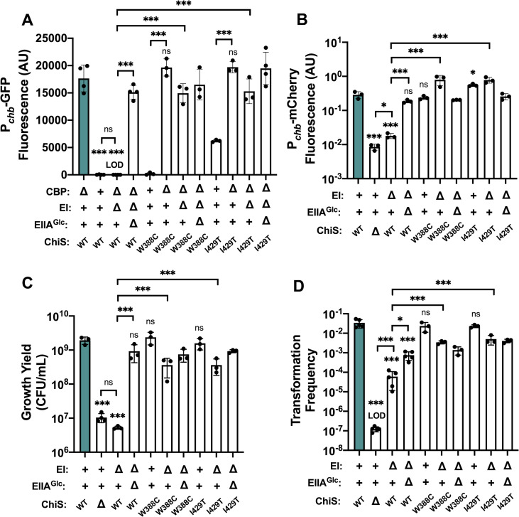
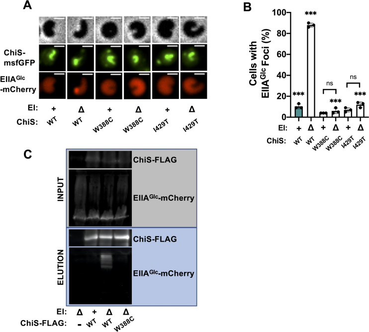
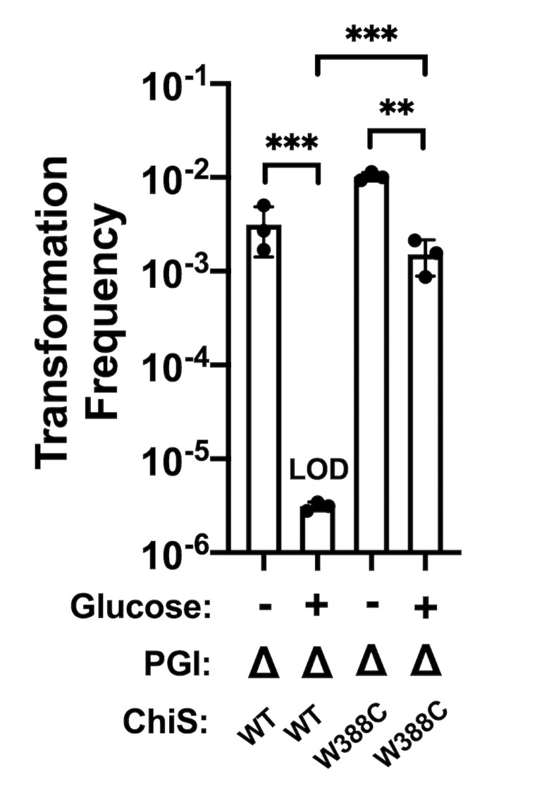
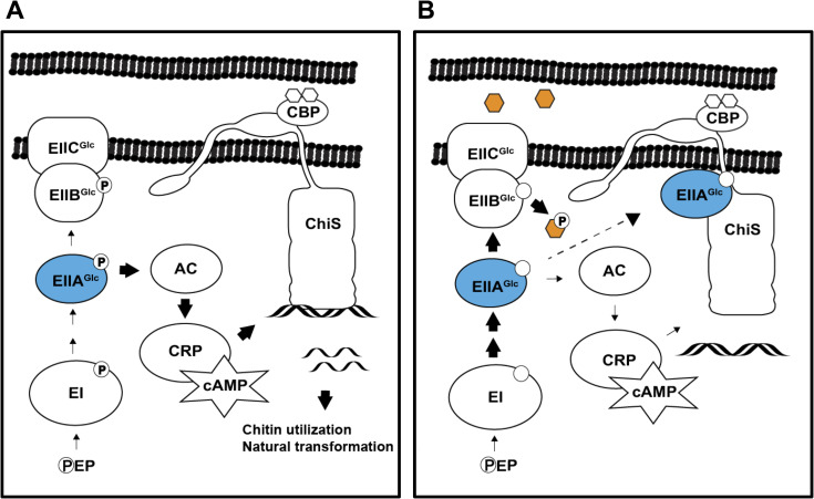
Vibrio cholerae is a facultative pathogen that primarily occupies marine environments. In this niche, V. cholerae commonly interacts with the chitinous shells of crustacean zooplankton. As a chitinolytic microbe, V. cholerae degrades insoluble chitin into soluble oligosaccharides. Chitin oligosaccharides serve as both a nutrient source and an environmental cue that induces a strong transcriptional response in V. cholerae. Namely, these oligosaccharides induce the chitin sensor, ChiS, to activate the genes required for chitin utilization and horizontal gene transfer by natural transformation. Thus, interactions with chitin impact the survival of V. cholerae in marine environments. Chitin is a complex carbon source for V. cholerae to degrade and consume, and the presence of more energetically favorable carbon sources can inhibit chitin utilization. This phenomenon, known as carbon catabolite repression (CCR), is mediated by the glucose-specific Enzyme IIA (EIIAGlc) of the phosphoenolpyruvate-dependent phosphotransferase system (PTS). In the presence of glucose, EIIAGlc becomes dephosphorylated, which inhibits ChiS transcriptional activity by an unknown mechanism. Here, we show that dephosphorylated EIIAGlc interacts with ChiS. We also isolate ChiS suppressor mutants that evade EIIAGlc-dependent repression and demonstrate that these alleles no longer interact with EIIAGlc. These findings suggest that EIIAGlc must interact with ChiS to exert its repressive effect. Importantly, the ChiS suppressor mutations we isolated also relieve repression of chitin utilization and natural transformation by EIIAGlc, suggesting that CCR of these behaviors is primarily regulated through ChiS. Together, our results reveal how nutrient conditions impact the fitness of an important human pathogen in its environmental reservoir.


Conflict of interest statement

The authors have declared that no competing interests exist.

Figures

Fig 1
Fig 1. Dephosphorylated EIIAGlc represses ChiS transcriptional activity and downstream behaviors.
(A) ChiS activation of the chb promoter (Pchb) was assessed in rich medium (in the absence of chitin) via a Pchb-GFP reporter in the indicated strain backgrounds. (B) ChiS activation of Pchb-mCherry was assessed in the indicated strains after cells were incubated on chitin for 48 hrs. mCherry signal was normalized to a constitutively expressed GFP construct. (C) Final growth yield of the indicated strains after a 72 hr incubation in M9 minimal medium containing chitin as the sole carbon source. (D) Chitin-induced natural transformation assays of the indicated strains. In all experiments, reactions were supplemented with 5 mM cAMP. Results are from at least three independent biological replicates and shown as the mean ± SD. Statistical comparisons were made by one-way ANOVA with Tukey’s multiple comparison test. NS, not significant. *** = p < 0.001, ** = p < 0.01, * = p < 0.05. LOD, limit of detection. Statistical identifiers directly above bars represent comparisons to the parent (teal, first bar).
Fig 2
Fig 2. Dephosphorylated EIIAGlc inhibits the DNA-binding activity of ChiSWT but not ChiSW388C or ChiSI429T in vivo.
ChIP-qPCR assays were performed on the indicated strains to assess ChiS binding to the chb promoter (Pchb) in vivo. Fold enrichment of Pchb relative to rpoB is shown. Results are from three independent biological replicates and shown as the mean ± SD. Statistical comparisons were made by one-way ANOVA with Tukey’s multiple comparison test. NS, not significant. *** = p < 0.001, ** = p < 0.01, * = p < 0.05. Statistical identifiers directly above bars represent comparisons to the parent (teal, first bar).
Fig 3
Fig 3. ChiS interacts with dephosphorylated EIIAGlc.
Colocalization of ChiS and EIIAGlc was assessed in cells either containing or lacking a Ptac-chiS-msfGFP construct and a functional EIIAGlc-mCherry fusion at the native locus. For strains containing a Ptac-chiS-msfGFP construct, cultures were supplemented with 20 μM IPTG. (A) Representative images of ChiS-msfGFP and EIIAGlc-mCherry localization in the indicated strain backgrounds. Phase (top) is shown to demarcate cell boundaries, GFP fluorescence (middle) shows ChiS-msfGFP localization, and mCherry fluorescence (bottom) shows EIIAGlc-mCherry localization. Scale bar, 1 μm. (B) Quantification of the percentage of cells containing EIIAGlc foci in the indicated strains (n = 300 cells analyzed per strain). All EIIAGlc foci observed colocalized with ChiS as shown in A. Data are from three independent biological replicates and shown as the mean ± SD. Statistical comparisons were made by one-way ANOVA with Tukey’s multiple comparison test. NS, not significant. *** = p < 0.001, ** = p < 0.01, * = p < 0.05. Statistical identifiers directly above bars represent comparisons to the parent (teal, first bar). (C) Representative heat maps showing the localization of EIIAGlc-mCherry and ChiS-H3H4-msfGFP foci in ΔEI cells in the presence and absence of PopZ expression. n ≥ 300 cells analyzed per condition. Data are representative of two independent experiments. (D) Representative image of pulldown assays. DSS crosslinked lysates from cells expressing EIIAGlc-mCherry and ChiS-FLAG were immunoprecipitated with anti-FLAG magnetic beads. Bound protein was eluted and protein input (grey box) and elution (blue box) samples were visualized by western blotting. Data are representative of two independent experiments.
Fig 4
Fig 4. Dephosphorylated EIIAGlc cannot inhibit the activity of ChiS suppressor alleles.
(A) ChiS activation of the chb promoter (Pchb) was assessed in rich medium (in the absence of chitin) using a Pchb-GFP reporter in the indicated strain backgrounds. (B) ChiS activation of Pchb-mCherry was assessed in the indicated strains after cells were incubated on chitin for 48 hrs. mCherry signal was normalized to a constitutively expressed GFP construct. (C) Final growth yield of the indicated strains after a 72 hr incubation in M9 minimal media containing chitin as the sole carbon source. (D) Chitin-induced natural transformation assays of the indicated strains. In all experiments, reactions were supplemented with 5 mM cAMP. Results are from at least three independent biological replicates and shown as the mean ± SD. Statistical comparisons were made by one-way ANOVA with Tukey’s multiple comparison test. NS, not significant. *** = p < 0.001, ** = p < 0.01, * = p < 0.05. LOD, limit of detection. Statistical identifiers directly above bars represent comparisons to the parent (teal, first bar). The first four bars in each panel are the same data presented in Fig 1 and are included here for ease of comparison.
Fig 5
Fig 5. ChiS suppressor alleles no longer interact with dephosphorylated EIIAGlc.
Colocalization of ChiS and EIIAGlc was assessed in cells containing a Ptac-chiS-msfGFP construct and a functional EIIAGlc-mCherry fusion at the native locus. Cultures were supplemented with 20 μM IPTG to induce expression of the indicated ChiS-msfGFP allele. (A) Representative images of cells to assess the localization of ChiS-msfGFP and EIIAGlc-mCherry in the indicated strains. Phase (top) is shown to demarcate cell boundaries, GFP fluorescence (middle) shows ChiS-msfGFP localization, and mCherry fluorescence (bottom) shows EIIAGlc-mCherry localization. Scale bar, 1 μm. (B) Quantification of the percent of cells containing EIIAGlc foci in the indicated strains (n = 300 cells analyzed per strain). All EIIAGlc foci observed colocalized with ChiS as shown in A. Data are from three independent biological replicates and shown as the mean ± SD. Statistical comparisons were made by one-way ANOVA with Tukey’s multiple comparison test. NS, not significant. *** = p < 0.001, ** = p < 0.01, * = p < 0.05. Statistical identifiers directly above bars represent comparisons to the parent (teal, first bar). Data for ChiSWT in B are the same data shown in Fig 2B and are included here for ease of comparison. (C) Representative image of pulldown assays. DSS crosslinked lysates from cells expressing EIIAGlc-mCherry and the indicated allele of ChiS-FLAG were immunoprecipitated with anti-FLAG magnetic beads. Bound protein was eluted and protein input (grey box) and elution (blue box) samples were visualized by western blotting. Data are representative of two independent experiments.
Fig 6
Fig 6. The ChiSW388C allele confers resistance to glucose-induced CCR of natural transformation.
Chitin-induced natural transformation assays of the indicated strains. Transformation reactions were supplemented with 0.5% glucose as indicated. All transformation reactions were supplemented with 5 mM cAMP. Results are from at least three independent biological replicates and shown as the mean ± SD. Statistical comparisons were made by one-way ANOVA with Tukey’s multiple comparison test. NS, not significant. *** = p < 0.001, ** = p < 0.01, * = p < 0.05. LOD, limit of detection.
Fig 7
Fig 7. Model for CCR of the V. cholerae chitin response.
(A) In the absence of glucose, phosphate (P) accumulates on PTS components due to the absence of a substrate. Thus, EIIAGlc (blue) is phosphorylated and unable to interact with ChiS. When activated by chitin-bound CBP, ChiS binds DNA and promotes the transcription of genes required for natural transformation and chitin utilization. Phospho-EIIAGlc also interacts with adenylate cyclase (AC) to strongly induce its activity, which results in the production of high levels of cAMP. This cAMP complexes with the cAMP receptor protein (CRP) to stimulate both ChiS activity and natural transformation. (B) In the presence of glucose (orange hexagon), rapid phosphotransfer through the PTS results in dephosphorylation of EIIAGlc. The absence of phosphorylated EIIAGlc reduces adenylate cyclase activity and resultant CRP-cAMP activity. However, our results demonstrate that CCR due to a reduction in cAMP production cannot account for repression of ChiS activity. Instead, we found that dephospho-EIIAGlc interacts with ChiS to inhibit its DNA-binding activity. Thus, ChiS is unable to activate its target genes, resulting in the loss of chitin utilization and natural transformation.

Similar articles

Cited by

References

    1. Silva AJ, Benitez JA. Vibrio cholerae Biofilms and Cholera Pathogenesis. PLoS Negl Trop Dis. 2016;10(2):e0004330. Epub 2016/02/06. doi: 10.1371/journal.pntd.0004330 ; PubMed Central PMCID: PMC4741415. - DOI - PMC - PubMed
    1. Faruque SM, Albert MJ, Mekalanos JJ. Epidemiology, genetics, and ecology of toxigenic Vibrio cholerae. Microbiol Mol Biol Rev. 1998;62(4):1301–14. Epub 1998/12/05. doi: 10.1128/MMBR.62.4.1301-1314.1998 ; PubMed Central PMCID: PMC98947. - DOI - PMC - PubMed
    1. Meibom KL, Li XB, Nielsen AT, Wu CY, Roseman S, Schoolnik GK. The Vibrio cholerae chitin utilization program. Proc Natl Acad Sci U S A. 2004;101(8):2524–9. Epub 2004/02/26. doi: 10.1073/pnas.0308707101 ; PubMed Central PMCID: PMC356983. - DOI - PMC - PubMed
    1. Watnick PI, Kolter R. Steps in the development of a Vibrio cholerae El Tor biofilm. Mol Microbiol. 1999;34(3):586–95. Epub 1999/11/17. doi: 10.1046/j.1365-2958.1999.01624.x ; PubMed Central PMCID: PMC2860543. - DOI - PMC - PubMed
    1. Meibom KL, Blokesch M, Dolganov NA, Wu CY, Schoolnik GK. Chitin induces natural competence in Vibrio cholerae. Science. 2005;310(5755):1824–7. Epub 2005/12/17. doi: 10.1126/science.1120096 . - DOI - PubMed

Publication types