. 2023 Jul:499:75-88.
doi: 10.1016/j.ydbio.2023.04.006. Epub 2023 May 10.

CFAP45, a heterotaxy and congenital heart disease gene, affects cilia stability

Affiliations

CFAP45, a heterotaxy and congenital heart disease gene, affects cilia stability

E Deniz et al. Dev Biol. 2023 Jul.

Abstract

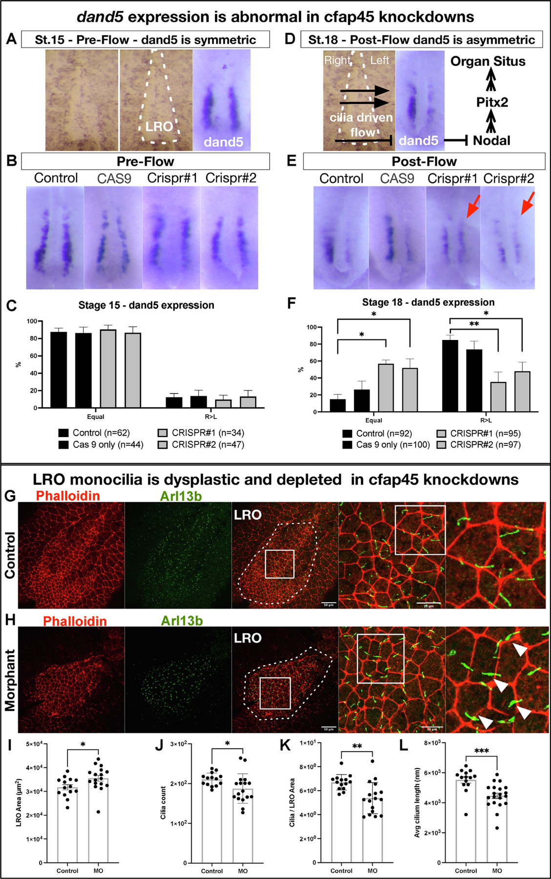
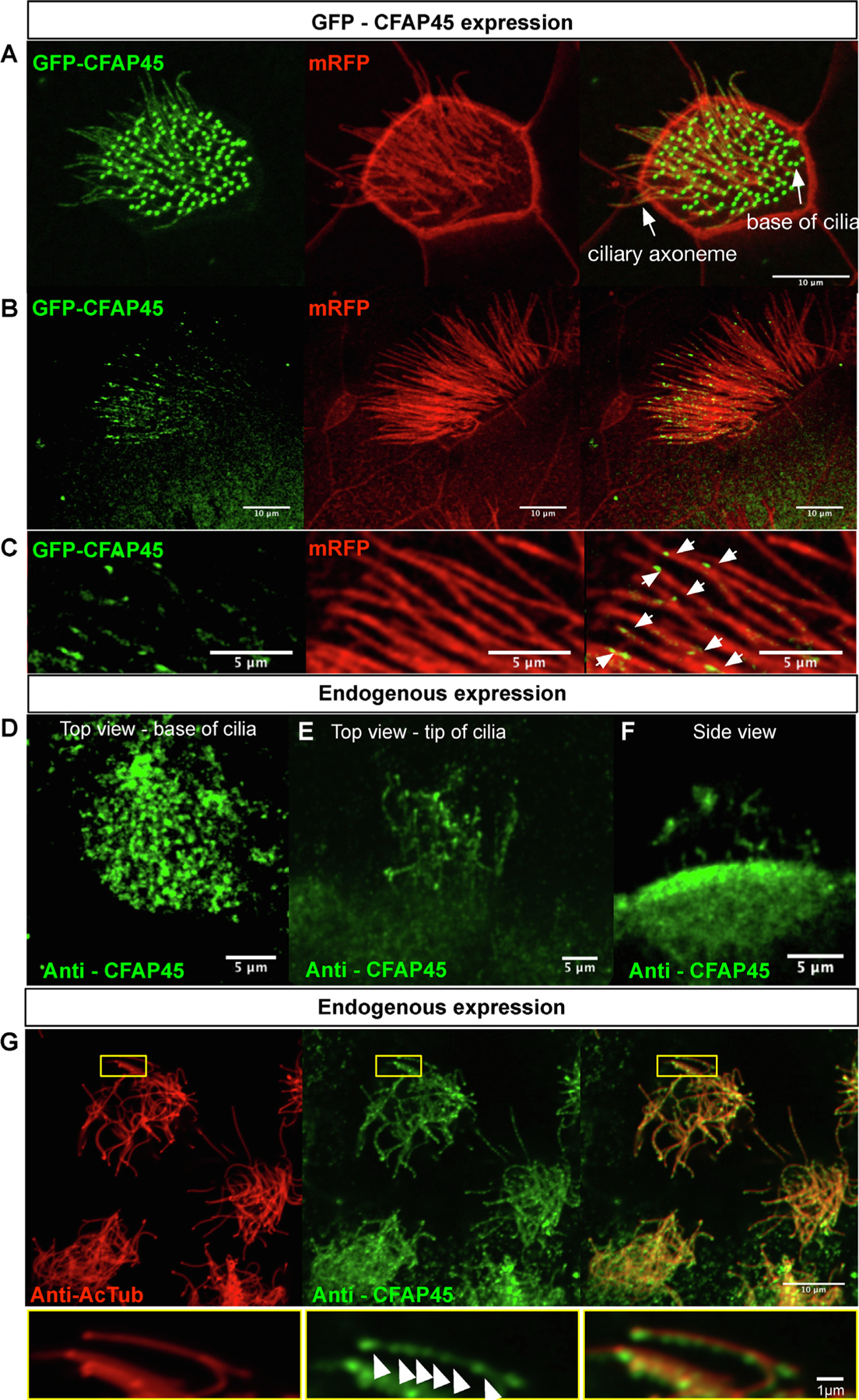
Congenital heart disease (CHD) is the most common and lethal birth defect, affecting 1.3 million individuals worldwide. During early embryogenesis, errors in Left-Right (LR) patterning called Heterotaxy (Htx) can lead to severe CHD. Many of the genetic underpinnings of Htx/CHD remain unknown. In analyzing a family with Htx/CHD using whole-exome sequencing, we identified a homozygous recessive missense mutation in CFAP45 in two affected siblings. CFAP45 belongs to the coiled-coil domain-containing protein family, and its role in development is emerging. When we depleted Cfap45 in frog embryos, we detected abnormalities in cardiac looping and global markers of LR patterning, recapitulating the patient's heterotaxy phenotype. In vertebrates, laterality is broken at the Left-Right Organizer (LRO) by motile monocilia that generate leftward fluid flow. When we analyzed the LRO in embryos depleted of Cfap45, we discovered "bulges" within the cilia of these monociliated cells. In addition, epidermal multiciliated cells lost cilia with Cfap45 depletion. Via live confocal imaging, we found that Cfap45 localizes in a punctate but static position within the ciliary axoneme, and depletion leads to loss of cilia stability and eventual detachment from the cell's apical surface. This work demonstrates that in Xenopus, Cfap45 is required to sustain cilia stability in multiciliated and monociliated cells, providing a plausible mechanism for its role in heterotaxy and congenital heart disease.

Keywords: CFAP45; Cilia; Congenital heart disease; Heterotaxy; Xenopus.


Conflict of interest statement

Declaration of competing interest SAL and MKK are co-founders of Victory Genomics, Inc.

Figures

Fig. 1.
Fig. 1.
cfap45 loss-of-function in humans and Xenopus leads to heterotaxy phenotype (A) Family pedigree showing the affected 6yo patient and the similarly affected sibling who died at 25 days of life. (B) Multiple alignments around the patient variant L522P demonstrate a remarkable degree of conservation of amino acids. (C) Chest X-ray shows the right gastric bubble. (D) CT-scan of the chest and (E) 3D reconstruction shows the right aortic arch indicating abnormal left-right patterning (F) The first row shows the tadpole heart in uninjected control (D-loop), and the second row shows the morphant with abnormal (L and A-loop) looping states at stage 46 Xenopus heart. The cardiac sac is magnified, the outflow tract border and the ventricle are outlined in white. OT: outflow tract; Ventricle. (G) The graph represents abnormal looping percentages in G0 mutants (Crispr#1, Crispr#2) and morphants. Each point represents one experimental group. (H) cfap45 knockdown leads to pitx2c abnormalities. The first column shows normal pitx2c expression in the left lateral mesoderm and is absent on the right of stage 28–31 Xenopus embryos, highlighted with a white arrow. The second and third columns show abnormal pitx2c expression as absent or bilateral, highlighted with red arrows following MO mediated cfap45 depletion. (I) The graph represents abnormal pitx2c expression percentages in G0 mutants (Crispr#1, Crispr#2) and morphants. Each point represents one experimental group. D: dextro, L: levo, A: ambiguous. *p < 0.05, **p < 0.01, ***p < 0.001, ****p < 0.0001.
Fig. 2.
Fig. 2.
cfap45 knockdown causes abnormal dand5 expression and defects in LRO cilia (A) dand5 is expressed bilaterally symmetrical in the LRO at stage 15 (pre-flow). (B) Pre-flow, normal bilateral expression of dand5 was observed in G0 mutants compared to uninjected or only CAS9 injected controls. (C) Percentages of normal (equal) and abnormal (R > L) dand5 expression in stage 15 (pre-flow) LROs. G0 mutants show no difference compared to uninjected and Cas9 injected controls. n = number of embryos. (D) At stage 18, right to left cilia-driven fluid flow has emerged, suppressing dand5 expression on the left side of the LRO. (E) Post-flow, normal reduced left side expression in uninjected and Cas9 injected controls show up as abnormal bilateral expression in G0 mutants indicated with red arrows. (F) Percentages of normal (R > L) and abnormal (equal) dand5 expression in stage 18 (post-flow) LROs. A significant number of G0 mutants show abnormal bilateral dand5 expression compared to uninjected and Cas9 injected controls—n = number of embryos. (G–H) LROs stained with phalloidin to label cell borders and anti - Arl13b antibody to label monocilia. The white dashed line indicates the LRO area. LRO monocilia were malformed and lost in morphants, also showing bulging at the tip and midshaft of the cilia, indicated by white arrows. White squares indicate the magnified areas (H). LRO area is slightly enlarged (I), and cilia numbers (J), cilia per LRO area (K), and cilia length are reduced in morphants. MO: Morpholino oligo, LRO: Left-right organizer.*p < 0.05, **p < 0.01.
Fig. 3.
Fig. 3.
cfap45 knockdown causes abnormalities in cilia-driven epidermal flow and leads to defects in epidermal multicilia. (A) Epidermal multicilia beating is planarly polarized, generating head-to-tail fluid flow along the surface. This flow can be visualized using Optical Coherence Tomography (OCT) imaging, where intravitelline endogenous particle movements can be tracked in vivo (Movie1). (B–E) We obtained real-time OCT movies and, using 25 frames, built particle trajectories with ImageJ. Temporal color coding depicts particle trajectory over time. The color bar represents color versus the corresponding frame number in the color-coded image. Based on the trajectory map over 25 frames, we classified the flow as normal (B), slow (C), or absent (D). When cfap45 is depleted, the epidermal flow is significantly slower or stationary. (E) Based on OCT imaging, percentages of embryos with normal, slow, and no-flow in uninjected controls and cfap45 depleted tadpoles—n = number of embryos. (F–G) Cilia are lost in multiciliated cells (MCCs) when Cfap45 is depleted with MO. Epidermal MCCs marked with anti-acetylated-tubulin (green) for cilia and phalloidin (white) for actin.
Fig. 4.
Fig. 4.
Epidermal MCCs demonstrate axonemal disorganization and instability when cfap45 is knocked down (A–C) Wide-field images of the Epidermal MCCs marked with membrane RFP in controls (A), G0 mutants (B), and morphants (C). When cfap45 is depleted, most MCCs display curled and shortened cilia (Movie 2,3). (D-F) Focused and magnified MCC images in control (D), G0 mutants (E), and morphants (F) show curling of the cilia leading to shortening and suggesting that the deformation leads to detached cilia. (G) Montaged images from live G0 mutant epidermal cilia imaging show the detached, free-floating cilium. The bottom row is colored black and white to track better the detached cilium encircled in yellow. (H) Electron Microscope image of the transverse section of the epidermal MCC shows 9 + 2 microtubule organization. A pair of microtubules is surrounded by nine pairs connected with radial spokes. This organization is abnormal when cfap45 is depleted using CRISPR.
Fig. 5.
Fig. 5.
Wild-type human cfap45 mRNA rescues loss of cilia phenotype, whereas the patient variant fails (A) Experimental setup. First, morpholino is injected at the one-cell stage, followed by human RNA (WT or patient variant) with membrane RFP tracer injection at the two-cell stage. Then, we raised tadpoles to stage 32 to assess epidermal MCCs. (B, D, F) Depletion of Cfap45 by MO results in MCC loss. (C) Wild-type RNA injection at the two-cell stage traced with membrane RFP shows the recovery of the MCCs on the injected side. (E) RNA with patient variation injections traced with membrane RFP fails to recover loss of cilia in MCCs. (G) Wild-type GFP-hCFAP45 RNA injection at the two-cell stage shows the recovery of the MCCs on the injected side. Phalloidin was used to label actin to show cell borders, and an anti-acetylated α-tubulin antibody was used to label cilia.
Fig. 6.
Fig. 6.
Cfap45 localization in X.tropicalis MCCs (A–C) X. tropicalis epidermis expressing GFP-CFAP45 and membrane RFP. (A) Focus is concentrated on the base of the cilia where GFP-CFAP45 is expressed, (B) Focus is concentrated on the cilia showing the dotty localization along the axonemes. (C) Magnified views. White arrows highlight GFP-CFAP45 localization. (D–F) X. tropicalis epidermal MCCs labeled with anti-CFAP45. (D) Top view focusing on the base of the cilia at the apical surface. (E) Top view focusing on the cilia axonemes. (F) Side view showing apical surface and cilia staining. (G) X. tropicalis epidermal MCCs labeled with anti-acetylated α-tubulin (red, cilia) and anti-CFAP45 (green). Anti-CFAP45 staining pattern is identical to the GFP-CFAP45 expression, showing CFAP45 localization to the cilia in a dotty fashion in the magnified views along the bottom (white arrows).
Fig. 7.
Fig. 7.
CFAP45 localized to the base of the cilia and the ciliary axoneme and, unlike IFT80 protein, is stationary within the cilia. (A) Cherry - CFAP45 expressed in the epidermal MCCs. Actin in MCCs stained with phalloidin (grey) delineates the cell borders and the apical actin network where the base of the cilia is located at the center. The magnified view delineates the circular actin network (blue). CFAP45 is enriched at the base of the cilia and situated in the center of the apical network. (B) GFP- CFAP45 expressed in MCCs and colocalized with Centrin at the base of the cilia. (C) IFT-80-GFP expressed in MCC. (D) z-projection of fifty frames. (E) CFAP45-Cherry expressed in MCC. (F) z-projection of fifty frames. (G) Kymograph presents the movement of the IFT80-GFP proteins along the single cilium. Multiple lines represent multiple IFTs. As the protein travels, intensity displaces along the cilium forming tracks over time. One particle tracked over time was marked with black arrows. (H) Kymograph presents stationary CFAP45-cherry protein. As no movement is observed, multiple straight lines form over time, representing the non-uniform punctuated pattern of cfap45. (Yellow rectangle encloses the single cilium analyzed.)

Similar articles

Cited by

References

    1. Antony D, et al., 2013. Mutations in CCDC39 and CCDC40 are the major cause of primary ciliary dyskinesia with axonemal disorganization and absent inner dynein arms. Hum. Mutat 34 (3), 462–472. - PMC - PubMed
    1. Basu B, Brueckner M, 2008. Cilia multifunctional organelles at the center of vertebrate left-right asymmetry. Curr. Top. Dev. Biol 85, 151–174. - PubMed
    1. Becker-Heck A, et al., 2011. The coiled-coil domain containing protein CCDC40 is essential for motile cilia function and left-right axis formation. Nat. Genet 43 (1), 79–84. - PMC - PubMed
    1. Bhattacharya D, et al., 2015. CRISPR/Cas9: an inexpensive, efficient loss of function tool to screen human disease genes in Xenopus. Dev. Biol 408 (2), 196–204. - PMC - PubMed
    1. Blum M, Vick P, 2015. Left-right asymmetry: cilia and calcium revisited. Curr. Biol 25 (5), R205–R207. - PubMed

Publication types

Substances