. 2023 Jun;299(6):104808.
doi: 10.1016/j.jbc.2023.104808. Epub 2023 May 11.

Structural basis of ELKS/Rab6B interaction and its role in vesicle capturing enhanced by liquid-liquid phase separation

Affiliations

Structural basis of ELKS/Rab6B interaction and its role in vesicle capturing enhanced by liquid-liquid phase separation

Gaowei Jin et al. J Biol Chem. 2023 Jun.

Abstract

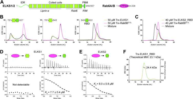
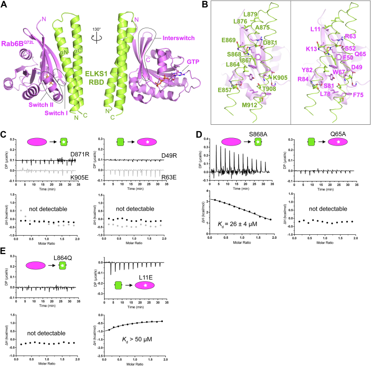
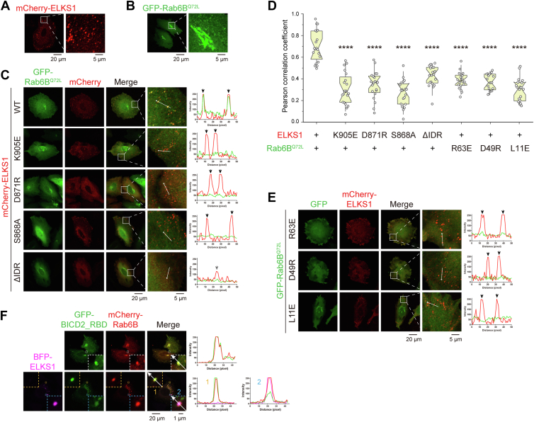
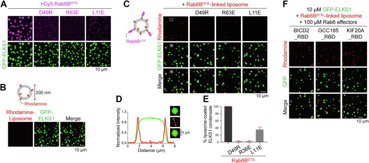
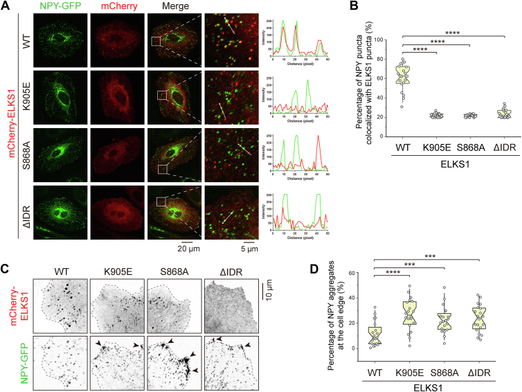
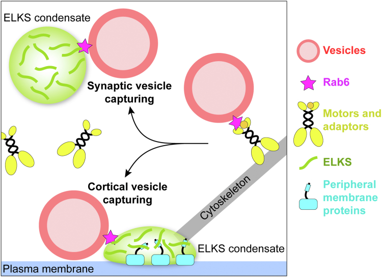
ELKS proteins play a key role in organizing intracellular vesicle trafficking and targeting in both neurons and non-neuronal cells. While it is known that ELKS interacts with the vesicular traffic regulator, the Rab6 GTPase, the molecular basis governing ELKS-mediated trafficking of Rab6-coated vesicles, has remained unclear. In this study, we solved the Rab6B structure in complex with the Rab6-binding domain of ELKS1, revealing that a C-terminal segment of ELKS1 forms a helical hairpin to recognize Rab6B through a unique binding mode. We further showed that liquid-liquid phase separation (LLPS) of ELKS1 allows it to compete with other Rab6 effectors for binding to Rab6B and accumulate Rab6B-coated liposomes to the protein condensate formed by ELKS1. We also found that the ELKS1 condensate recruits Rab6B-coated vesicles to vesicle-releasing sites and promotes vesicle exocytosis. Together, our structural, biochemical, and cellular analyses suggest that ELKS1, via the LLPS-enhanced interaction with Rab6, captures Rab6-coated vesicles from the cargo transport machine for efficient vesicle release at exocytotic sites. These findings shed new light on the understanding of spatiotemporal regulation of vesicle trafficking through the interplay between membranous structures and membraneless condensates.

Keywords: BICD; CAST; Rab-binding domain; Rab6A; active zone; biological condensate; cargo unloading; intracellular transport; neuropeptide secretion; protein-protein interaction.


Conflict of interest statement

Conflict of interest The authors declare no competing financial interests.

Figures

Figure 1
Figure 1
Biochemical characterization of the interaction between ELKS and Rab6B.A, schematic diagrams showing the domain organizations of ELKS and Rab6. The protein interacting regions in ELKS were indicated. The color coding for proteins was used throughout the entire manuscript except as otherwise indicated. B, analytical gel filtration chromatography shows that ELKS1_RBD contains the necessary region for binding to Rab6B. C, analytical gel filtration chromatography showing the interaction between ELKS2 and the active form of Rab6B. D, ITC-based analyses confirming that Rab6BQ72L binds to ELKS1_RBD but not the coiled-coil region at the N-terminal to the RBD. E, ITC-based analysis confirming the ELKS2_RBD/Rab6BQ72L interaction. F, multiangle light static scattering analysis indicating that the ELKS1_RBD forms a monomer in solution. ITC, Isothermal titration calorimetry.
Figure 2
Figure 2
Structural analysis of the interaction between ELKS1 and Rab6BQ72L.A, the overall structure of the ELKS1_RBD/Rab6BQ72L complex. The switch I/II and interswitch regions of Rab6B were indicated. B, molecular details of the ELKS1_RBD/Rab6BQ72L interaction with interface residues highlighted in sticks. Hydrogen bonds and salt bridges were indicated by dashed lines. C–E, ITC-based analyses of the disruptive interactions between ELKS1_RBD and Rab6BQ72L mutants, including the mutations of interface residues that interfere with charge–charge interactions (C), hydrogen bonding (D), and hydrophobic interactions (E).
Figure 3
Figure 3
Sequence and structural comparison of the Rab-binding modes of ELKS and other Rab effectors.A, amino acid sequence alignments of effector-binding regions in Rab proteins. The sequences of Rab6 proteins from mammals, Drosophila, C. elegans, and slime mold and those of other representative Rab family members from humans were aligned separately and then merged for comparison. Interface residues in Rabs that form contacts with their corresponding effectors were labeled with triangles. B, amino acid sequence alignment of the RBDs of ELKS proteins. Unique residues for Rab6 binding were indicated as purple circles. Residues involved in binding to the switch I, II, and interswitch regions of Rab6B are indicated. C, structural comparison of Rab6B/ELKS1_RBD to other Rab6/effector complexes, including the Rab6A/Rab6IP1 complex (PDB ID: 3CWZ), the Rab6A/KIF20A_RBD complex (5LEF), and the Rab6A/GCC185_RBD complex (3BBP). D, comparison of effector-binding surfaces on the Rab6 structures. E, superposition of the Rab6B/ELKS1_RBD structure with two Rab/Rabenosyn-5 structures (1Z0Q and 1Z0J). Compared to the two RBDs of Rabenosyn-5, ELKS1_RBD adopts a longer helical hairpin. The structural difference between them was highlighted by a dashed ellipse. F, comparison of Rabenosyn-5-binding surfaces on the Rab4A and Rab22 structures.
Figure 4
Figure 4
ELKS1 accumulates Rab6B by forming cellular condensates.A and B, cell imaging of exogenous expressed mCherry-tagged ELKS1 (A) and GFP-tagged Rab6BQ72L (B). The regions where ELKS1 or Rab6B can form puncta were selected as the ROIs, and the ROIs were boxed and enlarged. Scale bar was indicated at the bottom. C, cell imaging of exogenous co-expressed mCherry-tagged ELKS1 or its variants with GFP-tagged Rab6BQ72L. The regions where ELKS1 can form puncta were selected as the ROIs, and the ROIs were boxed and enlarged. Line analyses of fluorescent intensities were shown on the right side. Intensity peaks of ELKS puncta were indicated by arrows. D, Pearson correlation coefficients of GFP and mCherry signals were measured using the cell imaging data in (C) and (E). The whole cell fluorescence was used for analysis. Data were collected from 20 cells in each condition. Bars represent the means ± S.D. The unpaired Student’s t test analysis was used to define a statistically significant difference (∗∗∗∗p < 0.0001). E, cell imaging of exogenous co-expressed GFP-tagged Rab6BQ72L or its variants with mCherry-tagged ELKS1. The regions where ELKS1 was able to form puncta were selected as the ROIs, and the ROIs were boxed and enlarged. Line analyses of fluorescent intensities were shown on the right side. Intensity peaks of ELKS punta were indicated by arrows. F, cell imaging analysis of the potential competition between ELKS1 and BICD2 for accumulating Rab6 by the co-expression of GFP-tagged BICD2_RBD and mCherry-tagged Rab6BQ72L with or without BFP-tagged ELKS1. Two ROIs were boxed and enlarged in the condition with the expression of BFP-tagged ELKS1 to display different levels of BICD2_RBD in Rab6 enriched puncta formed with or without ELKS1. ROI, regions of interest.
Figure 5
Figure 5
Condensed ELKS1 recruits Rab6B-coated liposomes.A, fluorescence imaging of phase-separated GFP-ELKS1 in accumulating Rab6BQ72L. Rab6BQ72L was highly enriched in the droplets formed by ELKS1 but not its Rab6-binding deficient mutations (D49R, R63E, and L11E). Rab6BQ72L and ELKS1 were mixed at a 1:1 ratio at 10 μM concentrations. Rab6B was labeled with Cy5 at 5% level. B, fluorescence imaging of phase-separated GFP-ELKS1 in mixing with Rhodamine-labeled liposomes. C, fluorescence imaging of phase-separated GFP-ELKS1 in mixing with Rab6B-linked liposomes. Rab6BQ72L and its ELKS-binding deficient mutants were chemically linked to maleimide lipid by C-terminal cysteine and quenched by DTT. Rab6BQ72L-linked liposomes accumulated on the surface of ELKS1 droplets, which was disrupted by the ELKS-binding deficient mutations. A region of interest was boxed and enlarged as shown in (D). D, the zoom-in view of the boxed region in (C) shows that the Rab6BQ72L-liposome accumulates on the ELKS1 droplet surface. Line analysis of fluorescent intensities was shown below. E, quantification of the percentage of ELKS1 condensates coated with Rab6BQ72L-liposome. Eight views were selected for each condition and the number of liposome-coated droplets over the number of total droplets was calculated for each view. F, fluorescence imaging of the mixture of phase-separated GFP-ELKS1 and Rab6B-linked liposomes in the presence of Trx-tagged proteins of other Rab6 effectors, including BICD2_RBD, GCC185_RBD, and KIF20A_RBD.
Figure 6
Figure 6
ELKS1 promotes NPY secretion by recruiting Rab6.A, cell imaging of GFP-tagged NPY co-expressed with mCherry-tagged ELKS1 or its variants. The regions where the NPY-vesicles were relatively clear as the ROIs, and the ROIs were boxed, enlarged, and aligned at the right of each merged image. Line analysis of fluorescent intensities is shown on the right panel. B, quantifications of colocalization of the NPY and ELKS1 puncta in (A). Data were collected from 20 cells in each condition. Bars represent the means ± S.D. The unpaired Student’s t test analysis was used to define a statistically significant difference (∗∗∗∗p < 0.0001). C, representative cell images of peripherical NPY vesicles in the different conditions with the overexpressed mCherry-tagged ELKS or its variants. D, quantifications of peripherical NPY aggregates in (C). Data were collected from 20 cells in each condition. Bars represent the means ± SD. The unpaired Student’s t test analysis was used to define a statistically significant difference (∗∗∗p < 0.001; ∗∗∗∗p < 0.0001). ROI, regions of interest.
Figure 7
Figure 7
A proposed model of the phase-separated ELKS in capturing and unloading Rab6-coated vesicles from vesicle-transporting motors in the presynapse for vesicle deposition and in the cell cortex for vesicle secretion. The ELKS condensates can be attached to the plasma membrane aided by peripheral membrane proteins, like LL5β. After unloading cargos, motors may be recycled for the next run of transport.

References

    1. Cai H., Reinisch K., Ferro-Novick S. Coats, tethers, Rabs, and SNAREs work together to mediate the intracellular destination of a transport vesicle. Dev. Cell. 2007;12:671–682. - PubMed
    1. Munro S. The golgin coiled-coil proteins of the Golgi apparatus. Cold Spring Harb. Perspect. Biol. 2011;3 - PMC - PubMed
    1. Vukoja A., Rey U., Petzoldt A.G., Ott C., Vollweiter D., Quentin C., et al. Presynaptic biogenesis requires axonal transport of lysosome-related vesicles. Neuron. 2018;99:1216–1232.e7. - PubMed
    1. Wickner W., Schekman R. Membrane fusion. Nat. Struct. Mol. Biol. 2008;15:658–664. - PMC - PubMed
    1. Jahn R., Fasshauer D. Molecular machines governing exocytosis of synaptic vesicles. Nature. 2012;490:201–207. - PMC - PubMed

Publication types

MeSH terms

LinkOut - more resources