. 2023 May 12;13(1):7766.
doi: 10.1038/s41598-023-34676-w.

Ceramide present in cholangiocarcinoma-derived extracellular vesicle induces a pro-inflammatory state in monocytes

Affiliations

Ceramide present in cholangiocarcinoma-derived extracellular vesicle induces a pro-inflammatory state in monocytes

Barbara Oliviero et al. Sci Rep. .

Abstract

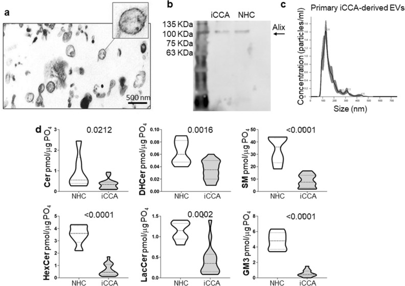
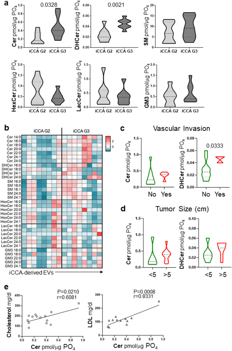
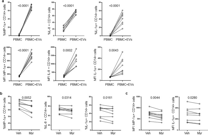
Cholangiocarcinoma (CCA) is a rare cancer characterized by a global increasing incidence. Extracellular vesicles (EV) contribute to many of the hallmarks of cancer through transfer of their cargo molecules. The sphingolipid (SPL) profile of intrahepatic CCA (iCCA)-derived EVs was characterized by liquid chromatography-tandem mass spectrometry analysis. The effect of iCCA-derived EVs as mediators of inflammation was assessed on monocytes by flow cytometry. iCCA-derived EVs showed downregulation of all SPL species. Of note, poorly-differentiated iCCA-derived EVs showed a higher ceramide and dihydroceramide content compared with moderately-differentiated iCCA-derived EVs. Of note, higher dihydroceramide content was associated with vascular invasion. Cancer-derived EVs induced the release of pro-inflammatory cytokines in monocytes. Inhibition of synthesis of ceramide with Myriocin, a specific inhibitor of the serine palmitoyl transferase, reduced the pro-inflammatory activity of iCCA-derived EVs, demonstrating a role for ceramide as mediator of inflammation in iCCA. In conclusion, iCCA-derived EVs may promote iCCA progression by exporting the excess of pro-apoptotic and pro-inflammatory ceramides.


Conflict of interest statement

The authors declare no competing interests.

Figures

Figure 1
Figure 1
SPL concentration was downregulated in primary iCCA cell-derived EVs. (a) Representative transmission electron micrograph of primary iCCA-derived EVs. Scale bar = 500 nm. (b) Representative image showing a Western blot of Alix (lane 1 = standard ladders; lane 2 = primary iCCA-derived EVs; lane 3 = NHC-derived EVs). The image has been cropped and the original, uncropped blots at multiple exposures (3, 10 and 15 s) are presented in Supplementary Fig. 6. (c) Size analysis of primary iCCA-derived EVs was determined by Nanoparticle Tracking Analysis. (d) Analysis of the different SPL classes in supernatant-derived EVs from iCCA and NHC cells by liquid chromatography-tandem mass spectrometry (iCCA = 16; NHC = 6). Median values and interquartile ranges are shown in the violin plots. The unpaired t test was used to compare data, except for Cer for which the Mann–Whitney U test was used.
Figure 2
Figure 2
Cer and DHCer expression in primary iCCA-derived EVs changed according to tumor grade. (a) Analysis of the SPLs in primary iCCA cell-derived EVs isolated from patients with iCCA stratified according to tumor grade. G2, moderately-differentiated tumors (n = 8); G3, poorly-differentiated tumors (n = 8). Median values and interquartile ranges are shown in the violin plots. The unpaired t test was used to compare data. (b) Heatmap of the main SPL species in G2- compared with G3-derived EVs. Each column represents EVs derived from primary iCCA cells obtained from each patient. The entity of the fold change has been visualized by color gradient from the smallest (cerulean) to the highest (red) values, passing through the baseline (white). Data has been log transformed and auto-scaled obtaining z-score prior to visualization. (c and d) Analysis of Cer and DHCer in EVs from patients stratified according to the presence of vascular invasion or tumor size, respectively. Vascular invasion was histologically defined by tumor cells infiltrating vessel walls with associated thrombi, or intravascular cancer cells mixed with thrombi. Median values and interquartile ranges are shown in the violin plots. The unpaired t test was used to compare data. (e) Significant correlations between cholesterol or LDL plasma levels and EV-Cer content.
Figure 3
Figure 3
iCCA-derived EVs induce pro-inflammatory cytokine expression in monocytes. (a) PBMC from healthy controls (n = 8) were treated with HuCCT-1-derived EVs for 16 h before flow cytometric analysis. Percentage (upper panels) and expression (lower panels, mean fluorescence intensity, MFI) of MIP-1α-, IL-8- and IL-1α-CD14 + cells are reported. The paired t test was used to compare data. (b) Percentage of MIP-1α-, IL-8- and IL-1α-CD14 + cells after treatment for 16 h of PBMC with EVs exhibiting a different Cer content. EVs were isolated from HuCCT-1 cells after treatment with myriocin (Myr) at 10 μM and DMSO (Veh) for 16 h before collecting supernatants. The paired t test was used to compare data. (c) Expression (MFI) of MIP-1α and IL-1α in CD14 + cells after treatment of PBMC with EVs. The paired t test was used to compare data.

References

    1. Banales JM, et al. Cholangiocarcinoma 2020: the next horizon in mechanisms and management. Nat. Rev. Gastroenterol. Hepatol. 2020;17:557–588. doi: 10.1038/s41575-020-0310-z. - DOI - PMC - PubMed
    1. Izquierdo-Sanchez L, et al. Cholangiocarcinoma landscape in Europe: Diagnostic, prognostic and therapeutic insights from the ENSCCA Registry. J Hepatol. 2022;76:1109–1121. doi: 10.1016/j.jhep.2021.12.010. - DOI - PubMed
    1. Théry C, et al. Minimal information for studies of extracellular vesicles 2018 (MISEV2018): a position statement of the International Society for Extracellular Vesicles and update of the MISEV2014 guidelines. J. Extracell. Vesicles. 2018;7:1535750. doi: 10.1080/20013078.2018.1535750. - DOI - PMC - PubMed
    1. Möller A, Lobb RJ. The evolving translational potential of small extracellular vesicles in cancer. Nat. Rev. Cancer. 2020;2:697–709. doi: 10.1038/s41568-020-00299-w. - DOI - PubMed
    1. Wang S, Sun J, Dastgheyb RM, Li Z. Tumor-derived extracellular vesicles modulate innate immune responses to affect tumor progression. Front. Immunol. 2022;13:1045624. doi: 10.3389/fimmu.2022.1045624. - DOI - PMC - PubMed

Publication types