. 2023 Aug;67(16):e2200601.
doi: 10.1002/mnfr.202200601. Epub 2023 Jun 30.

Bos d 13, A Novel Heat-Stable Beef Allergen

Affiliations

Bos d 13, A Novel Heat-Stable Beef Allergen

Patricia Román-Carrasco et al. Mol Nutr Food Res. 2023 Aug.

Abstract

Scope: Red meat, a staple food of Western diets, can also induce IgE-mediated allergic reactions. Yet, apart from the heat-labile protein serum albumin and the carbohydrate α-Gal, the molecules causing allergic reactions to red meat remain unknown.

Methods and results: IgE reactivity profiles of beef-sensitized individuals are analyzed by IgE-immunoblotting with protein extracts from raw and cooked beef. Two IgE-reactive proteins are identified by peptide mass fingerprinting as myosinlight chain 1 (MYL1) and myosin light chain 3 (MYL3) in cooked beef extract and are designated Bos d 13 isoallergens. MYL1 and MYL3 are produced recombinantly in Escherichia coli. ELISAs proved their IgE reactivity and circular dichroism analysis showed that they represent folded molecules with remarkable thermal stability. In vitro gastrointestinal digestion experiments showed the higher stability of rMYL1 as compared to rMYL3. Exposure of a monolayer of Caco-2 cells to rMYL1 indicated that the molecule is able to cross intestinal epithelial cells without disturbing the integrity of the tight junctions, suggesting the sensitizing capacity of MYL1.

Conclusion: MYLs are identified as novel heat-stable bovine meat allergens.

Keywords: allergens; heat stability; meat allergy; myosin light chain; recombinant allergens; red meat; stability to digestion.


Conflict of interest statement

The authors declare no conflicts of interest.

Figures
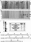
Figure 1
Figure 1
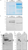
IgE reactivity patterns of patients to bovine meat extract. Extracts from raw (A) and cooked (B) beef separated by SDS‐PAGE blotted onto nitrocellulose membranes and exposed to sera from 31 patients sensitized to bovine meat. Bound IgE antibodies were detected with anti‐human IgE antibodies. Serum of a non‐meat allergic individual (NA) and PBST only (NC) were used as negative controls. Molecular weights (kDa) are indicated in the left margin. IgE reactive bands labeled as 1, 2, and 3 in raw beef and A, B, C, and D in cooked beef. C) Labeled IgE reactive bands were excised from a Coomassie stained gel (left) and the proteins identified by mass spectrometry (right). The number of confident peptides identified of each protein and the percentage sequence coverage are shown. Confident peptides are those peptides that have a 95% probability that they have been assigned correctly to the respective protein. D) Amino acid sequence alignment of bovine myosin light chain 1 (MYL1; UniProt accession number A0JNJ5) and 3 (MYL3; UniProt accession number P85100).
Figure 2
Figure 2
A) IgE binding capacity of rMYL1 and rMYL3. IgE binding to rMYL1 and rMYL3 was determined by ELISA. ELISA plate‐bound rMYL1 and rMYL3 were incubated with sera of beef sensitized patients (23–31). Mean OD values corresponding to IgE binding were measured by ELISA. The dashed line represents the mean value of the negative controls plus threefold the standard deviation. B and C) rMYL1 and rMYL3 represent folded proteins with mainly alpha‐helical secondary structure. Far‐UV circular dichroism analysis was performed with rMYL1 and rMYL3 in a wavelength range from 190 to 280 nm. The spectra are expressed as molar circular dichroism θ (deg cm2 dmol−1) at a given wavelength.
Figure 3
Figure 3
Effect of simulated gastric and duodenal digestion on the stability of rMYL1 and rMYL3. A) Coomassie stained SDS‐PAGE of the aliquots taken during in vitro digestion of rMYL1 (upper panel) and rMYL3 (lower panel) before being exposed to proteolytic enzymes (0) and after being exposed to pepsin for 1, 2, 4, 6, 8, 10, 15, 20, and 60 min (G1, G2, G4, G6, G8, G10, G15, G20, G60) and subsequently to trypsin and chymotrypsin for 5, 15, 30, and 60 min (D5, D15, D30, D60). B) Pepsin (gastric phase) cleavage sites identified by the ExPASy – PeptideCutter in the amino acid sequences of MYL1 (light blue triangles) and MYL3 (dark blue triangles). C) Aliquots 0, G1, G4, G8, G15, G60, D5, D15, and D30 of in vitro digestions of rMYL1 and rMYL3 were blotted onto nitrocellulose and either incubated with the rabbit anti‐rMYL1 antiserum (left) or with a pool of patients´ sera (23–27) (right). Molecular weights (kDa) are always indicated in the left margin.
Figure 4
Figure 4
The impact of food matrix on the stability of MYL1 and/or MYL3. A) Coomassie stained Tris‐tricine SDS‐PAGE gel of the aliquots taken during the oral phase of the in vitro digestion of cooked beef (O), and after 5, 10, 15, 30, and 60 min of simulated gastric digestion (G5, G10, G15, G30, G60) and 2, 5, 10, 15, and 60 min of simulated in vitro intestinal digestion (D2, D5, D10, D15, D60). B) Aliquots O, G5, G10, G15, G30, G60, D2, D5, D10, D15, and D60 of the in vitro digestions were blotted onto a nitrocellulose membrane and exposed to the anti‐rMYL1 antiserum. C) Aliquots O, G5, G15, G60, D2, D5, and D20 were blotted onto nitrocellulose and incubated with a pool of patients´ sera (patients 23, 24, 26, and 27). D) Aliquots 0, G15, and D5 were blotted onto a nitrocellulose membrane and then exposed to a pool of sera from patients 23, 24, 26, and 27 which was either pre‐incubated with PBS (sera) or with rMYL1 (sera+rMYL1). Molecular weights (kDa) are always indicated in the left margin. The molecular weight of MYL1 is indicated with arrows.
Figure 5
Figure 5
rMYL1 is transported intact through a monolayer of Caco‐2 cells. Anti‐rMYL1 immunoblot of the medium applied to the apical (AP) side and collected from the basolateral side (BL) of a Caco‐2 cell monolayer cultured on permeable supports. Cells were incubated with undigested rMYL1, with rMYL1 digested by pepsin for 60 min (G60) or with medium (CT) only. Molecular weights (kDa) are indicated in the left margin.
Figure 6
Figure 6
Bos d 13 cross‐reacts with pig myosin light chain. A) Anti‐rMYL1 immunoblot of raw (RP) and boiled (BP) pork. B) IgE reactivity of a pool of sera from sensitized patients incubated with blotted proteins of raw pork (RP) or boiled pork (BP). C) Immunoblot of boiled pork proteins incubated with a serum pool from beef sensitized patients (23, 25, 26, and 29) which was either pre‐incubated with buffer (sera) or with rMYL1 (sera+rMYL1). Molecular weights (kDa) are always indicated in the left margin. The molecular weight of MYL1 is indicated with an arrow.

Similar articles

Cited by

  • Treatment of Allergies to Fur Animals.
    Rosada T, Bartuzi Z, Grześk-Kaczyńska M, Rydzyńska M, Ukleja-Sokołowska N. Rosada T, et al. Int J Mol Sci. 2024 Jun 29;25(13):7218. doi: 10.3390/ijms25137218. Int J Mol Sci. 2024. PMID: 39000328 Free PMC article. Review.
  • Genomics of Shrimp Allergens and Beyond.
    Li S, Chu KH, Wai CYY. Li S, et al. Genes (Basel). 2023 Nov 27;14(12):2145. doi: 10.3390/genes14122145. Genes (Basel). 2023. PMID: 38136967 Free PMC article. Review.

References

    1. OECD/FAO , OECD‐FAO Agricultural Outlook 2018–2027, OECD Publishing, Paris/FAO, Rome: 2018.
    1. McNeill S. H., Meat Sci. 2014, 98, 452. - PubMed
    1. Uzhova I., Peñalvo J. L., Eur. J. Clin. Nutr. 2018, 72, 4. - PubMed
    1. Wilson J. M., Platts‐Mills T. A. E., Mol. Immunol. 2018, 100, 107. - PMC - PubMed
    1. Cabezas‐Cruz A., Hodžić A., Román‐Carrasco P., Mateos‐Hernández L., Duscher G. G., Sinha D. K., Hemmer W., Swoboda I., Estrada‐Peña A., De La Fuente J., Front. Immunol. 2019, 10, 1210. - PMC - PubMed

Publication types