. 2023 Apr 22;15(9):2415.
doi: 10.3390/cancers15092415.

PRAME Is a Novel Target of Tumor-Intrinsic Gas6/Axl Activation and Promotes Cancer Cell Invasion in Hepatocellular Carcinoma

Affiliations

PRAME Is a Novel Target of Tumor-Intrinsic Gas6/Axl Activation and Promotes Cancer Cell Invasion in Hepatocellular Carcinoma

Viola Hedrich et al. Cancers (Basel). .

Abstract

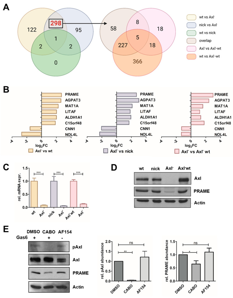
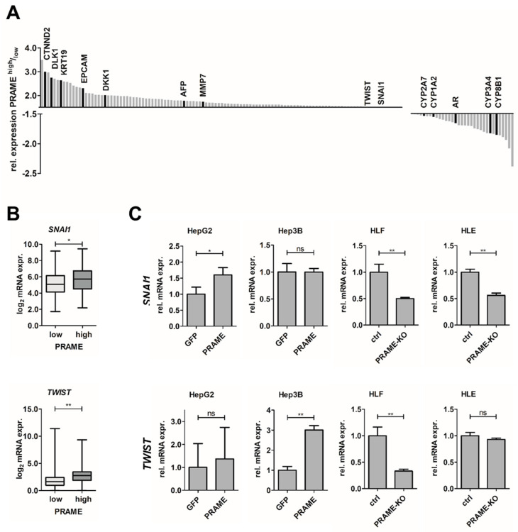
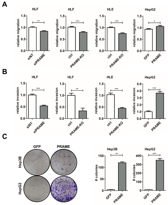
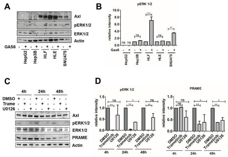
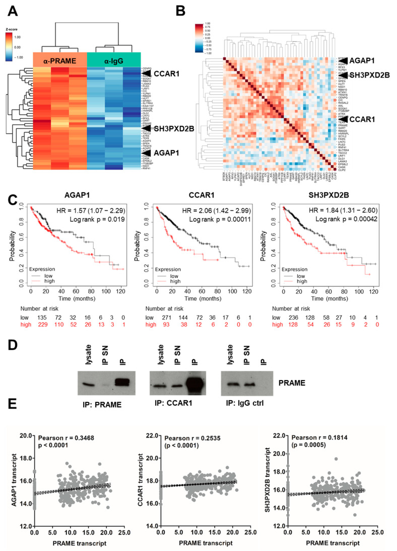
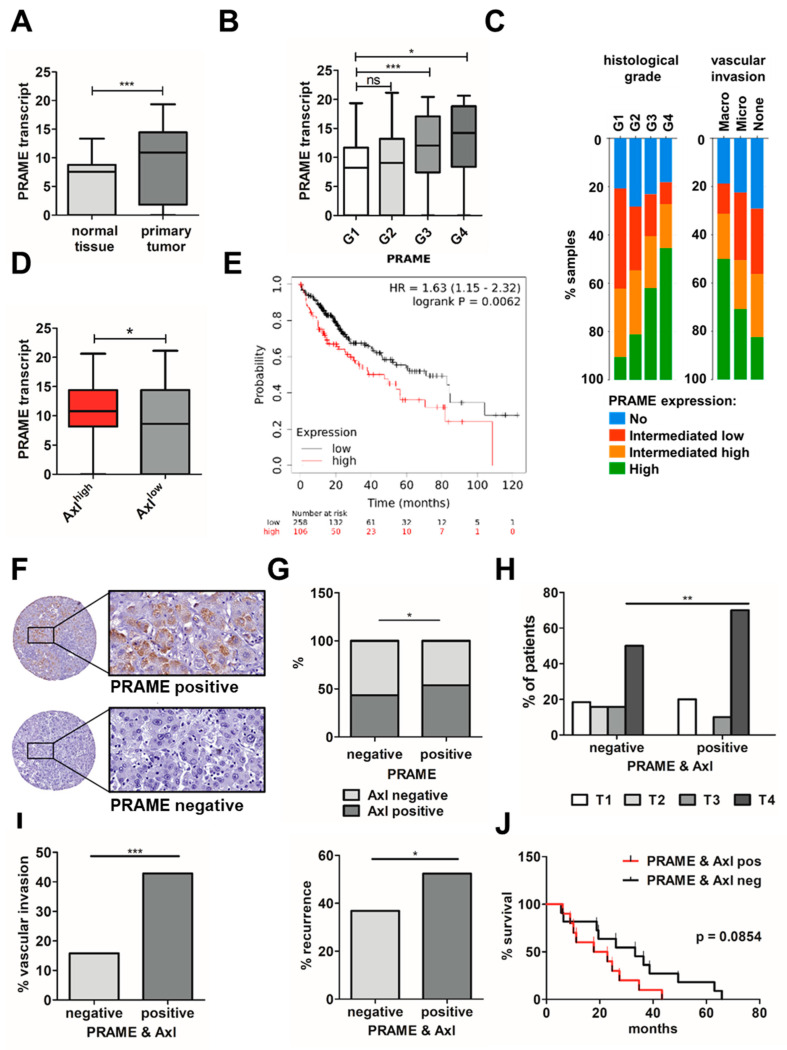
(1) Background: Activation of the receptor tyrosine kinase Axl by Gas6 fosters oncogenic effects in hepatocellular carcinoma (HCC), associating with increased mortality of patients. The impact of Gas6/Axl signaling on the induction of individual target genes in HCC and its consequences is an open issue. (2) Methods: RNA-seq analysis of Gas6-stimulated Axl-proficient or Axl-deficient HCC cells was used to identify Gas6/Axl targets. Gain- and loss-of-function studies as well as proteomics were employed to characterize the role of PRAME (preferentially expressed antigen in melanoma). Expression of Axl/PRAME was assessed in publicly available HCC patient datasets and in 133 HCC cases. (3) Results: Exploitation of well-characterized HCC models expressing Axl or devoid of Axl allowed the identification of target genes including PRAME. Intervention with Axl signaling or MAPK/ERK1/2 resulted in reduced PRAME expression. PRAME levels were associated with a mesenchymal-like phenotype augmenting 2D cell migration and 3D cell invasion. Interactions with pro-oncogenic proteins such as CCAR1 suggested further tumor-promoting functions of PRAME in HCC. Moreover, PRAME showed elevated expression in Axl-stratified HCC patients, which correlates with vascular invasion and lowered patient survival. (4) Conclusions: PRAME is a bona fide target of Gas6/Axl/ERK signaling linked to EMT and cancer cell invasion in HCC.

Keywords: Axl; EMT; Gas6; HCC; PRAME; dedifferentiation; immunotherapy; invasion.


Conflict of interest statement

The authors declare no conflict of interest.

Figures

Figure 1
Figure 1
Identification of Gas6/Axl-regulated transcripts in HCC cells. (A) Axl target genes were identified by RNA-seq of HCC cells stimulated with 500 ng/mL Gas6 for 24 h. p < 0.05 and an absolute log2-fold change of at least +1.0 were used for selection of targets. Axl dependencies were defined by comparisons of Axl-positive (wt, nick) versus Axl-negative (Axl) cells (left panel). As indicated by the arrow, 298 genes were further intersected with differentially expressed genes of Axl-reconstituted cells (Axl-wt). (B) Log2-fold changes (Log2FCs) of 8 Axl targets in Axl vs. wt (yellow), Axl vs. nick (blue) and Axl vs. Axl-wt (red). (C,D) Expression of PRAME transcript and protein levels as determined by qPCR and Western blotting, respectively. The expression of PRAME in Axl-expressing HCC cells was set to a value of 1. The expression of actin is shown as a loading control. (E) PRAME expression upon stimulation with 500 ng/mL Gas6 for 2 h in the absence (DMSO) or presence of 10 µM Axl inhibitor Cabozantinib (CABO) and upon administration of 900 ng/mL stimulatory Axl antibody (AF154) for 2 h. Protein levels were quantified by densitometry. wt, HLF; nick, HLF-nickase; Axl, HLF-Axl-KO; Axl-wt, HLF-Axl-KO-wt-Axl. Data are expressed as mean ± SD. ns: p > 0.05; *: p ≤ 0.05; **: p ≤ 0.01; ***: p ≤ 0.001.
Figure 2
Figure 2
Expression of PRAME associates with EMT. (A) Summary of differentially expressed genes in PRAME-stratified HCC patients in publicly available HCC samples from TCGA. n = 372. (B) Expression of the EMT-TFs SNAI1 (top) and TWIST (bottom) in PRAME−stratified HCC patient data from TCGA LIHC. n = 90 per group. (C) Modulation of EMT−TFs in PRAME-expressing epithelial cells (HepG2, Hep3B) and in mesenchymal cells (HLF, HLE) lacking PRAME expression (PRAME-KO). The expression of SNAI1 and TWIST in GFP-expressing HCC cells (HepG2, Hep3B) and control HLF/HLE cells (ctrl) were set to a value of 1. Data are expressed as mean ± SD. ns: p > 0.05; *: p ≤ 0.05; **: p ≤ 0.01.
Figure 3
Figure 3
PRAME raises migratory and invasive capabilities of HCC cells. (A) Cell migration of Gas6-stimulated HLF cells upon PRAME knockdown (siPRAME), PRAME-KO cells (HLF, HLE) and of PRAME-expressing HepG2 cells as analyzed by wound healing assays. (B) Invasion of HCC cells as described in (A) after spheroid formation and inclusion into collagen gels. The migration (A) and invasion (B) of non-target siRNA (siNT) treated cells, GFP-expressing HCC cells (HepG2) and control HLF/HLE cells (ctrl) were set to a value of 1. (C) Clonogenic growth behavior (left panels) of Hep3B-PRAME/Hep3B-GFP (top) and HepG2-PRAME/HepG2-GFP cells (bottom) and its quantification (right panels). Data are expressed as mean ± SD. *: p ≤ 0.05; **: p ≤ 0.01; ***: p ≤ 0.001.
Figure 4
Figure 4
Gas6/Axl induces MAPK signaling and PRAME expression. (A) Levels of phosho−ERK1/2 (pERK1/2) were assessed by Western blotting upon stimulation of HepG2, Hep3B, HLF, HLE and SNU475 cells with 500 ng/mL Gas6 for 15 min. (B) Quantification of pERK1/2 levels by densitometry (na: not applicable). The abundance of pERK1/2 in the absence of Gas6 was set to a value of 1 for each liver cancer cell line. (C) PRAME expression together with pERK1/2 and total ERK1/2 levels in HLF cells were assessed by Western blotting upon administration of 1 µM of either MEK inhibitors Trametinib or U0126. The expression of actin is shown as loading control (A,C). (D) pERK1/2 and PRAME expression were quantified by densitometry. The expression of DMSO−treated control cells was set to a value of 1. Data are expressed as mean ± SD. ns: p > 0.05; *: p ≤ 0.05; **: p ≤ 0.01; ***: p ≤ 0.001.
Figure 5
Figure 5
Immunoprecipitation reveals binding partners of PRAME. (A) Significantly enriched genes in anti−PRAME sample (orange) vs. IgG control (green) of anti−PRAME−IP−MS. (B) Positively and negatively correlated binding partners of PRAME in liver cancer cell lines using the cancer cell line encyclopedia. Higher resolution versions of (A,B) are included in the Supplemental Material. (C) Impact of the IP−MS-identified PRAME binding partners AGAP1, CCAR1 and SH3PXD2B on patient prognosis. AGAP1high, n = 229; AGAP1low, n = 135; CCAR1high, n = 93; CCAR1low, n = 271; SH3PXD2Bhigh, n = 128; SH3PXD2Blow, n = 236. (D) IP performed with anti-PRAME, anti-CCAR1 and IgG control antibody and subsequent assessment of PRAME expression via Western blotting. SN, supernatant. (E) Correlations between the expression of PRAME and the IP−MS−identified binding partners AGAP1, CCAR1 and SH3PXD2B in primary tumor samples using the publicly available patient dataset TCGA LIHC (n = 372). Each grey dot represents one sample. Regression lines are shown in solid black and the 95% confidence intervals as dashed black lines.
Figure 6
Figure 6
Correlation of PRAME and Axl expression levels with prognosis and survival of HCC patients. (A) PRAME transcript levels in normal liver tissue and primary HCC (n = 52) and (B) stratified according to different histological grades (G1–G4) from publicly available TCGA LIHC data. G1, n = 55; G2, n = 177, G3, n = 122, G4, n = 12. (C) Relative PRAME expression in percentage per histological grades (G1–G4; p-value: 1.839 × 10⁻⁴) and its contribution to invasiveness (p-value: 0.0429) in publicly available HCC patient samples using cBioPortal. n = 93 per group. (D) PRAME expression in Axl-stratified TCGA LIHC data. n = 93 per group. (E) Combined effect of PRAME and Axl on patient survival (red: PRAMEhigh + Axlhigh, n = 106; black: PRAMElow + Axllow, n = 258). (F) Representative images from immunohistochemical analysis of PRAME-positive and PRAME-negative HCC tissue samples. Rectangles represent a 30-fold magnification. (G) Distribution of Axl expression in PRAME-positive and -negative samples in percent. PRAME/Axl-negative, n = 38; PRAME/Axl-positive, n = 20; PRAME-positive, Axl-negative, n = 18; PRAME-negative, Axl-positive, n = 29. (H) Distribution of PRAME-positive samples per tumor stage. PRAME/Axl-negative: T1, n = 7, T2, n = 6, T3, n = 6, T4, n = 19; PRAME/Axl-positive: T1, n = 4, T2, n = 0, T3, n = 2, T4, n = 14, Tnd, n = 1. (I) Vascular invasion (left) and recurrence (right) in PRAME/Axl-positive and PRAME/Axl-negative HCC samples. PRAME/Axl-negative: vascular invasion, yes, n = 6, no, n = 32, recurrence, yes, n = 14, no, n = 24; PRAME/Axl-positive: vascular invasion, yes, n = 9, no, n = 11, recurrence, yes, n = 10, no, n = 10. (J) Combined effect of PRAME and Axl protein levels on patient survival (black: PRAME/Axl-negative, n = 38; red: PRAME/Axl-positive, n = 20). Data are expressed as mean ± SD. ns: p > 0.05; *: p ≤ 0.05; **: p ≤ 0.01; ***: p ≤ 0.001.

Similar articles

Cited by

References

    1. Singal A.G., Lampertico P., Nahon P. Epidemiology and surveillance for hepatocellular carcinoma: New trends. J. Hepatol. 2020;72:250–261. doi: 10.1016/j.jhep.2019.08.025. - DOI - PMC - PubMed
    1. Llovet J.M. Hepatocellular carcinoma. Nat. Rev. Dis. Primers. 2021;7:6. doi: 10.1038/s41572-020-00240-3. - DOI - PubMed
    1. Llovet J.M., De Baere T., Kulik L., Haber P.K., Greten T.F., Meyer T., Lencioni R. Locoregional therapies in the era of molecular and immune treatments for hepatocellular carcinoma. Nat. Rev. Gastroenterol. Hepatol. 2021;18:293–313. doi: 10.1038/s41575-020-00395-0. - DOI - PubMed
    1. Villanueva A. Hepatocellular Carcinoma. N. Engl. J. Med. 2019;380:1450–1462. doi: 10.1056/NEJMra1713263. - DOI - PubMed
    1. Craig A.J., Von Felden J., Garcia-Lezana T., Sarcognato S., Villanueva A. Tumour evolution in hepatocellular carcinoma. Nat. Rev. Gastroenterol. Hepatol. 2020;17:139–152. doi: 10.1038/s41575-019-0229-4. - DOI - PubMed