Review
. 2023 Apr 27;15(9):2504.
doi: 10.3390/cancers15092504.

Integrins and the Metastasis-like Dissemination of Acute Lymphoblastic Leukemia to the Central Nervous System

Affiliations
Review

Integrins and the Metastasis-like Dissemination of Acute Lymphoblastic Leukemia to the Central Nervous System

Signe Modvig et al. Cancers (Basel). .

Abstract

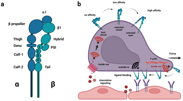
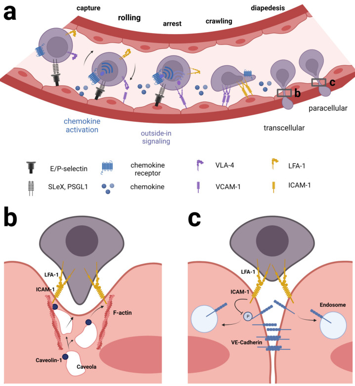
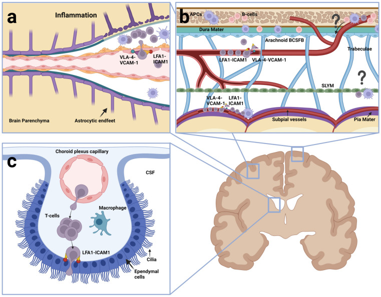
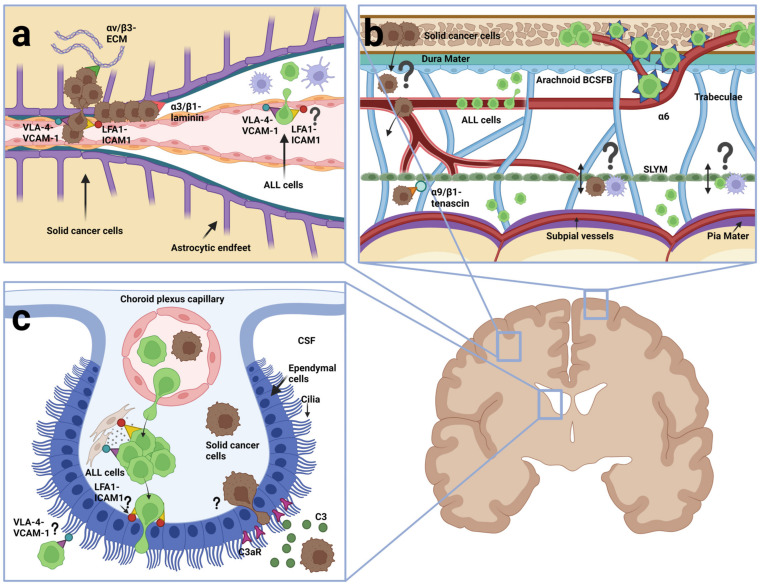
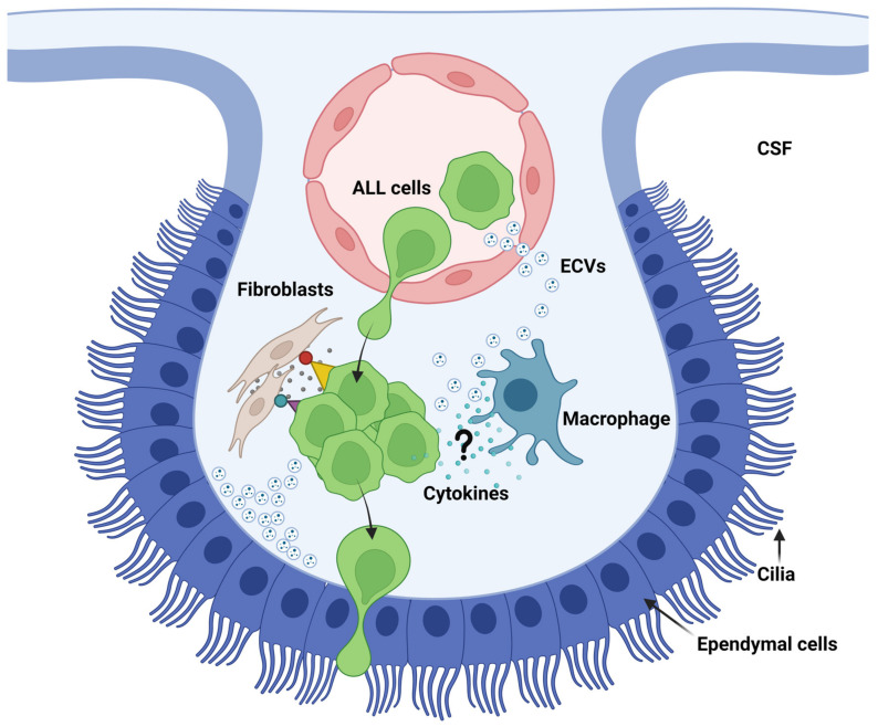
Acute lymphoblastic leukemia (ALL) disseminates with high prevalence to the central nervous system (CNS) in a process resembling aspects of the CNS surveillance of normal immune cells as well as aspects of brain metastasis from solid cancers. Importantly, inside the CNS, the ALL blasts are typically confined within the cerebrospinal fluid (CSF)-filled cavities of the subarachnoid space, which they use as a sanctuary protected from both chemotherapy and immune cells. At present, high cumulative doses of intrathecal chemotherapy are administered to patients, but this is associated with neurotoxicity and CNS relapse still occurs. Thus, it is imperative to identify markers and novel therapy targets specific to CNS ALL. Integrins represent a family of adhesion molecules involved in cell-cell and cell-matrix interactions, implicated in the adhesion and migration of metastatic cancer cells, normal immune cells, and leukemic blasts. The ability of integrins to also facilitate cell-adhesion mediated drug resistance, combined with recent discoveries of integrin-dependent routes of leukemic cells into the CNS, have sparked a renewed interest in integrins as markers and therapeutic targets in CNS leukemia. Here, we review the roles of integrins in CNS surveillance by normal lymphocytes, dissemination to the CNS by ALL cells, and brain metastasis from solid cancers. Furthermore, we discuss whether ALL dissemination to the CNS abides by known hallmarks of metastasis, and the potential roles of integrins in this context.

Keywords: CNS; acute lymphoblastic leukemia; immune surveillance; integrin; metastasis.


Conflict of interest statement

The authors declare no conflict of interest.

Figures

Figure 1
Figure 1
Structure of integrin heterodimers and regulation of integrin affinity on the cell surface. (a) heterodimer composed of α and β subunit. Shown is LFA-1 (αLβ2). Integrin α-subunits expressed in leukocytes (αL, αM, αD and αX) as well as α1, α2, α10 and α11 contain the so-called insertion or interaction domain (αI), which plays a vital part in ligand-binding of these integrins. All other α-subunits bind ligands in conjunction with the β-subunit, i.e., the head piece. (b) integrin heterodimers shift between bent conformation with closed head piece (no affinity for ligand), extended conformation with closed head piece (intermediate affinity) and extended conformation with open head piece (high affinity). Chemokine signaling and forces from the inside (e.g., actin-dynamics) can cause a shift between the conformations (inside-out activation), whereas binding to ligands or external forces (e.g., blood flow) can cause outside-in activation. This figure was created with BioRender.com (accessed on 21 April 2023).
Figure 2
Figure 2
Mechanism of leukocyte extravasation from blood vessels. (a) The process starts by capture/tethering, which mainly depends on endothelial E-and P-selectins binding sialyl-Le X ligands or P-selectin glycoprotein ligands on the leukocyte surface, leading to leukocyte rolling on the luminal surface of the endothelium. VLA-4 (α4β1) binding to VCAM-1 and LFA-1 (αLβ2) binding to ICAM-1 contribute to slowing down the rolling. A combination of chemokine activation and VCAM-1 and ICAM-1 binding increases the affinity of the integrins via inside-out and outside-in signaling, respectively. Additionally, the blood flow and the formation of catch bonds contribute to strengthening the LFA-1: ICAM-1 interaction. Eventually, this leads to arrest of the leukocyte. Following the formation of an LFA-1: ICAM-1 transmigratory cup, the leukocyte starts crawling and at the same time extends membrane protrusions to probe the endothelial surface to find a site permissive of transmigration. The last step of extravasation is diapedesis during which the leukocyte moves through the endothelium either through an individual EC cell (transcellularly) or through a space between neighboring EC (paracellularly) highlighted in (b,c), respectively. (b) transcellular diapedesis involves clusters of ICAM-1 on the endothelial cell, allegedly binding LFA-1 on the leukocyte. ICAM-1 is in physical contact with both F-actin filaments and the protein caveolin-1 on the surface of caveolae inside the endothelial cell. The caveolae gradually fuse to form a channel through which the leukocyte slides through. (c) In paracellular diapedesis, LFA-1 on the surface of the leukocyte binds ICAM-1 on two neighboring endothelial cells. ICAM-1 alters the phosphorylation status of key tyrosine residues in vascular-endothelial cadherin (VE-cadherin) causing VE-cadherin to be endocytosed by the endothelial cells. As a result, the adherens junctions between the endothelial cells are gradually unzipped as the leukocyte moves through. See text for further details. This figure was created with BioRender.com.
Figure 3
Figure 3
Entry routes and integrins used by normal lymphocytes (a) Postcapillary venule. In the presence of inflammation, the crossing of the BBB occurs in a manner dependent on VLA-4 (α4β1): VCAM-1 and LFA-1 (αLβ2): ICAM-1. (b) Section showing meningeal layers between the calvaria and the brain parenchyma. Lymphocytes may adhere and extravasate from subpial and meningeal vessels, independently of inflammation, using VLA-4: VCAM-1 and LFA-1: ICAM-1. T cells and other immune cells may migrate from calvarial bone marrow into the dura. Whether lymphocytes migrate on emissary vessels and how they cross the arachnoid BCSFB (light blue cell layer) is not known. Whether they interact with cells of the SLYM (green) is also unknown (indicated by question marks). (c) Choroid plexus (CP). Lymphocytes cross the fenestrated capillaries underneath the CP. LFA-1-ICAM-1 has been shown to be involved when T cells cross the ependymal cells (blue) and enter CSF (white). This figure was created with BioRender.com.
Figure 4
Figure 4
Entry routes and integrins used by ALL cells (green) and metastatic cells from solid cancers (brown) (a) Postcapillary venule. Metastatic cells extravasate from venules in a manner dependent on VLA-4 (α4β1): VCAM-1 and LFA-1 (αLβ2)-ICAM-1. Once across, they either engage in vascular co-optive growth to which α3 and β1 integrins may contribute, or they establish metastasis in the parenchyma, which involves αVβ3. Although presumed to involve α4β1: VCAM-1 and αLβ2-ICAM-1, little is known of integrins involved in ALL extravasation from postcapillary venules. (b) section showing meningeal layers between the calvaria and the brain parenchyma. ALL cells use α6 integrin: laminin binding to migrate on the basement membrane of emissary vessels connecting calvarial bone marrow and the meninges. It is unknown if and how metastatic cells from solid cancers enter the meninges from calvarial bone marrow. Both ALL cells and metastatic cells are found in the dura and leptomeninges. Different integrins may be involved in the binding to meningeal components. It is unknown whether cancer cells of both types traverse or otherwise interact with the SLYM (indicated by a question mark). (c) choroid plexus (CP). ALL cells use α4β1: VCAM-1 and αLβ2: ICAM-1 to interact with stromal fibroblasts and studies show that they can cross the BCSFB. Metastatic cancer cells in the CSF use the complement component C3 binding the receptor C3aR on the ependymal cells to disrupt the BCSFB. This figure was created with BioRender.com.
Figure 5
Figure 5
Model of ALL interaction with stromal cells of the choroid plexus. ALL cells have been shown to bind stromal fibroblasts through α4β1: VCAM-1 and αLβ2: ICAM-1 converting them to cancer-associated fibroblasts and instructing them to secrete cytokines in the process. The result is increased chemoresistance of the ALL cells. Furthermore, in vitro studies have shown that extracellular vesicles produced by ALL cells can prepare ependymal cells for ALL transmigration. The role of integrins in this process has been shown for some ALL cells. It remains to be shown whether communication takes place in the CP stroma between macrophages and ALL cells, whether cytokines and exosomal integrins are involved, and whether the macrophages are converted to tumor-associated macrophages during such a process (indicated by question mark). See text for further details. This figure was created with BioRender.com.

References

    1. Inaba H., Greaves M., Mullighan C.G. Acute lymphoblastic leukaemia. Lancet. 2013;381:1943–1955. doi: 10.1016/S0140-6736(12)62187-4. - DOI - PMC - PubMed
    1. Hunger S.P., Mullighan C.G. Acute Lymphoblastic Leukemia in Children. N. Engl. J. Med. 2015;373:1541–1552. doi: 10.1056/NEJMra1400972. - DOI - PubMed
    1. Toft N., Birgens H., Abrahamsson J., Griškevičius L., Hallböök H., Heyman M., Klausen T.W., Jónsson Ó.G., Palk K., Pruunsild K., et al. Results of NOPHO ALL2008 treatment for patients aged 1-45 years with acute lymphoblastic leukemia. Leukemia. 2018;32:606–615. doi: 10.1038/leu.2017.265. - DOI - PubMed
    1. Arber D.A., Orazi A., Hasserjian R., Thiele J., Borowitz M.J., Le Beau M.M., Bloomfield C.D., Cazzola M., Vardiman J.W. The 2016 revision to the World Health Organization classification of myeloid neoplasms and acute leukemia. Blood. 2016;127:2391–2405. doi: 10.1182/blood-2016-03-643544. - DOI - PubMed
    1. Cunningham I. Extramedullary sites of leukemia relapse after transplant. Leuk. Lymphoma. 2006;47:1754–1767. doi: 10.1080/10428190600632857. - DOI - PubMed

LinkOut - more resources