Review
. 2023 Apr 29;15(9):2546.
doi: 10.3390/cancers15092546.

Comments on and Illustrations of the EFSUMB CEUS Guidelines: Transabdominal and Endoscopic Ultrasound Features of Intrapancreatic Metastases and the Role of Multiparametric Imaging and EUS-Guided Sampling in Rare Pancreatic Tumors

Affiliations
Review

Comments on and Illustrations of the EFSUMB CEUS Guidelines: Transabdominal and Endoscopic Ultrasound Features of Intrapancreatic Metastases and the Role of Multiparametric Imaging and EUS-Guided Sampling in Rare Pancreatic Tumors

Kathleen Möller et al. Cancers (Basel). .

Abstract

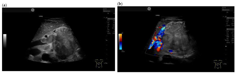
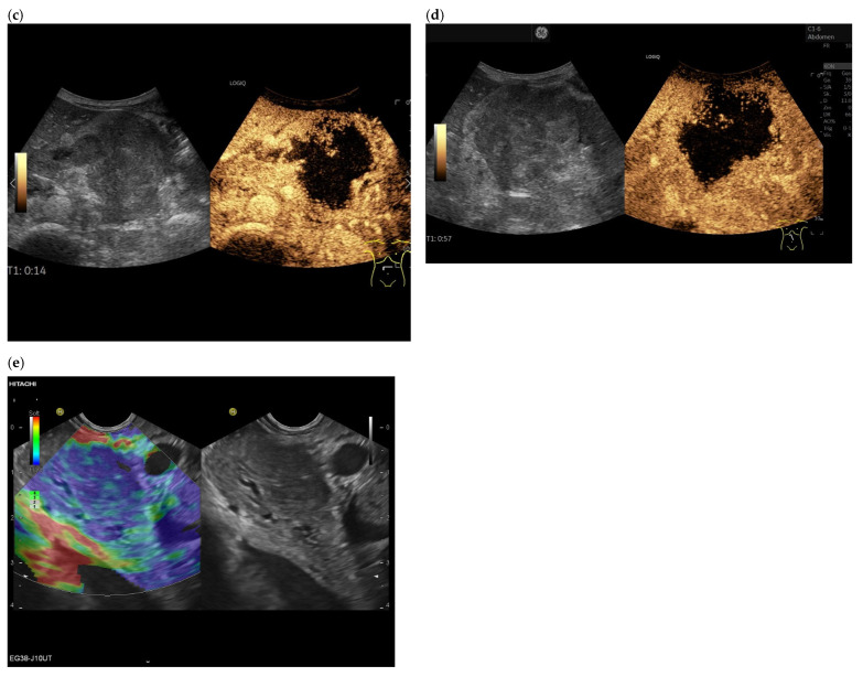
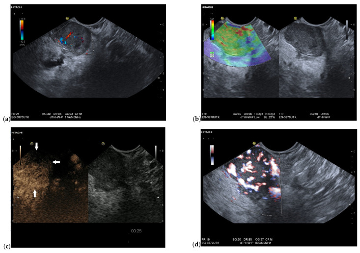
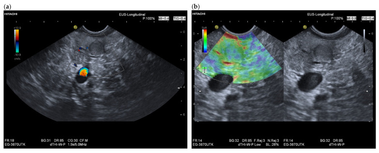
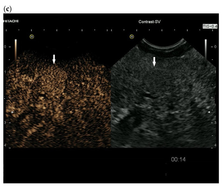
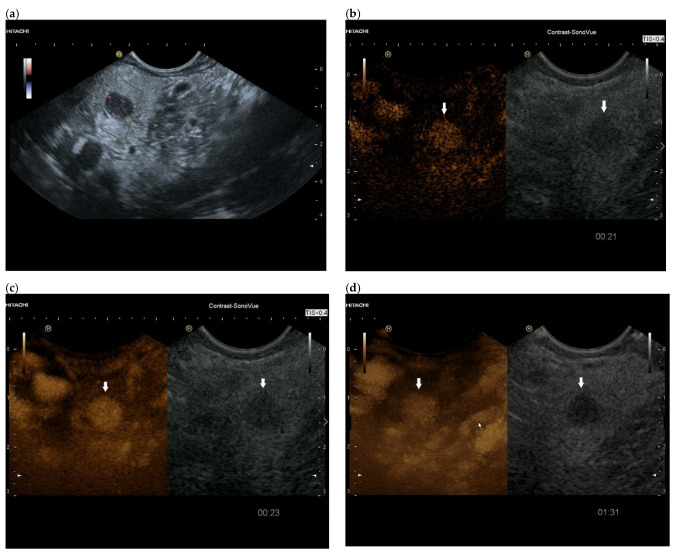
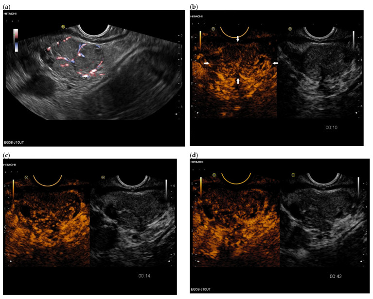
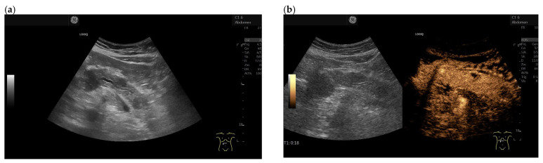
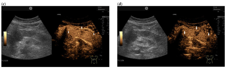
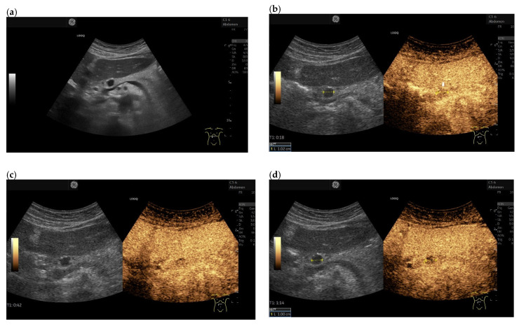
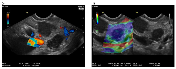
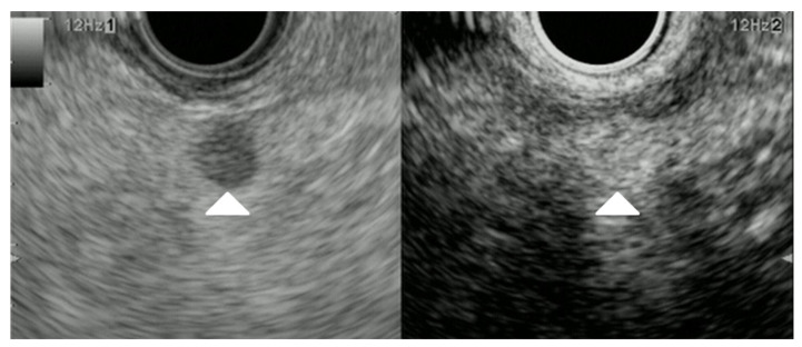
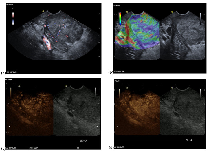
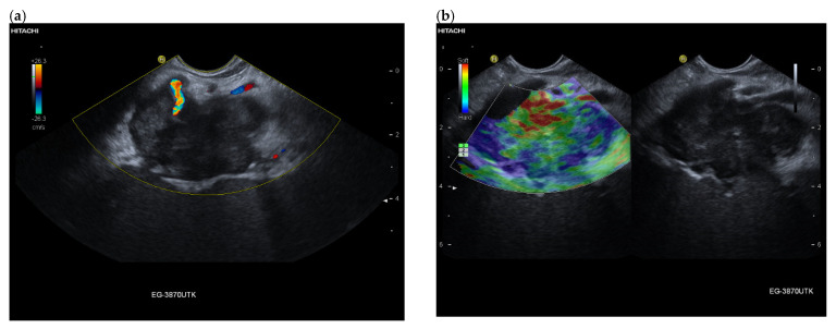
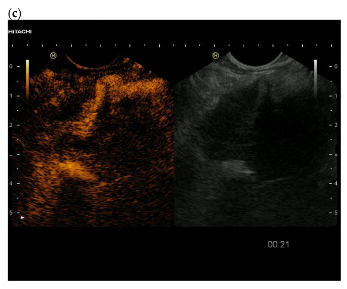
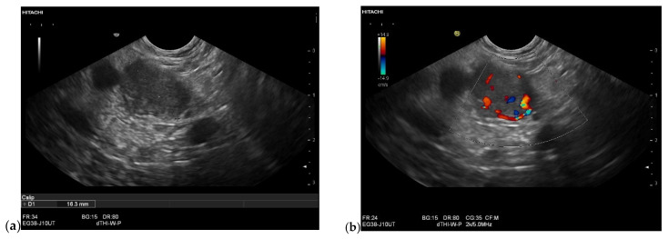
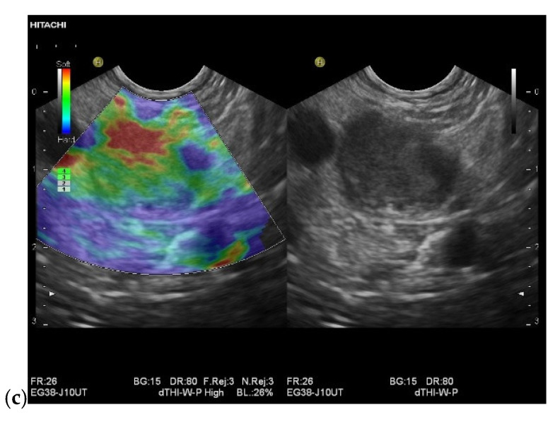
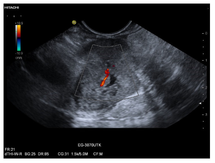
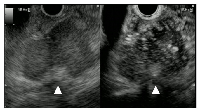
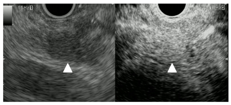
A definite pathologic diagnosis of intrapancreatic metastasis is crucial for the management decision, i.e., curative or palliative surgery versus chemotherapy or conservative/palliative therapy. This review focuses on the appearance of intrapancreatic metastases on native and contrast-enhanced transabdominal ultrasound and endoscopic ultrasound. Differences and similarities in relation to the primary tumor, and the differential diagnosis from pancreatic carcinoma and neuroendocrine neoplasms are described. The frequency of intrapancreatic metastases in autopsy studies and surgical resection studies will be discussed. Further emphasis is placed on endoscopic ultrasound-guided sampling to confirm the diagnosis.

Keywords: CEUS; CH-EUS; EUS-guided sampling; intrapancreatic metastases; prevalence.


Conflict of interest statement

The authors declare no conflict of interest.

Figures

Figure 1
Figure 1
Intrapancreatic metastasis of a colon carcinoma in the transverse colon. Female, 42 years old, with severe abdominal pain and weight loss. Large inhomogeneous, hypoechoic lesion in the pancreatic body with invasion into the portal vein. Percutaneous US, 9 MHz linear (a). The lesion is without vessels on duplex US (b). Percutaneous CEUS shows rim sign in the arterial phase. All other parts are without enhancement (c). In the venous phase, the rim-enhanced areas show washout (d). In strain elastography on EUS, the lesion is stiffer (e). EUS-FNA obtaining histologic material confirmed metastasis from a colon cancer.
Figure 1
Figure 1
Intrapancreatic metastasis of a colon carcinoma in the transverse colon. Female, 42 years old, with severe abdominal pain and weight loss. Large inhomogeneous, hypoechoic lesion in the pancreatic body with invasion into the portal vein. Percutaneous US, 9 MHz linear (a). The lesion is without vessels on duplex US (b). Percutaneous CEUS shows rim sign in the arterial phase. All other parts are without enhancement (c). In the venous phase, the rim-enhanced areas show washout (d). In strain elastography on EUS, the lesion is stiffer (e). EUS-FNA obtaining histologic material confirmed metastasis from a colon cancer.
Figure 2
Figure 2
Intrapancreatic metastasis of a RCC in a male, 76 years old. EUS finding of a 25 mm well-circumscribed lesion in the head of the pancreas. The pancreatic duct is not dilated. Vessels are already detectable in the native duplex (a). In the EUS strain elastography, the lesion is softer (b). On CH-EUS with 4.8 mL SonoVue i.v., the lesion is hyperenhanced with a peripheral emphasis and sparing of the central parts (arrows) (c). In contrast-enhanced Power Doppler EUS according to CH-EUS, a large number of vessels are again visible (d).
Figure 3
Figure 3
Intrapancreatic metastasis of an RCC. Male, 69 years old, with a small lesion in the body of the pancreas on surveillance MRI. Tumor nephrectomy 17 years ago, pancreatic tail resection with splenectomy for intrapancreatic metastasis 7 years ago. Endosonographically 10 mm, slightly hypoechoic lesion in the body of the pancreas. No evidence of vessels in the lesion in the native duplex EUS (a). In the EUS strain elastography, the lesion is not stiffer, rather soft (b). In the CH-EUS with 4.8 mL SonoVue, the lesion in the arterial phase shows homogeneous hyperenhancement. The arrow marks the hyperenhanced lesion in the left image, and an arrow marks the lesion in B-mode US on the right. (c). The RCC metastasis was diagnosed using EUS-FNP. The patient underwent pancreatic resection for second time.
Figure 3
Figure 3
Intrapancreatic metastasis of an RCC. Male, 69 years old, with a small lesion in the body of the pancreas on surveillance MRI. Tumor nephrectomy 17 years ago, pancreatic tail resection with splenectomy for intrapancreatic metastasis 7 years ago. Endosonographically 10 mm, slightly hypoechoic lesion in the body of the pancreas. No evidence of vessels in the lesion in the native duplex EUS (a). In the EUS strain elastography, the lesion is not stiffer, rather soft (b). In the CH-EUS with 4.8 mL SonoVue, the lesion in the arterial phase shows homogeneous hyperenhancement. The arrow marks the hyperenhanced lesion in the left image, and an arrow marks the lesion in B-mode US on the right. (c). The RCC metastasis was diagnosed using EUS-FNP. The patient underwent pancreatic resection for second time.
Figure 4
Figure 4
Intrapancreatic metastasis of RCC in a female, 69 years old. During post-RCC surveillance, MRI revealed a small pancreatic lesion. EUS depicts a 7.5 mm, smooth-bordered, low-echo lesion with small vessels on power Doppler in the pancreatic head (a). The pancreatic duct was not dilated. On CH-EUS with 4.8 mL SonoVue, the mass was homogeneously hyperenhanced in the arterial phase after 21 s (b) and in the accumulation after 24 s (c). The lesion showed no washout in the venous phase, as seen here in the venous phase after 1.31 min accumulation (d). The lesion is marked by an arrow. Surgical histology revealed metastasis of RCC.
Figure 5
Figure 5
Intrapancreatic metastasis of a rectal cancer in a male, 72 years old. During follow-up after rectal carcinoma, a 29 × 15 mm mass was noted in the pancreatic body. The proximal pancreatic duct was dilated. On EUS, the lesion was multi-nodular, smooth-edged, slightly hypoechoic. On EUS power Doppler, annular vessels presented (a). On CH-EUS, the lesion had numerous vessels in the arterial phase at 10 s but was heterogeneous and slightly hypoenhanced. The arrows mark the border of the hypoenhanced lesion in the arterial phase of CH-EUS. (b). A progressive washout began as early as 14 s (c), which continued in the venous phase, here 42 s (d). Rectal carcinoma metastasis was confirmed by EUS-FNP. Immunohistochemistry proved necessary for differentiation from a PDAC. The patient underwent resection.
Figure 6
Figure 6
Multiple intrapancreatic metastasis of malignant melanoma in a male, 55 years old. Upper abdominal pain with elevated lipase on blood tests. A malignant melanoma on the arm had been removed months earlier. B-mode ultrasonography revealed multiple hypoechoic, well-confined lesions measuring up to 13 × 23 mm. The pancreatic duct was slightly dilated (a). On CEUS with 1.2 mL SonoVue, the lesions were primarily hypoenhanced, 18 s in the arterial phase (b). Thereby, they presented slightly less hypoenhanced at the end of the arterial phase at 29 s (c) than at the beginning of the arterial phase (b) and in the venous phase (d), shown by arrows. Differentially, autoimmune pancreatitis was considered. Contrast behavior on CEUS argued against this. The anamnesis was helpful. The diagnosis was confirmed by EUS-FNP. Histologically evaluable particles were obtained.
Figure 6
Figure 6
Multiple intrapancreatic metastasis of malignant melanoma in a male, 55 years old. Upper abdominal pain with elevated lipase on blood tests. A malignant melanoma on the arm had been removed months earlier. B-mode ultrasonography revealed multiple hypoechoic, well-confined lesions measuring up to 13 × 23 mm. The pancreatic duct was slightly dilated (a). On CEUS with 1.2 mL SonoVue, the lesions were primarily hypoenhanced, 18 s in the arterial phase (b). Thereby, they presented slightly less hypoenhanced at the end of the arterial phase at 29 s (c) than at the beginning of the arterial phase (b) and in the venous phase (d), shown by arrows. Differentially, autoimmune pancreatitis was considered. Contrast behavior on CEUS argued against this. The anamnesis was helpful. The diagnosis was confirmed by EUS-FNP. Histologically evaluable particles were obtained.
Figure 7
Figure 7
Intrapancreatic metastases of malignant melanoma in a male, 75 years old. Peripheral lymph node increasing for diagnosis. No abdominal complaints. B-mode ultrasonography shows a highly hypoechoic or anechoic lesion in the pancreatic body (a). In duplex, no vessels, no aneurysm. In CEUS, with 1.2 mL SonoVue in arterial phase at 18 s iso- to slight hypoenhancement (arrow) (b). In venous phase, slow washout at 42 s (c) with heterogeneous pattern at 1.14 min (d). EUS revealed additional multiple small hypoechoic lesions that had escaped percutaneous sonography. The lesions were highly hypoechoic, almost anechoic, smoothly confined, without evidence of vessels in the EUS duplex (e). Lesions were stiffer on strain elastography in EUS (f). The diagnosis of metastatic malignant melanoma was confirmed percutaneously, ultrasound-guided from the lymph nodes and by EUS-FNP from the pancreas.
Figure 7
Figure 7
Intrapancreatic metastases of malignant melanoma in a male, 75 years old. Peripheral lymph node increasing for diagnosis. No abdominal complaints. B-mode ultrasonography shows a highly hypoechoic or anechoic lesion in the pancreatic body (a). In duplex, no vessels, no aneurysm. In CEUS, with 1.2 mL SonoVue in arterial phase at 18 s iso- to slight hypoenhancement (arrow) (b). In venous phase, slow washout at 42 s (c) with heterogeneous pattern at 1.14 min (d). EUS revealed additional multiple small hypoechoic lesions that had escaped percutaneous sonography. The lesions were highly hypoechoic, almost anechoic, smoothly confined, without evidence of vessels in the EUS duplex (e). Lesions were stiffer on strain elastography in EUS (f). The diagnosis of metastatic malignant melanoma was confirmed percutaneously, ultrasound-guided from the lymph nodes and by EUS-FNP from the pancreas.
Figure 8
Figure 8
Metastasis of malignant melanoma. Pancreatic lesion 6mm in size without main pancreatic duct dilation; it was detected as a hypoechoic lesion (arrowhead) with B-mode EUS (left). CE-EUS detected a pancreatic lesion, surprisingly, with hyperintensity of enhancement (arrowhead) as compared to that of surrounding pancreatic tissue (right).
Figure 9
Figure 9
Metastasis of an endometroid adenocarcinoma in a female, 68 years old. Incidental finding of a mass at the pancreatic head, 1 year before an endometroid uterine carcinoma had been diagnosed and surgically operated. Endosonographically well vascularized lesion at the pancreatic head. Power Doppler imaging (a). In strain elastography of EUS, the lesion was predominantly stiffer, but also with softer portions (b). In CH -EUS with 4.8 mL SonoVue, the lesion was well vascularized in the arterial phase: first with an increased peripheral rim-like enhancement at 12 s and central nonenhancement (c), then heterogeneously hyperenhanced with emphasis of the periphery and small area of central nonenhancement at 14 s (d).
Figure 10
Figure 10
Intrapancreatic metastasis of poorly differentiated serous adenocarcinoma in a female, 80 years old. Two years earlier, she had undergone total gynecological surgery for a carcinoma of the uterus. Recently, a syncope diagnostic was performed. Incidental finding of a 45 mm hypoechoic lesion with indistinct borders on the pancreatic tail in the splenic hilum. On EUS, the lesion surrounds the splenic artery and is indistinguishable from the spleen (a). In strain elastography of EUS, the lesion shows softer and stiffer parts (b). In the arterial phase of CH-EUS, the lesion is hypoenhanced. Assignment was made by EUS-FNP (c).
Figure 10
Figure 10
Intrapancreatic metastasis of poorly differentiated serous adenocarcinoma in a female, 80 years old. Two years earlier, she had undergone total gynecological surgery for a carcinoma of the uterus. Recently, a syncope diagnostic was performed. Incidental finding of a 45 mm hypoechoic lesion with indistinct borders on the pancreatic tail in the splenic hilum. On EUS, the lesion surrounds the splenic artery and is indistinguishable from the spleen (a). In strain elastography of EUS, the lesion shows softer and stiffer parts (b). In the arterial phase of CH-EUS, the lesion is hypoenhanced. Assignment was made by EUS-FNP (c).
Figure 11
Figure 11
Intrapancreatic metastasis of RCC in a male, 70 years old. Incidental finding of a 16 mm lesion on the pancreatic tail. Native EUS showed the lesion to be hypoechoic and smoothly confined (a), the pancreatic duct was not dilated, duplex EUS showed good vascularization with vessels in the lesion (b). Elastographically, the lesion was stiffer (c). Anamnestically, a nephrectomy had been performed due to trauma-related hemorrhage. This had occurred 20 years previously. The patient was not explicitly aware of a tumor. EUS-FNP (19 G) was performed, the aspirates of which were very bloody and yielded no result. It was not until the repeat (19 G) that the metastasis of an RCC could be diagnosed, although a renal tumor was not known.
Figure 11
Figure 11
Intrapancreatic metastasis of RCC in a male, 70 years old. Incidental finding of a 16 mm lesion on the pancreatic tail. Native EUS showed the lesion to be hypoechoic and smoothly confined (a), the pancreatic duct was not dilated, duplex EUS showed good vascularization with vessels in the lesion (b). Elastographically, the lesion was stiffer (c). Anamnestically, a nephrectomy had been performed due to trauma-related hemorrhage. This had occurred 20 years previously. The patient was not explicitly aware of a tumor. EUS-FNP (19 G) was performed, the aspirates of which were very bloody and yielded no result. It was not until the repeat (19 G) that the metastasis of an RCC could be diagnosed, although a renal tumor was not known.
Figure 12
Figure 12
Intrapancreatic metastasis of breast carcinoma in a female, 71 years old. Incidental finding of a 25 mm mass on the pancreatic body during ultrasound examination. On EUS, the lesion was mildly polycyclic but smoothly circumscribed. A tiny anechoic area was seen centrally. Vessels were demonstrable on duplex. A neuroendocrine tumor was suspected. In the EUS-FNP, this was not confirmed at first. The medical history was taken again, and it was found out that a breast carcinoma had been treated many years ago. Knowing this history, additional immunohistological examinations were performed, which revealed the metastasis of the breast cancer.
Figure 13
Figure 13
Lung cancer. Pancreatic lesion of 40 mm in size without main pancreatic duct dilation was detected as a low echoic lesion (arrowhead) with B-mode EUS (left). CE-EUS detected a pancreatic lesion with hypo-intensity enhancement (arrowhead) as compared to that of surrounding pancreatic tissue (right).
Figure 14
Figure 14
Merkel cell carcinoma. Pancreatic lesion 10mm in size without main pancreatic duct dilation was detected as a low echoic lesion (arrowhead) with B-mode EUS (left). CE-EUS detected a pancreatic lesion with hyper-intensity of enhancement (arrowhead) as compared to that of surrounding pancreatic tissue (right).

Similar articles

Cited by

References

    1. Dietrich C.F., Averkiou M., Nielsen M.B., Barr R.G., Burns P.N., Calliada F., Cantisani V., Choi B., Chammas M.C., Clevert D.A., et al. How to perform Contrast-Enhanced Ultrasound (CEUS) Ultrasound Int. Open. 2018;4:E2–E15. doi: 10.1055/s-0043-123931. - DOI - PMC - PubMed
    1. Claudon M., Dietrich C.F., Choi B.I., Cosgrove D.O., Kudo M., Nolsoe C.P., Piscaglia F., Wilson S.R., Barr R.G., Chammas M.C., et al. Guidelines and good clinical practice recommendations for Contrast Enhanced Ultrasound (CEUS) in the liver-update 2012: A WFUMB-EFSUMB initiative in cooperation with representatives of AFSUMB, AIUM, ASUM, FLAUS and ICUS. Ultrasound Med. Biol. 2013;39:187–210. doi: 10.1016/j.ultrasmedbio.2012.09.002. - DOI - PubMed
    1. Dietrich C.F., Nolsoe C.P., Barr R.G., Berzigotti A., Burns P.N., Cantisani V., Chammas M.C., Chaubal N., Choi B.I., Clevert D.A., et al. Guidelines and Good Clinical Practice Recommendations for Contrast-Enhanced Ultrasound (CEUS) in the Liver-Update 2020 WFUMB in Cooperation with EFSUMB, AFSUMB, AIUM, and FLAUS. Ultrasound Med. Biol. 2020;46:2579–2604. doi: 10.1016/j.ultrasmedbio.2020.04.030. - DOI - PubMed
    1. Piscaglia F., Nolsoe C., Dietrich C.F., Cosgrove D.O., Gilja O.H., Bachmann Nielsen M., Albrecht T., Barozzi L., Bertolotto M., Catalano O., et al. The EFSUMB Guidelines and Recommendations on the Clinical Practice of Contrast Enhanced Ultrasound (CEUS): Update 2011 on non-hepatic applications. Ultraschall Med. 2012;33:33–59. doi: 10.1055/s-0031-1281676. - DOI - PubMed
    1. Sidhu P.S., Cantisani V., Dietrich C.F., Gilja O.H., Saftoiu A., Bartels E., Bertolotto M., Calliada F., Clevert D.A., Cosgrove D., et al. The EFSUMB Guidelines and Recommendations for the Clinical Practice of Contrast-Enhanced Ultrasound (CEUS) in Non-Hepatic Applications: Update 2017 (Long Version) Ultraschall Med. 2018;39:e2–e44. doi: 10.1055/a-0586-1107. - DOI - PubMed

LinkOut - more resources