. 2023 May 4;15(9):2601.
doi: 10.3390/cancers15092601.

Extracellular Vesicles Potentiate Medulloblastoma Metastasis in an EMMPRIN and MMP-2 Dependent Manner

Affiliations

Extracellular Vesicles Potentiate Medulloblastoma Metastasis in an EMMPRIN and MMP-2 Dependent Manner

Hannah K Jackson et al. Cancers (Basel). .

Abstract

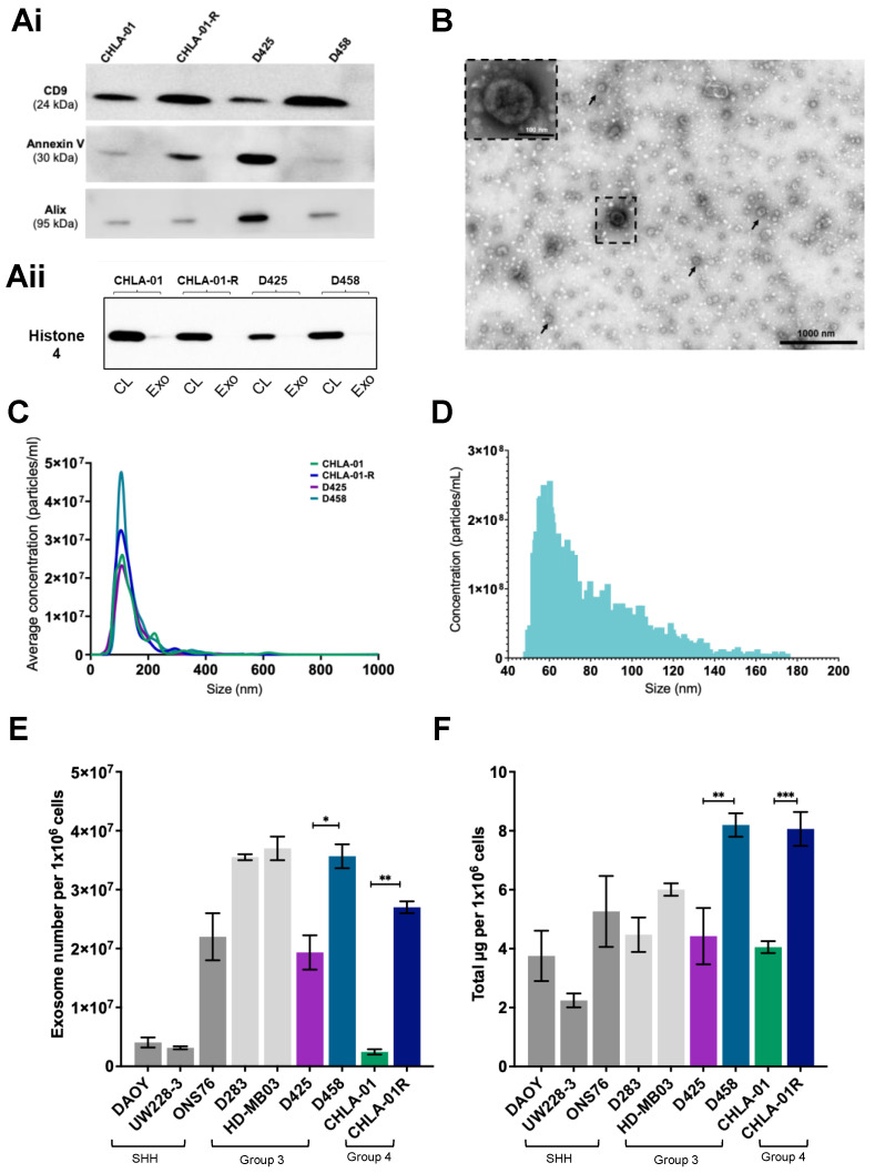
Extracellular vesicles (EVs) have emerged as pivotal mediators of communication in the tumour microenvironment. More specifically, nanosized extracellular vesicles termed exosomes have been shown to contribute to the establishment of a premetastatic niche. Here, we sought to determine what role exosomes play in medulloblastoma (MB) progression and elucidate the underlying mechanisms. Metastatic MB cells (D458 and CHLA-01R) were found to secrete markedly more exosomes compared to their nonmetastatic, primary counterparts (D425 and CHLA-01). In addition, metastatic cell-derived exosomes significantly enhanced the migration and invasiveness of primary MB cells in transwell migration assays. Protease microarray analysis identified that matrix metalloproteinase-2 (MMP-2) was enriched in metastatic cells, and zymography and flow cytometry assays of metastatic exosomes demonstrated higher levels of functionally active MMP-2 on their external surface. Stable genetic knockdown of MMP-2 or extracellular matrix metalloproteinase inducer (EMMPRIN) in metastatic MB cells resulted in the loss of this promigratory effect. Analysis of serial patient cerebrospinal fluid (CSF) samples showed an increase in MMP-2 activity in three out of four patients as the tumour progressed. This study demonstrates the importance of EMMPRIN and MMP-2-associated exosomes in creating a favourable environment to drive medulloblastoma metastasis via extracellular matrix signalling.

Keywords: EMMPRIN; MMP-2; exosomes; extracellular vesicles; medulloblastoma; metastasis.


Conflict of interest statement

The authors declare no conflict of interest. The funders had no role in the design of the study; in the collection, analyses, or interpretation of data; in the writing of the manuscript; or in the decision to publish the results.

Figures

Figure 1
Figure 1
Isolation and characterisation of exosomes from medulloblastoma cell lines. Western blot confirmation that exosomes (Ai) express the marker proteins CD9, Annexin V, and Alix, and show no cross contamination with the nuclear protein histone 4 which is only detected in cell lysate (CL) rather than exosome (Exo) fractions (Aii). The uncropped blots are shown in Figure S1. (B) Exosomes from CHLA-01 cells were imaged by transmission electron microscopy (TEM) and identified as multiple cup-shaped structures ranging from 30–150 nm in size (arrowheads). Scale bar 1000 nm. (C,D) Exosomal particle concentrations were measured by NTA (C) and NanoFCM (D). NTA data represent the average of at least three independent repeats. Representative NanoFCM data is shown for CHLA-01-derived exosomes. (E,F) Metastatic cell lines (D283, HD-MB03, D458, and CHLA-01-R) typically secrete more exosomes than the primary cell lines as determined by exosomal number (E) and exosomal protein content (F), both corrected for cell density. SHH (sonic hedgehog). Significant differences were calculated using one-way ANOVA analyses with Sidak’s multiple-comparisons test, (* p ≤ 0.05, ** p ≤ 0.01, *** p ≤ 0.005). Data represent the average of three independent experiments with error bars indicating the standard error of the mean (SEM).
Figure 2
Figure 2
Exosomes from metastatic cell lines can confer a proinvasive phenotype on recipient cells. (A) Experimental setup for migration through an uncoated chamber insert, or invasion and migration through a collagen IV- and laminin I-coated insert. (B,C) Exosomes derived from the metastatic cell lines CHLA-01R and D458 were applied (+Exo) to matched primary cell lines CHLA-01 (B) and D425 (C), respectively, and led to an increase in cell invasion behaviour (grey bars) compared to exosome-free supernatant control cells (coloured bars). (D,E) Exosomes from metastatic cell lines conferred only a modest effect on cell proliferation determined by Presto Blue viability assays. (F,G) Exosomes from the metastatic CHLA-01R cell line were able to confer an invasive phenotype on the noncancerous FB83 cell line (F), independent of an effect on cell proliferation (G). Significant differences in migration were calculated using one-way ANOVA analyses with Dunnett’s multiple-comparisons post hoc test, (* p ≤ 0.05, ** p ≤ 0.01, *** p ≤ 0.005) (ns = not significant). Data represent the average of three independent experiments with error bars indicating the standard error of the mean (SEM).
Figure 3
Figure 3
MMP-2 and EMMPRIN are expressed on exosomes released by medulloblastoma cell lines. Exosomal EMMPRIN and MMP2 protein expression was determined by Western blotting (representative image in panel (Ai) and was quantified relative to Alix protein expression (Aii,Aiii). (B). Flow-cytometry analysis of EMMPRIN (B,C) and MMP-2 (D,E) in medulloblastoma-derived exosomes. Exosomes from the metastatic and primary matched pair of cell lines (D458 and D425) were labelled with anti-EMMPRIN or anti-MMP-2 antibodies and secondary antibodies conjugated to either PE (EMMPRIN) or APC (MMP-2). Gating was identical across cell lines and the percentage of cells in populations representing high and low expression is shown. One representative experiment out of two performed with similar results is shown. Data in (AiAiii) represent the average of three independent experiments with error bars indicating SEM. Significant differences in protein expression between exosomes derived from the matched cell lines were calculated using the Kruskal–Wallis test with Dunn’s multiple-comparisons post hoc test (* p ≤ 0.05, ns = not significant). The uncropped blots are shown in Supplementary Figure S6.
Figure 4
Figure 4
Exosomes contain functionally active MMP-2 and can transfer gelatinase activity onto recipient cell lines. (A) Gelatin zymography was used for the detection of MMP-2 activity in medulloblastoma-derived exosomes. The 5 μg of exosomes suspended in PBS were loaded onto gelatin-incorporated zymography gels and the functional activity of the gelatinase and MMP-2 was determined. Data are representative of three independent experiments. (B) Schematic representation of the stimulation of a low MMP-2 expressing cell line (CHLA-01) with 10 μg of exosomes from a high MMP-2 expressing cell lines (CHLA-01R). After 24 and 48 h, supernatant from the recipient cell line was collected and loaded onto the gelatin-containing gel. Proteolysis was detected as a white band. MMP-2 levels shown in (C) were quantified and displayed graphically in (D). Data represent the average of two independent experiments with error bars indicating the standard error of the mean (SEM). Significant differences were calculated using one-way ANOVA analyses with Dunnett’s multiple-comparisons post hoc test, (* p ≤ 0.05, ** p ≤ 0.01). The uncropped blots are shown in File S1.
Figure 5
Figure 5
Knockdown of MMP-2 and EMMPRIN appears to reduce exosome-mediated migration and invasion in group four medulloblastoma cell lines. (A) Group four cell invasion through a collagen and laminin IV coated transwell chamber insert was quantified by PrestoBlue metabolic staining and compared to cell lines transduced with nontargeting (NT). (B) Exosomes from the CHLA-01R knockdown cell lines and nontransduced cells were isolated and their size and concentration were measured by NanoFCM. (Ci) Western blot analysis and concurrent densitometry revealed both EMMPRIN (Cii) and MMP-2 (Ciii) protein levels to be significantly depleted in exosomes isolated from the knockdown cell lines. Densitometry data are presented relative to the Alix loading control and compared to the appropriate nonsilencing control cell line. (D) Exosomes from the CHLA-01R knockdown cell lines were applied to the matched parental cell line CHLA-01 (+NT exo: nontargetting, +shBSG exo, knockdown BSG, or shMMP-2 exo knockdown MMP-2) following which MMP-2 mRNA expression levels in recipient cells was determined by qRT-PCR. (E) The ability of these recipient cell lines to invade through a laminin and collage IV matrix was determined by metabolic assay. MMP-2 activity in the medium of CHLA-01 cells receiving exosomes was determined by gelatin zymography. Proteolysis was detected as a white band. MMP-2 levels shown in (Fi) were quantified and displayed graphically in (Fii). Significance was assessed by ordinary one-way ANOVA analysis with Sidak’s multiple-comparison tests (* p ≤ 0.05, ** p ≤ 0.01) (ns = not significant). Data represent the average of at least two independent experiments with error bars indicating the standard error of the mean (SEM). The uncropped blots are shown in File S1.
Figure 6
Figure 6
Determination of MMP-2 activity in clinical CSF samples from medulloblastoma patients indicates MMP-2 activity as a possible marker of disease progression. (A) Clinical characteristics of 4 medulloblastoma patients. (B) Axial MRI with gadolinium T1 and T2 images taken at presentation (top row) and at disease progression (bottom row) with tumours highlighted with white arrows. (C) Paired CSF sampled from the patients was examined and the levels of functional activity of MMP-2 determined and normalised to recombinant MMP-2, at their presentation and disease progression. The uncropped blots are shown in File S1.

References

    1. Hill R.M., Richardson S., Schwalbe E.C., Hicks D., Lindsey J.C., Crosier S., Rafiee G., Grabovska Y., Wharton S.B., Jacques T.S., et al. Time, Pattern, and Outcome of Medulloblastoma Relapse and Their Association with Tumour Biology at Diagnosis and Therapy: A Multicentre Cohort Study. Lancet Child Adolesc. Health. 2020;4:865–874. doi: 10.1016/S2352-4642(20)30246-7. - DOI - PMC - PubMed
    1. Ramaswamy V., Remke M., Bouffet E., Bailey S., Clifford S.C., Doz F., Kool M., Dufour C., Vassal G., Milde T., et al. Risk Stratification of Childhood Medulloblastoma in the Molecular Era: The Current Consensus. Acta Neuropathol. 2016;131:821–831. doi: 10.1007/s00401-016-1569-6. - DOI - PMC - PubMed
    1. Van Niel G., D’Angelo G., Raposo G. Shedding Light on the Cell Biology of Extracellular Vesicles. Nat. Rev. Mol. Cell Biol. 2018;19:213–228. doi: 10.1038/nrm.2017.125. - DOI - PubMed
    1. Banks W.A., Sharma P., Bullock K.M., Hansen K.M., Ludwig N., Whiteside T.L. Transport of Extracellular Vesicles across the Blood-Brain Barrier: Brain Pharmacokinetics and Effects of Inflammation. Int. J. Mol. Sci. 2020;21:4407. doi: 10.3390/ijms21124407. - DOI - PMC - PubMed
    1. Magaña S.M., Peterson T.E., Evans J.E., Decker P.A., Simon V., Eckel-Passow J.E., Daniels D.J., Parney I.F. Pediatric Brain Tumor Cell Lines Exhibit MiRNA-Depleted, Y RNA-Enriched Extracellular Vesicles. J. Neurooncol. 2022;156:269–279. doi: 10.1007/s11060-021-03914-4. - DOI - PubMed

LinkOut - more resources