

Retrospective Analysis of Artifacts in Cone Beam Computed Tomography Images Used to Diagnose Chronic Rhinosinusitis
- PMID: 37174903
- PMCID: PMC10177128
- DOI: 10.3390/diagnostics13091511
Retrospective Analysis of Artifacts in Cone Beam Computed Tomography Images Used to Diagnose Chronic Rhinosinusitis
Abstract
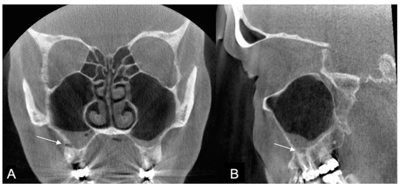
Background: Cone beam computed tomography (CBCT) is frequently used to corroborate the signs and symptoms of chronic rhinosinusitis (CRS). However, artifacts induced by dental restorations might complicate the diagnosis of CRS. Here, we assessed the frequency and location of artifacts in CBCT images taken to confirm the CRS.
Methods: All CBCT images of the patients referred to the Emergency Radiology unit, Turku University Hospital, with an indication of CRS in 2017 were re-examined. The prevalence of the artifacts was analyzed in three cross-sectional views and three horizontal levels delimited by anatomical landmarks.
Results: In total, 214 CBCT images of patients with CRS were evaluated. The diagnosis of apical periodontitis (AP) was impaired by artifacts present in 150/214 images (70%). The diagnosis of CRS was impaired in 5 of the 214 images (2.3%). The main origins of the artifacts were large dental fillings or crowns, and endodontic fillings were present in 95% (203/214) and 52% (111/214) of the images, respectively.
Conclusions: AP as an etiology of CRS is possible to miss because of artifacts originating from dental and endodontic fillings in the CBCT images of the paranasal sinuses.
Keywords: apical periodontitis; artifact; cone beam CT; dental restorations; maxilla; rhinosinusitis.
Conflict of interest statement
The authors declare no conflict of interest.
Figures


Similar articles
-
Curved Planar Reformation: A Useful Method for Screening Dental Pathologies in Chronic Rhinosinusitis via Paranasal Sinus Computed Tomography.Tomography. 2022 Sep 16;8(5):2330-2338. doi: 10.3390/tomography8050194. Tomography. 2022. PMID: 36136890 Free PMC article.
-
Cone-beam Computed Tomographic-based Assessment of Filled C-shaped Canals: Artifact Expression of Cone-beam Computed Tomography as Opposed to Micro-computed Tomography and Nano-computed Tomography.J Endod. 2020 Nov;46(11):1702-1711. doi: 10.1016/j.joen.2020.07.010. Epub 2020 Jul 16. J Endod. 2020. PMID: 32682791
-
Clinical Factors Associated with Apical Periodontitis Visible on Cone-beam Computed Tomography but Missed with Periapical Radiographs: A Retrospective Clinical Study.J Endod. 2020 Jun;46(6):832-838. doi: 10.1016/j.joen.2020.03.005. Epub 2020 Apr 12. J Endod. 2020. PMID: 32295704
-
Measurement Accuracy in Cone Beam Computed Tomography in the Presence of Metal Artifact.Int J Oral Maxillofac Implants. 2022 Jan-Feb;37(1):143-152. doi: 10.11607/jomi.9079. Int J Oral Maxillofac Implants. 2022. PMID: 35235633
-
Metal Artifact Reduction in Cone-Beam Computed Tomography for Head and Neck Radiotherapy.Technol Cancer Res Treat. 2016 Dec;15(6):NP88-NP94. doi: 10.1177/1533034615618319. Epub 2015 Nov 26. Technol Cancer Res Treat. 2016. PMID: 26614780
References
-
- Lu Y., Liu Z., Zhang L., Zhou X., Zheng Q., Duan X., Zheng G., Wang H., Huang D. Associations between Maxillary Sinus Mucosal Thickening and Apical Periodontitis Using Cone-Beam Computed Tomography Scanning: A Retrospective Study. J. Endod. 2012;38:1069–1074. doi: 10.1016/j.joen.2012.04.027. - DOI - PubMed
LinkOut - more resources
Full Text Sources
