Review
. 2023 Apr 25;13(9):1541.
doi: 10.3390/diagnostics13091541.

Point-of-Care Ultrasound in Airway Evaluation and Management: A Comprehensive Review

Affiliations
Review

Point-of-Care Ultrasound in Airway Evaluation and Management: A Comprehensive Review

Judy Lin et al. Diagnostics (Basel). .

Abstract

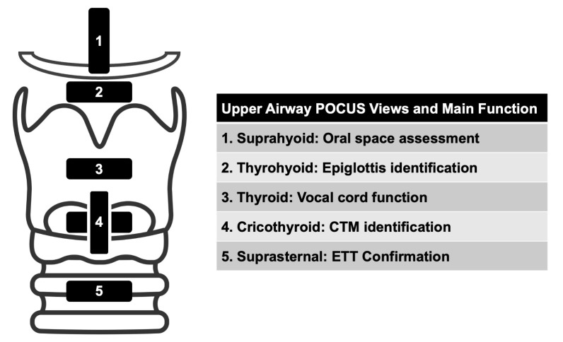
Airway management is a common and critical procedure in acute settings, such as the Emergency Department (ED) or Intensive Care Unit (ICU) of hospitals. Many of the traditional physical examination methods have limitations in airway assessment. Point-of-care ultrasound (POCUS) has emerged as a promising tool for airway management due to its familiarity, accessibility, safety, and non-invasive nature. It can assist physicians in identifying relevant anatomy of the upper airway with objective measurements of airway parameters, and it can guide airway interventions with dynamic real-time images. To date, ultrasound has been considered highly accurate for assessment of the difficult airway, confirmation of proper endotracheal intubation, prediction of post-extubation laryngeal edema, and preparation for cricothyrotomy by identifying the cricothyroid membrane. This review aims to provide a comprehensive overview of the key evidence on the use of ultrasound in airway management. Databases including PubMed and Embase were systematically searched. A search strategy using a combination of the term "ultrasound" combined with several search terms, i.e., "probe", "anatomy", "difficult airway", "endotracheal intubation", "laryngeal edema", and "cricothyrotomy" was performed. In conclusion, POCUS is a valuable tool with multiple applications ranging from pre- and post-intubation management. Clinicians should consider using POCUS in conjunction with traditional exam techniques to manage the airway more efficiently in the acute setting.

Keywords: cricothyrotomy; difficult airway; endotracheal intubation; point-of-care; ultrasound.


Conflict of interest statement

The authors declare no conflict of interest.

Figures

Figure 1
Figure 1
Views to Assess the Upper Airway.
Figure 2
Figure 2
Suprahyoid View. (A) Suprahyoid probe placement on the subject’s neck in sagittal orientation. (B) Suprahyoid view of anterior neck with curvilinear probe in sagittal orientation and probe indicator directed cranially. The mentum of the mandible is indicated by the solid arrow and the hyoid bone is indicated by the dashed arrow. Deep to the hypoechoic mylohyoid and geniohyoid muscles (single star) lies the tongue (double star). The hyomental distance (HMD) is spanned by the double-headed arrow.
Figure 3
Figure 3
Thyrohyoid View. (A) Thyrohyoid probe placement on subject’s neck. (B) Thyrohyoid view of anterior neck with linear probe in transverse orientation. The pre-epiglottic space (solid, double-headed arrow) appears between the thyrohyoid membrane (solid, single-headed arrow) and the epiglottis (dashed arrows). The strap muscles (stars) are again visible superficially to the thyrohyoid membrane. The distance from skin to epiglottis (DSE) is spanned by the two dashed lines.
Figure 4
Figure 4
Thyroid View. (A) Thyroid probe placement on subject’s neck. (B) Thyroid view of anterior neck with linear probe in transverse orientation. The vocal cords (dashed arrows) join together at the anterior commissure (solid arrow). The thyroid cartilage (dashed lines) appears lateral to the vocal cords, and the strap muscles (stars) just superficial to the thyroid cartilage. Although not well visualized in this image, the arytenoids will usually appear at the posterior aspect of the bilateral vocal cords.
Figure 5
Figure 5
Cricothyroid View: Transverse and Sagittal Orientations. (A) Cricothyroid probe placement on subject’s neck in transverse orientation. (B) Cricothyroid view of anterior neck with linear probe in transverse orientation. The cricothyroid membrane (solid arrow) overlies the trachea with prominent reverberation artifact (dashed arrow) in the tracheal lumen. (C) Cricothyroid probe placement on subject’s neck in sagittal orientation. (D) Cricothyroid view of anterior neck with linear probe in sagittal orientation. The thyroid cartilage (solid arrow) appears superior to the hypoechoic cricoid cartilage (star). The cricothyroid membrane (dashed arrow) spans between the two cartilages.
Figure 6
Figure 6
Suprasternal View. (A) Suprasternal probe placement on subject’s neck. (B) Suprasternal view of anterior neck with curvilinear probe. The tracheal cartilage (solid arrow) appears hyperechoic with reverberation artifact noted in the air-filled tracheal lumen posteriorly. The common carotid arteries (stars) and internal jugular veins (dashed arrow) appear laterally on each side of the trachea.
Figure 7
Figure 7
Difficult Airway Evaluation with Sonography (DARES) Protocol. Abbreviations: DSE, distance to epiglottis; HMD, hyomental distance; TT, tongue thickness; HMDR2, HMD extended/HMD neutral; HMDR1, HMD ramped/HMD neutral. * Falcetta, 2018 [16]; Wu, 2022 [61]; Yao, 2017 [51]; § Rana, 2018 [44]; Petrisor, 2018 [60].
Figure 8
Figure 8
Confirmation of Endotracheal Intubation with Ultrasound. (A) Place the transducer transversely on the anterior neck at the level of the suprasternal notch for the best visualization and diagnostic accuracy. (B) Tracheal ETT position: only one A–M interface (arrow) with comet tail artifact (arrowhead) and posterior shadowing is observed (C) Esophageal ETT position: two A–M interfaces (arrows) with comet-tail artifacts (arrowheads) and posterior shadowing are noted (the “double tract” sign).
Figure 9
Figure 9
Air column width (double-headed arrow). (A) With ETT cuff up. (B) With ETT cuff down.
Figure 10
Figure 10
The longitudinal “string of pearls” technique and the transverse “thyroid-airline-cricoid-airline” (TACA) protocol to identify the CTM. (A) String of pearls with cricoid cartilage (asterisk). (B) “T” in “TACA” with outline thyroid cartilage (dashed lines). (C) “A” in “TACA” with hyperechoic CTM (solid arrow). (D) “C” in “TACA” with hypoechoic cricoid cartilage (solid arrow). (E) “A” in “TACA” with hyperechoic CTM (solid arrow).

References

    1. Nee P.A., Benger J., Walls R.M. Airway management. Emerg. Med. J. 2008;25:98–102. doi: 10.1136/emj.2005.030635. - DOI - PubMed
    1. O’Dell K. Predictors of difficult intubation and the otolaryngology perioperative consult. Anesth. Clin. 2015;33:279–290. doi: 10.1016/j.anclin.2015.02.002. - DOI - PubMed
    1. Brown C.A., 3rd, Bair A.E., Pallin D.J., Walls R.M., Near III Investigators Techniques, success, and adverse events of emergency department adult intubations. Ann. Emerg. Med. 2015;65:363–370.e1. doi: 10.1016/j.annemergmed.2014.10.036. - DOI - PubMed
    1. Diaz-Gomez J.L., Mayo P.H., Koenig S.J. Point-of-Care Ultrasonography. N. Engl. J. Med. 2021;385:1593–1602. doi: 10.1056/NEJMra1916062. - DOI - PubMed
    1. Ultrasound Guidelines: Emergency, Point-of-Care and Clinical Ultrasound Guidelines in Medicine. Ann. Emerg. Med. 2017;69:e27–e54. doi: 10.1016/j.annemergmed.2016.08.457. - DOI - PubMed

LinkOut - more resources