Review
. 2023 May 1;13(9):1607.
doi: 10.3390/diagnostics13091607.

Pulmonary Hypertension in Chronic Lung Diseases: What Role Do Radiologists Play?

Affiliations
Review

Pulmonary Hypertension in Chronic Lung Diseases: What Role Do Radiologists Play?

Adele Valentini et al. Diagnostics (Basel). .

Abstract

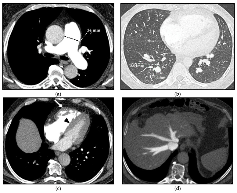
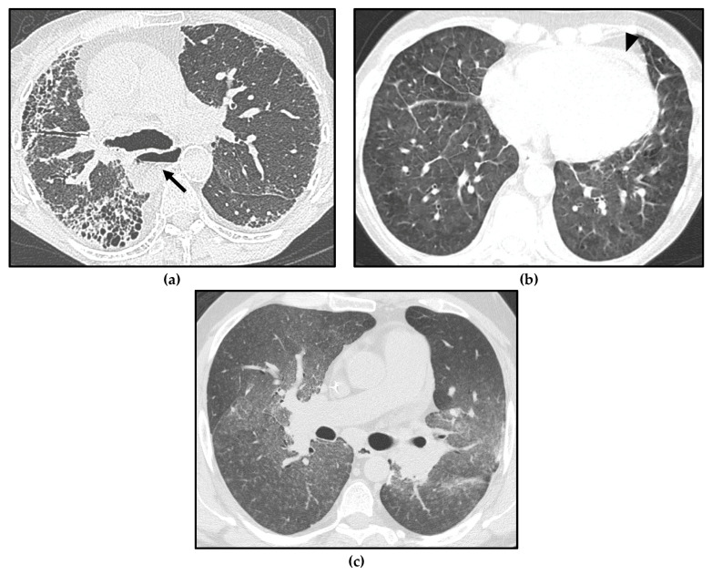
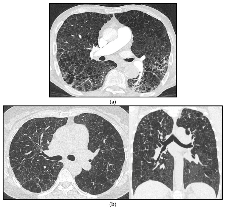
Pulmonary hypertension (PH) is a pathophysiological disorder, defined by a mean pulmonary arterial pressure (mPAP) > 20 mmHg at rest, as assessed by right heart catheterization (RHC). PH is not a specific disease, as it may be observed in multiple clinical conditions and may complicate a variety of thoracic diseases. Conditions associated with the risk of developing PH are categorized into five different groups, according to similar clinical presentations, pathological findings, hemodynamic characteristics, and treatment strategy. Most chronic lung diseases that may be complicated by PH belong to group 3 (interstitial lung diseases, chronic obstructive pulmonary disease, combined pulmonary fibrosis, and emphysema) and are associated with the lowest overall survival among all groups. However, some of the chronic pulmonary diseases may develop PH with unclear/multifactorial mechanisms and are included in group 5 PH (sarcoidosis, pulmonary Langerhans' cell histiocytosis, and neurofibromatosis type 1). This paper focuses on PH associated with chronic lung diseases, in which radiological imaging-particularly computed tomography (CT)-plays a crucial role in diagnosis and classification. Radiologists should become familiar with the hemodynamical, physiological, and radiological aspects of PH and chronic lung diseases in patients at risk of developing PH, whose prognosis and treatment depend on the underlying disease.

Keywords: chronic lung diseases; computed tomography; imaging; pulmonary hypertension.


Conflict of interest statement

The authors declare no conflict of interest.

Figures

Figure 1
Figure 1
Chest X-ray (CXR) signs of pulmonary hypertension: enlarged central pulmonary arteries, with the prominence of the main pulmonary artery (MPA) (black arrow); the left pulmonary artery seen within the cardiac silhouette (dotted line); tapering of peripheral pulmonary arteries (so-called pruning); prominence of the right heart border, represented by the right atrium (black arrowhead); and filling of the retrosternal space in the lateral view due to right ventricle dilation (white arrow).
Figure 2
Figure 2
Cardiac magnetic resonance (CMR) in a patient affected by pulmonary arterial hypertension (PAH) in congenital heart disease with anomalous pulmonary venous return and intracardiac shunt. The Steady-state free precession (SSFP) in the four-chamber orientation in the diastolic phase shows marked dilation of the right ventricle (RV) and the compressed left ventricle (LV) (a). The SSFP in the short-axis orientation in the systolic phase depicts right ventricle dilation and leftward ventricular septal bowing (arrow) (b). The delayed enhancement CMR image (10 min post-gadolinium infusion) demonstrates mid-wall septal fibrosis and fibrosis at the anterior and posterior junctions between the septum and the RV free wall (arrowheads) (c).
Figure 2
Figure 2
Cardiac magnetic resonance (CMR) in a patient affected by pulmonary arterial hypertension (PAH) in congenital heart disease with anomalous pulmonary venous return and intracardiac shunt. The Steady-state free precession (SSFP) in the four-chamber orientation in the diastolic phase shows marked dilation of the right ventricle (RV) and the compressed left ventricle (LV) (a). The SSFP in the short-axis orientation in the systolic phase depicts right ventricle dilation and leftward ventricular septal bowing (arrow) (b). The delayed enhancement CMR image (10 min post-gadolinium infusion) demonstrates mid-wall septal fibrosis and fibrosis at the anterior and posterior junctions between the septum and the RV free wall (arrowheads) (c).
Figure 3
Figure 3
Cardiac magnetic resonance (CMR) phase contrast imaging in the same patient as in Figure 2. Magnitude (a,d) and velocity (b,e) images in phase contrast imaging through the main pulmonary artery (MPA) and aorta (Ao) for flow quantification; note the increase of the main pulmonary artery caliber, larger than the aortic one. Graphical representation of flow velocity in the MPA (c) and in the Ao (f) (x-axis: time in msec; y-axis: flow velocity in mL/s) demonstrates a markedly increased flow in the MPA compared to the Ao, with a calculated pulmonary blood flow (Qp) of 9.96 L/min and a systemic blood flow (Qs) of 3.45 L/min, with a Qp/Qs ratio of 2.8, indicative of left-to-right shunt.
Figure 4
Figure 4
Computed tomography (CT) indirect signs of pulmonary hypertension (PH). Vascular CT signs: main pulmonary artery (MPA) dilation (34 mm) (a) and increased segmental artery-to-bronchus ratio (>1) (b). Cardiac CT signs: right ventricle (RV) hypertrophy, with free wall thickness > 4 mm and trabecular hypertrophy (arrow), and flattening of the ventricular septum (arrowhead) (c). Extensive reflux of contrast medium into the inferior vena cava and hepatic veins (d).
Figure 5
Figure 5
Different forms of pulmonary hypertension (PH) in systemic sclerosis (SSc) patients. Interstitial lung disease (ILD) showing a usual interstitial pneumonia (UIP) pattern, with asymmetric distribution of fibrotic abnormalities and honeycombing foci in the subpleural zones, especially in the right middle lobe and right lower lobe (a); note massive esophageal dilation, with the air–fluid level (arrow) and the enlargement of the main pulmonary artery (PA). Pulmonary veno-occlusive disease/ pulmonary capillary hemangiomatosis (PVOD/PCH), with smooth interlobular septal thickening and diffuse ground-glass opacities in the lung bases (b); note the mild pericardial effusion, which may be linked either to serositis or to PH (arrowhead). Pulmonary arterial hypertension (PAH) characterized by profuse bilateral tiny centrilobular ground-glass micronodules (c).
Figure 6
Figure 6
Pulmonary hypertension (PH) in a chronic obstructive pulmonary disease (COPD) patient with confluent centrilobular pulmonary emphysema (a). Multiple confluent bilateral irregular cysts in a patient affected by advanced pulmonary Langerhans’ cell histiocytosis (PLCH), complicated by PH (note the increased mean pulmonary artery diameter) (b); in advanced cases, the distribution of parenchymal abnormalities shows a rather diffuse longitudinal distribution, as seen in the coronal view, and the differential diagnosis with pulmonary emphysema is not straightforward.

References

    1. Humbert M., Kovacs G., Hoeper M.M., Badagliacca R., Berger R.M.F., Brida M., Carlsen J., Coats A.J.S., Escribano-Subias P., Ferrari P., et al. ESC/ERS Scientific Document Group 2022 ESC/ERS Guidelines for the diagnosis and treatment of pulmonary hypertension. Eur. Heart J. 2022;43:3618–3731. doi: 10.1093/eurheartj/ehac237. - DOI - PubMed
    1. Remy-Jardin M., Ryerson C.J., Schiebler M.L., Leung A.N.C., Wild J.M., Hoeper M.M., Alderson P.O., Goodman L.R., Mayo J., Haramati L.B., et al. Imaging of pulmonary hypertension in adults: A position paper from the Fleischner Society. Eur. Respir. J. 2021;57:2004455. doi: 10.1183/13993003.04455-2020. - DOI - PubMed
    1. Maron B.A., Hess E., Maddox T.M., Opotowsky A.R., Tedford R.J., Lahm T., Joynt K.E., Kass D.J., Stephens T., Stanislawski M.A., et al. Association of borderline pulmonary hypertension with mortality and hospitalization in a large patient cohort: Insights from the veterans affairs clinical assessment, reporting, and tracking program. Circulation. 2016;133:1240–1248. doi: 10.1161/CIRCULATIONAHA.115.020207. - DOI - PMC - PubMed
    1. Kolte D., Lakshmanan S., Jankowich M.D., Brittain E.L., Maron B.A., Choudhary G. Mild Pulmonary Hypertension Is Associated With Increased Mortality: A Systematic Review and Meta-Analysis. J. Am. Heart Assoc. 2018;7:e009729. doi: 10.1161/JAHA.118.009729. - DOI - PMC - PubMed
    1. Sharma M., Burns A.T., Yap K., Prior D.L. The role of imaging in pulmonary hypertension. Cardiovasc. Diagn. Ther. 2021;11:859–880. doi: 10.21037/cdt-20-295. - DOI - PMC - PubMed