Review
. 2023 May 2;13(9):1612.
doi: 10.3390/diagnostics13091612.

The Role of Ultrasound in the Diagnosis of Pulmonary Infection Caused by Intracellular, Fungal Pathogens and Mycobacteria: A Systematic Review

Affiliations
Review

The Role of Ultrasound in the Diagnosis of Pulmonary Infection Caused by Intracellular, Fungal Pathogens and Mycobacteria: A Systematic Review

Mariaclaudia Meli et al. Diagnostics (Basel). .

Abstract

Background: Lung ultrasound (LUS) is a widely available technique allowing rapid bedside detection of different respiratory disorders. Its reliability in the diagnosis of community-acquired lung infection has been confirmed. However, its usefulness in identifying infections caused by specific and less common pathogens (e.g., in immunocompromised patients) is still uncertain.

Methods: This systematic review aimed to explore the most common LUS patterns in infections caused by intracellular, fungal pathogens or mycobacteria.

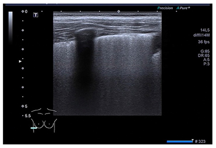
Results: We included 17 studies, reporting a total of 274 patients with M. pneumoniae, 30 with fungal infection and 213 with pulmonary tuberculosis (TB). Most of the studies on M. pneumoniae in children found a specific LUS pattern, mainly consolidated areas associated with diffuse B lines. The typical LUS pattern in TB consisted of consolidation and small subpleural nodes. Only one study on fungal disease reported LUS specific patterns (e.g., indicating "halo sign" or "reverse halo sign").

Conclusions: Considering the preliminary data, LUS appears to be a promising point-of-care tool, showing patterns of atypical pneumonia and TB which seem different from patterns characterizing common bacterial infection. The role of LUS in the diagnosis of fungal disease is still at an early stage of exploration. Large trials to investigate sonography in these lung infections are granted.

Keywords: lung ultrasound; pneumonia; pulmonary infection.


Conflict of interest statement

The authors declare no conflict of interest.

Figures

Figure 1
Figure 1
PRISMA flow-chart [22].
Figure 2
Figure 2
Cosolidation and air bronchogram in Mycoplasma pneumonia [31].
Figure 3
Figure 3
Subpleural effusions in Mycoplasma pneumonia [32].
Figure 4
Figure 4
B lines in Mycoplasma pneumonia [31].
Figure 5
Figure 5
Consolidation with air bronchogram in fungal pneumonia [37].
Figure 6
Figure 6
B-lines (AC), Atelectasias, consolidation with air bronchogram, shred signs in fungal pneumonioa [37].
Figure 7
Figure 7
Comparison between LUS images and CT images in fungal pneumonia [40].
Figure 8
Figure 8
(A,B) Multiple nodular lesions with peripheral ground-glass opacity or halo sign, reverse halo sign, and air crescent sign. (C) The target lesions; a hyper-echoic central nodule with a hypo-echoic rim. (D) Hyper-echoic nodule (fungus ball) within consolidaion. (E) Hyper-echoic fungus ball with air exension at the peripheral hup-echoic rim [36].
Figure 9
Figure 9
(A) Multiple nodular lesions with ground-glass opacity or halo sign, without air crescent sign. Two target lesions in ultrasound. (B) Hyper-echoic central nodule with a hypo-echoic rim. Figure (C) Hypo-echoic center with a hyper-echoic rim. (D,E) A nodular lesion of the right lung that has developed and air crescent sign in control HRCT [36].
Figure 10
Figure 10
(A,B) Multiple nodular lesions with cavitation. (C) The target lesion in ultrasound has a Hypo-echoic center with a hyper-echoic rim [36].
Figure 11
Figure 11
Subpleural consolidation (thick arrow), characterized by subpleural hypo-echoic region < 1 × 1 cm, with distinct borders and trailing artifact (arrowhead), next to the normal white pleural line (thin arrow) [44].
Figure 12
Figure 12
Consolidation (thick arrow), characterized by echo-poor region > 1 × 1 cm, with air bronchograms [44].
Figure 13
Figure 13
Cavitation in mycobacterial pneumonia [45].

References

    1. Joyner C., Herman R.J., Reid J.M. Reflected ultrasound in the detection and localization of pleural effusion. JAMA. 1967;200:399–402. doi: 10.1001/jama.1967.03120180087013. - DOI - PubMed
    1. Lichtenstein D., Mézière G., Biderman P., Gepner A., Barrè O. The comettail artifact. An ultrasound sign of alveolar-interstitial syndrome. Am. J. Respir. Crit. Care Med. 1997;156:1640–1646. doi: 10.1164/ajrccm.156.5.96-07096. - DOI - PubMed
    1. Soldati G., Testa A., Silva F.R., Carbone L., Portale G., Silveri N.G. Chest ultrasonography in lung contusion. Chest. 2006;130:533–538. doi: 10.1378/chest.130.2.533. - DOI - PubMed
    1. Chavez M.A., Shams N., Ellington L.E., Naithani N., Gilman R.H., Steinhoff M.C., Santosham M., Black R.E., Price C., Gross M., et al. Lung ultrasound for the diagnosis of pneumonia in adults: A systematic review and meta-analysis. Respir. Res. 2014;15:50. doi: 10.1186/1465-9921-15-50. - DOI - PMC - PubMed
    1. Iorio G., Capasso M., De Luca G., Prisco S., Mancusi C., Laganà B., Comune V. Lung Ultrasound in the Diagnosis of Pneumonia in Children: Proposal for a New Diagnostic Algorithm. Peer J. 2015;3:e1374. doi: 10.7717/peerj.1374. - DOI - PMC - PubMed

LinkOut - more resources