Review
. 2023 May 7;13(9):1647.
doi: 10.3390/diagnostics13091647.

Pitfalls of Diffusion-Weighted Imaging: Clinical Utility of T2 Shine-through and T2 Black-out for Musculoskeletal Diseases

Affiliations
Review

Pitfalls of Diffusion-Weighted Imaging: Clinical Utility of T2 Shine-through and T2 Black-out for Musculoskeletal Diseases

Yuri Kim et al. Diagnostics (Basel). .

Abstract

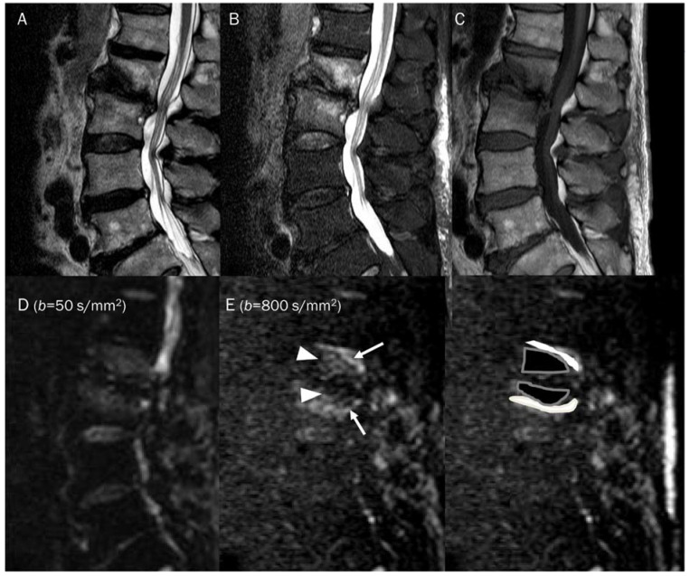
Diffusion-weighted imaging (DWI) with an apparent diffusion coefficient (ADC) value is a relatively new magnetic resonance imaging (MRI) sequence that provides functional information on the lesion by measuring the microscopic movement of water molecules. While numerous studies have evaluated the promising role of DWI in musculoskeletal radiology, most have focused on tumorous diseases related to cellularity. This review article aims to summarize DWI-acquisition techniques, considering pitfalls such as T2 shine-through and T2 black-out, and their usefulness in interpreting musculoskeletal diseases with imaging. DWI is based on the Brownian motion of water molecules within the tissue, achieved by applying diffusion-sensitizing gradients. Regardless of the cellularity of the lesion, several pitfalls must be considered when interpreting DWI with ADC values in musculoskeletal radiology. This review discusses the application of DWI in musculoskeletal diseases, including tumor and tumor mimickers, as well as non-tumorous diseases, with a focus on lesions demonstrating T2 shine-through and T2 black-out effects. Understanding these pitfalls of DWI can provide clinically useful information, increase diagnostic accuracy, and improve patient management when added to conventional MRI in musculoskeletal diseases.

Keywords: apparent diffusion coefficient; diffusion-weighted imaging; magnetic resonance imaging; musculoskeletal diseases; pitfall.


Conflict of interest statement

The authors declare no conflict of interest.

Figures

Figure 1
Figure 1
A 60-year-old female with giant-cell tumor with secondary aneurysmal bone cyst change in the distal femur. (A) Coronal T2-weighted image and (B) axial T2-weighted fat-suppressed image show secondary aneurysmal bone cyst change with fluid–fluid level. (C) The DWI with b-value of 0 s/mm2 appears similar to the heavily T2-weighted fat-suppressed image (B).
Figure 2
Figure 2
Progressive increase in b-values (same patient in Figure 1). As b-values increase on DWI (AC), the perfusion effect is gradually suppressed and only hypercellular tissues remain bright at high b-value images with a darkening background.
Figure 3
Figure 3
DWI of a pathologic lymph node, a normal submandibular gland, and a normal spinal cord. (A) Axial T1-weighted enhanced image of the neck shows a pathologic lymph node (arrow), a normal submandibular gland (arrowhead), and a normal spinal cord (curved arrow). These hypercellular tissues remain bright on DWI with increasing b-value images (B,C).
Figure 4
Figure 4
A 25-year-old female with a soft tissue hemangioma in the proximal thigh. (A) Axial T1-weighted enhanced image shows hemangioma (arrowhead) at the quadratus femoris muscle and femoral artery and vein (arrow) at the inguinal area. (B) DWI with a b-value of 50 s/mm2 image still exhibits a signal of the hemangioma (arrowhead) due to slow blood flow within the vessels, while the femoral artery and vein (arrow) are invisible due to fast-flowing blood within the vessels, creating a “black blood” effect.
Figure 5
Figure 5
A 15-year-old female with intramuscular myxoma in the sartorius. (A) Coronal T2-weighted image shows a lobulated mass with T2 high signal intensity in the sartorius muscle. (B) Axial T1-weighted enhanced image shows peripheral and septal enhancement in the mass. (C,D) DWI with b-values of 50 and 800 s/mm2 exhibit progressive signal loss of the tumor with (E) no reduction in the ADC map, suggesting “free” diffusivity.
Figure 6
Figure 6
A 39-year-old male with foot dorsum pain. (A) Axial T2-weighted fat-suppressed image shows soft tissue swelling at the dorsal surface of the foot and interosseous muscle (arrow). (B,C) Axial and sagittal T1-weighted enhanced images show heterogeneous enhancement at the affected areas (arrows), suggesting phlegmon or abscess. (D,E) DWI with b-values of 50 and 800 s/mm2 exhibit progressive signal increase in the interosseous lesion (arrows) with (F) a reduced signal in the ADC map, suggesting “restricted” diffusivity, interpreted as an abscess.
Figure 7
Figure 7
An 87-year-old female identified as pathologic compression fracture with renal cell carcinoma at 11th thoracic vertebra. Conventional sagittal MRIs (A: T2-weighted fat-suppressed, B: T1-weighted, C: T1-weighted enhanced) show severe vertebral collapse with posterior bulging contour, as well as frame-like enhancement around the fluid cleft. (D,E) DWI with b-values of 50 and 800 s/mm2 exhibit progressive signal increase in the enhancing cellular portion with (F) a reduced signal in the ADC map with mean ADC value of 1193.83 μm2/s, suggesting “restricted” diffusivity and interpreting it as bone metastasis. Note that the largest ROI is selected to encompass most of the lowest signal intensity on the ADC map.
Figure 8
Figure 8
A 55-year-old male with a ganglion cyst in the popliteal fossa. (A) Sagittal and (B) axial T2-weighted fat-suppressed images show a mass with a bright signal intensity mass in the popliteal fossa. (C) Axial T1-weighted enhanced image shows peripheral thin wall enhancement. (D,E) DWI with b-values of 50 and 800 s/mm2 exhibit persistent hyperintensity with (F) no reduction in the ADC map, suggesting a T2 “shine-through” effect, possibly due to a high T2 relaxation time.
Figure 9
Figure 9
A 46-year-old male with a hematoma on his elbow caused by being struck with a baseball three months ago. Conventional axial MRIs (A: T2-weighted, B: T1-weighted, C: T1-weighted enhanced) show a well-circumscribed mass (arrow) with T2 high and T1 intermediate signal intensity, and heterogeneous enhancement in the elbow. (D,E) DWI with b-values of 50 and 800 s/mm2 exhibit persistent low signal (arrow) with different b-values, while (F) the ADC map shows a signal drop (arrow), indicating T2 “black-out” effect, possibly due to susceptibility artifacts.
Figure 10
Figure 10
Guidelines for interpreting DWI [2,69]. The table provides interpretation based on the increase in b-values and the corresponding ADC map. The direction of the arrow indicates increase and decrease of signal intensity.
Figure 11
Figure 11
A 38-year-old female with cystic degeneration of fibrous dysplasia in the proximal femur. (A) Coronal T2-weighted fat-suppressed and (B) T1-weighted enhanced images show an intramedullary, lobulated bony mass with a bright signal intensity mass and heterogeneous enhancement in the proximal femur. (C,D) DWI with b-values of 50 and 800 s/mm2 exhibit slight signal loss of the tumor as the b-value increases. However, hyperintensity remains in high b-values, raising the possibility of “restricted” diffusivity. (E) ADC map shows little signal reduction in the ADC map, indicating a T2 “shine-through” effect, possibly due to the high T2 relaxation time of the cystic change of the tumor.
Figure 12
Figure 12
The differentiation between chronic expanding hematoma (AE) and hemorrhagic malignant soft tissue tumor (FJ). Conventional axial MRIs of chronic expanding hematoma (A: T1-weighted, B: T2-weighted fat-suppressed, C: T1-weighted enhanced) show a lobulated mass with heterogeneously high T2 signal intensity and iso-to-high T1 signal intensity, located in the pelvic cavity, containing nodular enhancement. (D,E) DWI with b-value of 800 s/mm2 and ADC map exhibit an area of restricted diffusivity (arrow), and the mean ADC is 1320 μm2/s. Conventional axial MRIs of hemorrhagic high-grade sarcoma (F: T1-weighted, G: T2-weighted fat-suppressed, H: T1-weighted enhanced) show a lobulated mass with heterogeneously high T2 signal intensity and iso-to-high T1 signal intensity, located in the proximal thigh, containing nodular enhancement. (I,J) DWI with b-value of 800 s/mm2 and ADC map exhibit an area of restricted diffusivity (arrow) and the mean ADC is 1128 μm2/s.
Figure 13
Figure 13
A 30-year-old male with a GCT of bone in the femoral head. Conventional MRIs (A: coronal proton-density, B: axial T1-weighted, C: axial T1-weighted enhanced) show a well-circumscribed mass (arrow) with proton-density high and T1 intermediate signal intensity, as well as heterogeneous enhancement in the femoral head, accompanied by extensive peritumoral edema, raising the possibility of a malignant bone tumor. (D,E) DWI with b-values of 50 and 800 s/mm2 exhibit persistent low signal (arrow) with different b-values, while (F) the ADC map shows a signal drop (arrow), indicating a T2 “black-out” effect, which could be attributed to susceptibility artifacts of hemosiderin.
Figure 14
Figure 14
A 47-year-old male with gouty tophi around the knee. Conventional axial MRIs (A: T2-weighted fat-suppressed, B: T1-weighted, C: T1-weighted enhanced) show an infiltrative soft tissue mass (arrow) with heterogeneously high T2 signal intensity and intermediate T1 signal intensity, as well as heterogeneous enhancement at the medial side of the tibia, accompanied by extrinsic erosion of the tibia, raising the possibility of a malignant soft tissue tumor. (D,E) DWI with b-values of 50 and 800 s/mm2 exhibit persistent low signal (arrow) with different b-values, while (F) the ADC map shows a signal drop (arrow), indicating a T2 “black-out” effect, which could be attributed to susceptibility artifacts caused by the mineralization of monosodium urate crystals.
Figure 15
Figure 15
A 81-year-old male with type 1 Modic change in the endplates of L2-3. Conventional sagittal MRIs (A: T2-weighted, B: T2-weighted fat-suppressed, C: T1-weighted) show an indistinct bone marrow edema in the adjoining endplates of L2-3. (D,E) DWI with b-values of 50 and 800 s/mm2 exhibit persistent outer high signal bands (arrows) with inner low signal areas (arrowheads), displaying a ‘claw sign’, indicative of type 1 Modic change. The inner low signal areas may be due to the T2 black-out effect caused by fatty marrow.

Similar articles

Cited by

References

    1. Baur A., Reiser M.F. Diffusion-weighted imaging of the musculoskeletal system in humans. Skelet. Radiol. 2000;29:555–562. doi: 10.1007/s002560000243. - DOI - PubMed
    1. Khoo M.M., Tyler P.A., Saifuddin A., Padhani A.R. Diffusion-weighted imaging (DWI) in musculoskeletal MRI: A critical review. Skelet. Radiol. 2011;40:665–681. doi: 10.1007/s00256-011-1106-6. - DOI - PubMed
    1. Lim H.K., Jee W.H., Jung J.Y., Paek M.Y., Kim I., Jung C.K., Chung Y.G. Intravoxel incoherent motion diffusion-weighted MR imaging for differentiation of benign and malignant musculoskeletal tumours at 3 T. Br. J. Radiol. 2018;91:20170636. doi: 10.1259/bjr.20170636. - DOI - PMC - PubMed
    1. Choi Y.J., Lee I.S., Song Y.S., Kim J.I., Choi K.U., Song J.W. Diagnostic performance of diffusion-weighted (DWI) and dynamic contrast-enhanced (DCE) MRI for the differentiation of benign from malignant soft-tissue tumors. J. Magn. Reson. Imaging. 2019;50:798–809. doi: 10.1002/jmri.26607. - DOI - PubMed
    1. Lee S.K., Jee W.H., Jung C.K., Chung Y.G. Multiparametric quantitative analysis of tumor perfusion and diffusion with 3T MRI: Differentiation between benign and malignant soft tissue tumors. Br. J. Radiol. 2020;93:20191035. doi: 10.1259/bjr.20191035. - DOI - PMC - PubMed

LinkOut - more resources