

Comparison between Two-Dimensional and Point Shear Wave Elastography Techniques in Evaluating Liver Fibrosis Using Histological Staging as the Reference Standard: A Prospective Pilot Study
- PMID: 37175039
- PMCID: PMC10178401
- DOI: 10.3390/diagnostics13091646
Comparison between Two-Dimensional and Point Shear Wave Elastography Techniques in Evaluating Liver Fibrosis Using Histological Staging as the Reference Standard: A Prospective Pilot Study
Abstract
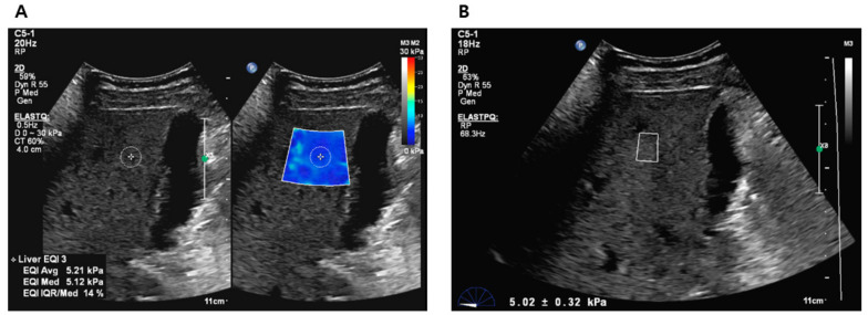
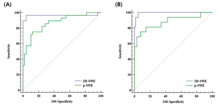
Evaluation of hepatic fibrosis is essential to prevent liver-related morbidity and mortality. Although various types of ultrasound shear wave elastography (SWE) have been used and validated, there are limited studies on the relatively newer technique, two-dimensional SWE (2D-SWE). Therefore, this study aimed to compare the diagnostic performances of 2D-SWE and point SWE (p-SWE) for evaluating liver fibrosis using histology as the reference standard. To measure liver stiffness (LS) values, 87 patients underwent 2D-SWE and p-SWE using the same machine. Technical failures and unreliable measurements were also evaluated. The diagnostic performances of 2D-SWE and p-SWE were compared using area under the receiver operating characteristic (AUROC) curve analysis. No technical failures were observed in either method; however, unreliable measurements were less frequent in 2D-SWE (1/87 [1.1%]) than in p-SWE (8/87 [9.2%]) (p < 0.001). The AUROC of the LS values of 2D-SWE were significantly higher than those of p-SWE for diagnosing significant fibrosis (0.965 vs. 0.872, p = 0.022) and cirrhosis (0.994 vs. 0.886, p = 0.042). In conclusion, 2D-SWE is more reliable and accurate than p-SWE for diagnosing hepatic fibrosis.
Keywords: chronic liver disease; histology; liver fibrosis; point shear wave elastography; two-dimensional shear wave elastography.
Conflict of interest statement
The authors declare no conflict of interest.
Figures


Similar articles
-
Hepatic fibrosis: prospective comparison of MR elastography and US shear-wave elastography for evaluation.Radiology. 2014 Dec;273(3):772-82. doi: 10.1148/radiol.14132000. Epub 2014 Jul 7. Radiology. 2014. PMID: 25007047
-
Performance of 2-Dimensional Ultrasound Shear Wave Elastography in Liver Fibrosis Detection Using Magnetic Resonance Elastography as the Reference Standard: A Pilot Study.J Ultrasound Med. 2016 Feb;35(2):401-12. doi: 10.7863/ultra.15.03036. Epub 2016 Jan 18. J Ultrasound Med. 2016. PMID: 26782164 Free PMC article.
-
Ultrasound Shear Wave Elastography for Liver Disease. A Critical Appraisal of the Many Actors on the Stage.Ultraschall Med. 2016 Feb;37(1):1-5. doi: 10.1055/s-0035-1567037. Epub 2016 Feb 12. Ultraschall Med. 2016. PMID: 26871407
-
Assessment of biopsy-proven liver fibrosis by two-dimensional shear wave elastography: An individual patient data-based meta-analysis.Hepatology. 2018 Jan;67(1):260-272. doi: 10.1002/hep.29179. Epub 2017 Nov 15. Hepatology. 2018. PMID: 28370257 Free PMC article. Review.
-
Technical performance of shear wave elastography for measuring liver stiffness in pediatric and adolescent patients: a systematic review and meta-analysis.Eur Radiol. 2019 May;29(5):2560-2572. doi: 10.1007/s00330-018-5900-6. Epub 2019 Jan 7. Eur Radiol. 2019. PMID: 30617493
Cited by
-
A comprehensive evaluation system for ultrasound-guided infusion of human umbilical cord-derived MSCs in liver cirrhosis patients.Stem Cells Transl Med. 2025 Jan 17;14(1):szae081. doi: 10.1093/stcltm/szae081. Stem Cells Transl Med. 2025. PMID: 39520328 Free PMC article.
-
Role of 2D shear wave elastography in assessing chronic kidney disease and its correlation with point shear wave elastography and eGFR.Int Urol Nephrol. 2025 Aug;57(8):2697-2704. doi: 10.1007/s11255-025-04473-7. Epub 2025 Mar 31. Int Urol Nephrol. 2025. PMID: 40163268
-
Application and Diagnostic Performance of Two-Dimensional Shear Wave Elastography and Liver Fibrosis Scores in Adults with Class 3 Obesity.Nutrients. 2023 Dec 25;16(1):74. doi: 10.3390/nu16010074. Nutrients. 2023. PMID: 38201904 Free PMC article.
-
Modern ultrasound techniques for diagnosing liver steatosis and fibrosis: A systematic review with a focus on biopsy comparison.World J Hepatol. 2025 Feb 27;17(2):100033. doi: 10.4254/wjh.v17.i2.100033. World J Hepatol. 2025. PMID: 40027573 Free PMC article.
References
LinkOut - more resources
Full Text Sources
