. 2023 Apr 26;28(9):3724.
doi: 10.3390/molecules28093724.

Molecular Basis for the Selectivity of DHA and EPA in Sudlow's Drug Binding Sites in Human Serum Albumin with the Combined Use of NMR and Docking Calculations

Affiliations

Molecular Basis for the Selectivity of DHA and EPA in Sudlow's Drug Binding Sites in Human Serum Albumin with the Combined Use of NMR and Docking Calculations

Eleni Alexandri et al. Molecules. .

Abstract

Medium- and long-chain saturated and unsaturated free fatty acids (FFAs) are known to bind to human serum albumin (HSA), the main plasma carrier protein. Atomic-level structural data regarding the binding mode in Sudlow's sites I (FA7) and II (FA4, FA3) of the polyunsaturated ω-3 fatty acids docosahexaenoic acid (DHA) and eicosapentaenoic acid (EPA), however, are largely unknown. Herein, we report the combined use of saturation transfer difference (STD) and Interligand NOEs for Pharmacophore Mapping (INPHARMA) NMR techniques and molecular docking calculations to investigate the binding mode of DHA and EPA in Sudlow's sites Ι and ΙΙ of HSA. The docking calculations and the significant number of interligand NOEs between DHA and EPA and the drugs warfarin and ibuprofen, which are stereotypical ligands for Sudlow's sites I and II, respectively, were interpreted in terms of competitive binding modes and the presence of two orientations of DHA and EPA at the binding sites FA7 and FA4. The exceptional flexibility of the long-chain DHA and EPA and the formation of strongly folded structural motives are the key properties of HSA-PUFA complexes.

Keywords: DHA; EPA; HSA; INPHARMA NMR; STD NMR; docking calculations.


Conflict of interest statement

The authors declare no conflict of interest.

Figures

Figure 1
Figure 1
Molecular structures of EPA, DHA, warfarin, and ibuprofen with the numbering of atoms.
Figure 2
Figure 2
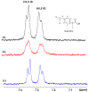
Selected regions of the STD NMR spectra: (a) of warfarin (W) (1.25 mM) with HSA (25 μΜ) in 50 mM PBS buffer in D2O with 10% DMSO-d6; (b) after the addition in solution (a) of DHA (2.5 mM); (c) after the addition in solution (a) of EPA (2.5 mM) (T = 310 K, number of scans = 80, saturation time = 2 s, experimental time = 52 min for (ac).
Figure 3
Figure 3
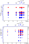
Selected regions of interligand 2D Tr-NOESY NMR spectra for warfarin (W) (2.5 mM) with HSA (25 μΜ) in 50 mM PBS buffer in D2O with 10% DMSO-d6 after the addition of DHA (2.5 mM) (A) and EPA (2.5 mM) (B) (mixing time = 300 ms, number of scans = 112, experimental time = 17 h). The red cross-peaks denote interligand NOE connectivities. The blue cross-peaks denote intraligand NOE connectivities of warfarin (W).
Figure 4
Figure 4
Selected regions of the STD NMR spectra: (a) of ibuprofen (IB) (2.5 mM) with HSA (25 μΜ) in 50 mM PBS buffer in D2O with 10% DMSO-d6; (b) after the addition in solution (a) of DHA (2.5 mM); (c) after the addition in solution (a) of EPA (2.5 mM) (T = 310 K, number of scans = 80, saturation time = 2 s, experimental time = 52 min for (ac).
Figure 5
Figure 5
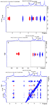
Selected regions of interligand 2D Tr-NOESY NMR spectra for DHA (2.5 mM) with HSA (25 μΜ) in 50 mM PBS buffer in D2O with 10% DMSO-d6 after the addition of ibuprofen (IB) (2.5 mM) (mixing time = 300 ms, number of scans = 112, experimental time = 17 h). The red cross-peaks denote the interligand NOE connectivities of H5,9, H6,8 (A), H2 (B), and H10,11,3 (C) of ibuprofen with DHA. The blue cross-peaks denote the intraligand NOE connectivities of ibuprofen (IB) and DHA.
Figure 6
Figure 6
Poses for binding site FA7 of HSA with DHA (a) and EPA (b) for the two anchoring sites (up and down) and their superpositions (c).
Figure 7
Figure 7
The superposition of poses one and two for DHA and EPA in panels (A) and (B), respectively, generated via molecular docking in the current work and the crystallographic structure of ARA bound in FA7 of HSA (PDB code: 1GNJ.pdb) [23]. Color code: DHA—blue, EPA—yellow, ARA—gray. In the crystallographic structure of ARA, the carboxylate group is absent due to low density.
Figure 8
Figure 8
Poses of the electrostatic and hydrogen bond interactions between the carboxylate groups of DHA/EPA (a,b) and Arg-410/Tyr-411 (up) and Ser-419/Tyr-422 (down) in the binding site FA4 of HSA. The superposition of molecular docking poses and the crystallographic structure of ARA in the same binding site of HSA [23] are shown in (c). Color code for (c): DHA—blue, EPA—yellow, ARA—gray.
Figure 9
Figure 9
Poses with best scores for the binding site FA3 of HSA with DHA (a) and EPA (b), superposition of molecular docking poses, and crystallographic structure of ARA bound in FA3 of HSA (c) [22,23].
Figure 10
Figure 10
Poses of the electrostatic interactions between the carboxylate groups of DHA and EPA and Arg-209 in the binding site FA6 of HSA (a,b) and superposition of molecular docking poses and the crystallographic structure of ARA for the same site. For (a,b), the crystal structure with the PDB code 1GNI.pdb was employed [23]. The superposition was generated based on the crystal structure of HSA with ARA with the PDB code 1GNJ.pdb. Color code for (c): DHA—blue, EPA—yellow, ARA—gray.

References

    1. Vance D.E., Vance J.E. Biochemistry of Lipids, Lipoproteins, and Membranes. 5th ed. Elsevier; Amsterdam, The Netherlands: 2008.
    1. Akoh C.C., Min D.B. Food Lipids, Chemistry, Nutrition and Biochemistry. 1st ed. Marcel Dekker Inc.; New York, NY, USA: 2002.
    1. Leray C. Dietary Lipids for Healthy Brain Function. CRC Press; Boca Ratan, FL, USA: 2017.
    1. Alexandri E., Ahmed R., Siddiqui H., Choudhary M.I., Tsiafoulis C.G., Gerothanassis I.P. High resolution NMR spectroscopy as a structural and analytical tool for unsaturated lipids in solution. Molecules. 2017;22:1663. doi: 10.3390/molecules22101663. - DOI - PMC - PubMed
    1. Stillwell W., Wassall S.R. Docosahexaenoic acid: Membrane properties of a unique fatty acid. Chem. Phys. Lipids. 2003;126:1–27. doi: 10.1016/S0009-3084(03)00101-4. - DOI - PubMed