Review
. 2023 Apr 28;28(9):3798.
doi: 10.3390/molecules28093798.

Radiolabeled Liposomes for Nuclear Imaging Probes

Affiliations
Review

Radiolabeled Liposomes for Nuclear Imaging Probes

Ho Ying Low et al. Molecules. .

Abstract

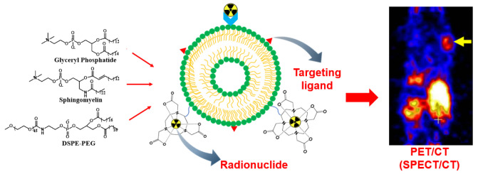
Quantitative nuclear imaging techniques are in high demand for various disease diagnostics and cancer theranostics. The non-invasive imaging modality requires radiotracing through the radioactive decay emission of the radionuclide. Current preclinical and clinical radiotracers, so-called nuclear imaging probes, are radioisotope-labeled small molecules. Liposomal radiotracers have been rapidly developing as novel nuclear imaging probes. The physicochemical properties and structural characteristics of liposomes have been elucidated to address their long circulation and stability as radiopharmaceuticals. Various radiolabeling methods for synthesizing radionuclides onto liposomes and synthesis strategies have been summarized to render them biocompatible and enable specific targeting. Through a variety of radionuclide labeling methods, radiolabeled liposomes for use as nuclear imaging probes can be obtained for in vivo biodistribution and specific targeting studies. The advantages of radiolabeled liposomes including their use as potential clinical nuclear imaging probes have been highlighted. This review is a comprehensive overview of all recently published liposomal SPECT and PET imaging probes.

Keywords: liposomes; nuclear imaging probes; radiolabeling; radiopharmaceutical; theranostics.


Conflict of interest statement

The authors declare no conflict of interest.

Figures

Scheme 1
Scheme 1
Typical liposome modifications for nuclear imaging.
Scheme 2
Scheme 2
Cell internalization pathways of liposomes.
Figure 1
Figure 1
MicroSPECT/CT and WBAR images of 188Re-DXR-liposome targeting C26 tumors in BALB/c mice. 188Re-DXR-liposome, containing 22.2 MBq of 188Re, was administered to each mouse via intravenous injection. The microSPECT images were acquired at 1, 4, 24, 48, and 72 h after injection. The WBAR image was followed by microSPECT image acquisition at 72 h after injection of 188Re- DXR-liposome with the animal in the same position. Reprinted with permission from Ref. [114]. Copyright 2010 Elsevier.
Figure 2
Figure 2
Collagen-induced arthritis in a DBA/1 mouse model using chicken type II collagen. (A) Representative images of (left) joints displaying arthritis 30 days after immunization, including swelling and erythema of the right tarsus, and (right) lucigenin BLI illustrating the inflammatory condition of the right tarsus on day 30 after immunization (same mouse). (B) Coronal whole-body distribution of 64Cu-liposomes in a CIA mouse. Three-hour and twenty-four-hour PET scans with liposomal activity (%Max ID/g) in the inflamed foci. Carpal and tarsal joints were clinically scored 4 and 3, respectively. Values for blood, liver, and spleen (%ID/g) radio-labeled liposomal activity after 3 h and 24 h are shown. (C) Mean joint accumulation (%ID/lesion ± SEM) and (D) maximum accumulation in the lesion (Max %ID/g ± SEM). After iv. injection of 64Cu-liposomes, 3 h and 24 h scans were performed, and results show significantly increased amounts of 64Cu-liposomes accumulated in joints with a high clinical score. Data are presented as mean ± SEM. Significant differences represented as * p < 0.05; ** p < 0.01; *** p < 0.001; **** p < 0.0001; paired t-test. Reprinted with permission from Ref. [127]. Copyright 2020 Elsevier.
Figure 3
Figure 3
64Cu-loaded liposome PET/CT of a S. aureus Xen29 infectious mouse model. (A) BLI of female NMRI mice sc. or im. injected with S. aureus Xen29. Mice were scanned on day 0, 1, 3, and 6 after bacterial inoculation. (B) PET/CT images of S. aureus Xen29 infection 24 h after iv. injection with 64Cu-liposomes. White rings encircle lesion sites and 64Cu-liposome activity. (C,D) Activity levels of 64Cu-liposomes on PET/CT scans performed on day 4 and 5 (acute) and on day 6 and 7 (chronic) after bacterial inoculation. All mice were scanned 10 min and 24 h after injection with 64Cu-liposomes. Data are presented as mean ± SEM. Significant differences presented as * p < 0.05; ** p < 0.01; *** p < 0.001; paired t-test. sc.: subcutaneous; im.: intramuscular. Reprinted with permission from Ref. [127]. Copyright 2020 Elsevier.

References

    1. Bangham A.D., Standish M.M., Weissmann G. The action of steroids and streptolysins on the permeability of phospholipid structures to cations. J. Mol. Biol. 1965;13:253–259. doi: 10.1016/S0022-2836(65)80094-8. - DOI - PubMed
    1. Allen T.M. Liposomen. Drugs. 1997;54((Suppl. 4)):8–14. doi: 10.2165/00003495-199700544-00004. - DOI - PubMed
    1. Liu P., Chen G., Zhang J. A Review of liposomes as a drug delivery system: Current status of approved products, regulatory environments, and future perspectives. Molecules. 2022;27:1372. doi: 10.3390/molecules27041372. - DOI - PMC - PubMed
    1. Danhier F., Feron O., Préat V. To exploit the tumor microenvironment: Passive and active tumor targeting of nanocarriers for anti-cancer drug delivery. J. Control. Release. 2010;148:135–146. doi: 10.1016/j.jconrel.2010.08.027. - DOI - PubMed
    1. Gabizon A., Tzemach D., Gorin J., Mak L., Amitay Y., Shmeeda H., Zalipsky S. Improved therapeutic activity of folate-targeted liposomal doxorubicin in folate receptor-expressing tumor models. Cancer Chemother. Pharmacol. 2010;66:43–52. doi: 10.1007/s00280-009-1132-4. - DOI - PubMed

MeSH terms

LinkOut - more resources