. 2023 May 5;28(9):3908.
doi: 10.3390/molecules28093908.

Different In Silico Approaches Using Heterocyclic Derivatives against the Binding between Different Lineages of SARS-CoV-2 and ACE2

Affiliations

Different In Silico Approaches Using Heterocyclic Derivatives against the Binding between Different Lineages of SARS-CoV-2 and ACE2

Federica Sipala et al. Molecules. .

Abstract

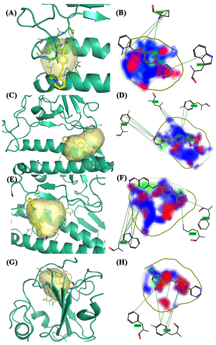
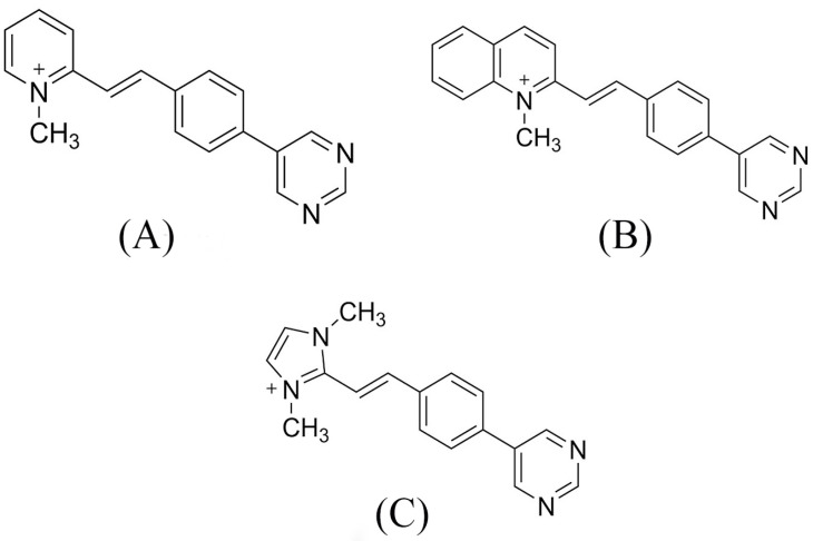
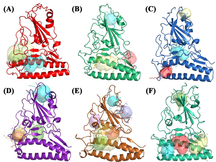
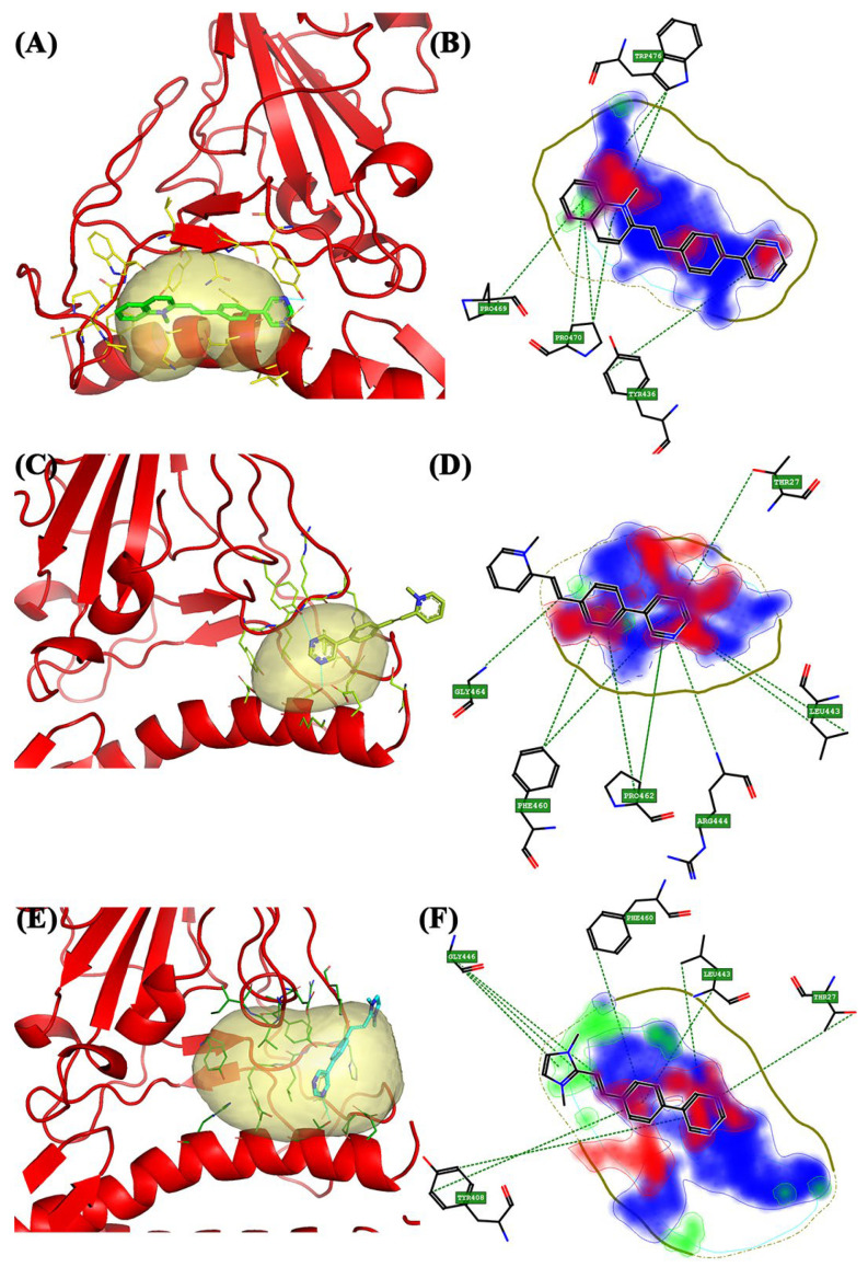
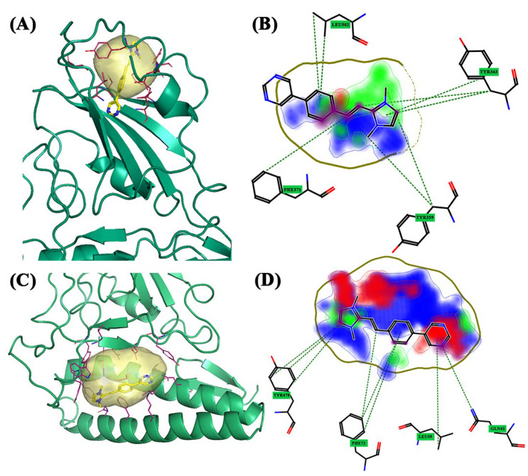
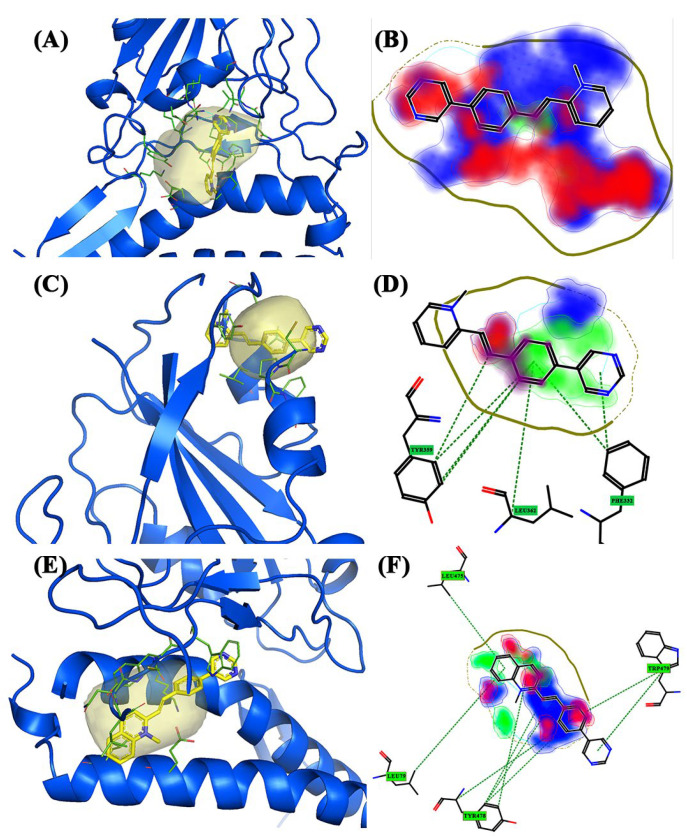
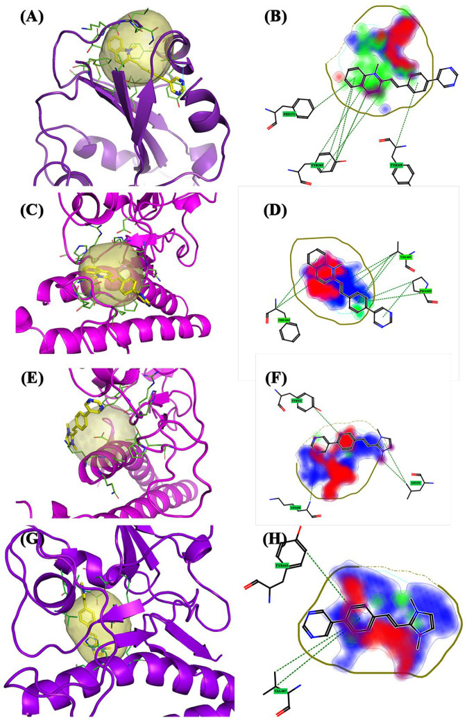
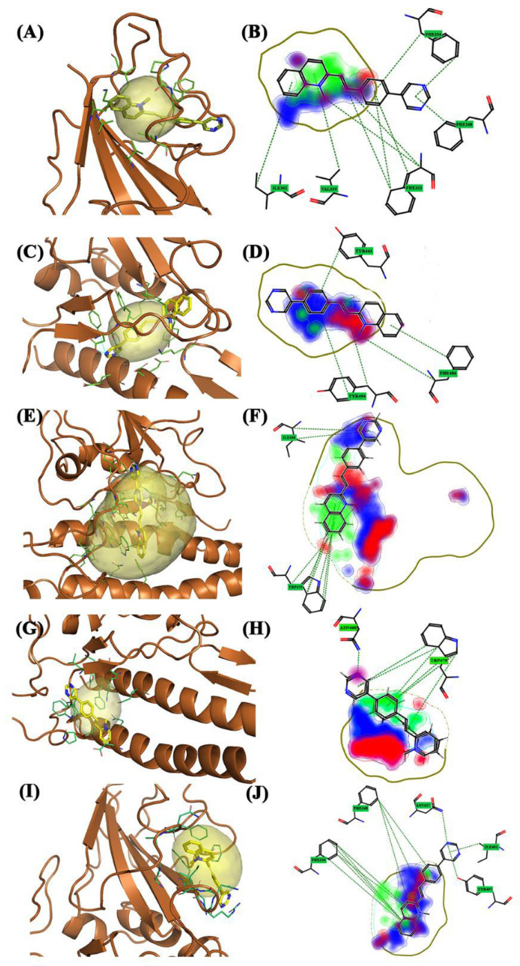
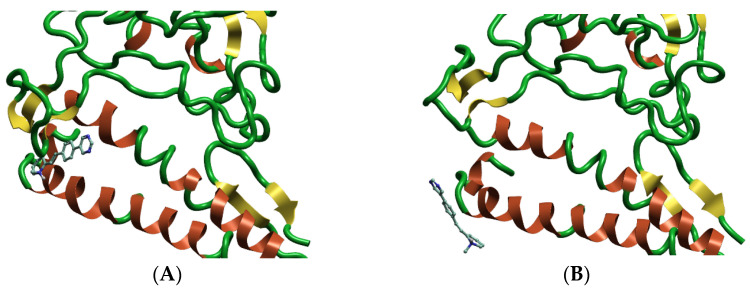
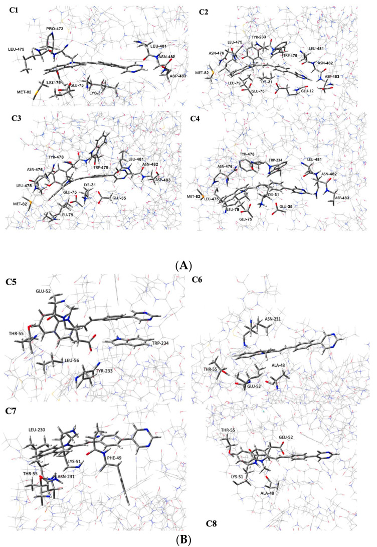
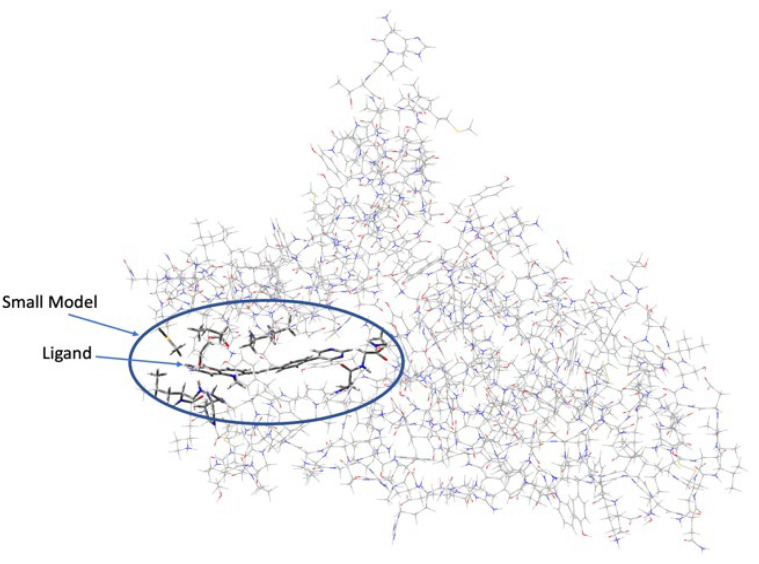
Over the last few years, the study of the SARS-CoV-2 spike protein and its mutations has become essential in understanding how it interacts with human host receptors. Since the crystallized structure of the spike protein bound to the angiotensin-converting enzyme 2 (ACE2) receptor was released (PDB code 6M0J), in silico studies have been performed to understand the interactions between these two proteins. Specifically, in this study, heterocyclic compounds with different chemical characteristics were examined to highlight the possibility of interaction with the spike protein and the disruption of the interaction between ACE2 and the spike protein. Our results showed that these compounds interacted with the spike protein and interposed in the interaction zone with ACE2. Although further studies are needed, this work points to these heterocyclic push-pull compounds as possible agents capable of interacting with the spike protein, with the potential for the inhibition of spike protein-ACE2 binding.

Keywords: SARS-CoV-2; heterocyclic derivatives; molecular modeling.


Conflict of interest statement

The authors declare no conflict of interest.

Figures

Figure 8
Figure 8
The 3D and 2D docking Omicron variant: (A) 3D pocket 1 with BCC1; (B) 2D pocket 1 with BCC1; (C) 3D pocket 2 with BCC1; (D) 2D pocket 2 with BCC1; (E) 3D pocket 3 with BCC2; (F) 2D pocket 3 with BCC2; (G) 3D pocket 4 with BCC3; and (H) 2D pocket 4 with BCC3. Water and the accessory parts of the spike protein and ACE2 receptor were omitted for clarity. The text in the green square of the image describes the amino acids involved in interaction with the ligand.
Figure 1
Figure 1
Push–pull compounds structures: (A) (E)-1-methyl-2-4-(pyrimidin-5-yl)styryl) pyridin-1-ium (BCC1), (B) (E)-1-methyl-2-4-(pyrimidin-5-yl)styryl) quinolin-1-ium (BCC2), and (C) (E)-1,3-dimethyl-2-4-(pyrimidin-5-yl)styryl)-1-h-imidazol-3-ium (BCC3).
Figure 2
Figure 2
(A) Wild-type: pocket 1 (blue), pocket 2 (green), and pocket 2.5 (yellow); (B) Alpha: pocket 1 (blue), pocket 2 (yellow), and pocket 3 (red); (C) Beta: pocket 1 (blue), pocket 2 (yellow), and pocket 3 (red); (D) Delta: pocket 1 (blue), pocket 2 (yellow), pocket 3 (red), and pocket 4 (green); (E) Gamma: pocket 1 (blue), pocket 2 (violet), pocket 3 (red), pocket 3.5 (yellow), pocket 4 (green), and pocket 5 (dark blue); and (F) Omicron: pocket 1 (blue), pocket 2 (yellow), pocket 3 (red), and pocket 4 (green). Water and the accessory parts of the spike protein and ACE2 receptor were omitted for clarity.
Figure 3
Figure 3
The 3D and 2D docking wild-type: (A) 3D pocket 1 with BCC2; (B) 2D pocket 1 with BCC2; (C) 3D pocket 2 with BCC1; (D) 2D pocket 2 with BCC1; (E) 3D pocket 2.5 with BCC3; and (F) 2D pocket 2.5 with BCC3. Water and the accessory parts of the spike protein and ACE2 receptor were omitted for clarity. The text in the green square of the image describes the amino acids involved in interaction with the ligand.
Figure 4
Figure 4
The 3D and 2D docking Alpha variant: (A) 3D pocket 1 with BCC3; (B) 2D pocket 1 with BCC3; (C) 3D pocket 2 with BCC3; and (D) 2D pocket 2 with BCC3. Water and the accessory parts of the spike protein and ACE2 receptor were omitted for clarity. The text in the green square of the image describes the amino acids involved in interaction with the ligand.
Figure 5
Figure 5
The 3D and 2D docking Beta variant: (A) 3D pocket 1 with BCC1; (B) 2D pocket 1 with BCC1; (C) 3D pocket 2 with BCC1; (D) 2D pocket 2 with BCC1; (E) 3D pocket 3 with BCC2; and (F) 2D pocket 3 with BCC2. Water and the accessory parts of the spike protein and ACE2 receptor were omitted for clarity. The text in the green square of the image describes the amino acids involved in interaction with the ligand.
Figure 6
Figure 6
The 3D and 2D docking Delta variant: (A) 3D pocket 1 with BCC2; (B) 2D pocket 1 with BCC2; (C) 3D pocket 2 with BCC2; (D) 2D pocket 2 with BCC2; (E) 3D pocket 3 with BCC3; (F) 2D pocket 3 with BCC3; (G) 3D pocket 4 with BCC3; and (H) 2D pocket 4 with BCC3. Water and the accessory parts of the spike protein and ACE2 receptor were omitted for clarity. The text in the green square of the image describes the amino acids involved in interaction with the ligand.
Figure 7
Figure 7
The 3D and 2D docking Gamma variant: (A) 3D pocket 1 with BCC2; (B) 2D pocket 1 with BCC2; (C) 3D pocket 3 with BCC2; (D) 2D pocket 3 with BCC2; (E) 3D pocket 3.5 with BCC2; (F) 2D pocket 3.5 with BCC2; (G) 3D pocket 4 with BCC1; (H) 2D pocket 4 with BCC1; (I) 3D pocket 5 with BCC2; and (J) 2D pocket 5 with BCC2. Water and the accessory parts of the spike protein and ACE2 receptor were omitted for clarity. The text in the green square of the image describes the amino acids involved in interaction with the ligand.
Figure 9
Figure 9
MD simulations of BCC1 in pocket 3 of the Beta variant: (A) Conformation of the loop from amino acid Val461 to Leu481; and (B) conformational change in the loop from amino acid Val461 to Leu481 leads to the exit of the ligand from the pocket. Water and the accessory parts of the spike protein and ACE2 were omitted for clarity.
Figure 10
Figure 10
(A) ONIOM-optimized geometries of C1–C4. C1: geometry of the BCC1-S1AS complex and interactions in conformation 1; C2: geometry of the BCC1-S1AS complex and interactions in conformation 2; C3: geometry of the BCC1-S1AS complex and interactions in conformation 3; C4: geometry of the BCC1-S1AS complex and interactions in conformation 4. (B) ONIOM-optimized geometries of C5–C8. C5: geometry of the BCC1-S1AS complex and interactions in conformation 5; C6: geometry of the BCC1-S1AS complex and interactions in conformation 6; C7: geometry of the BCC1-S1AS complex and interactions in conformation 7; C8: geometry of the BCC1-S1AS complex and interactions in conformation 8.
Figure 11
Figure 11
ONIOM model of the BCC-1-S1AS complex. The high layer is represented by the tube model, and in the wireframe, the low layer.

Similar articles

Cited by

References

    1. Wang M., Zhao R., Gao L., Gao X., Wang D., Cao J. SARS-CoV-2: Structure, Biology, and Structure Based Therapeutics Development. Front. Cell. Infect. Microbiol. 2020;10:587269. doi: 10.3389/fcimb.2020.587269. - DOI - PMC - PubMed
    1. He F., Deng Y., Li W. Coronavirus disease 2019: What we know? J. Med. Virol. 2020;92:719–725. doi: 10.1002/jmv.25766. - DOI - PMC - PubMed
    1. Cui J., Li F., Shi Z.L. Origin and evolution of pathogenic coronaviruses. Nat. Rev. Microbiol. 2019;17:181–192. doi: 10.1038/s41579-018-0118-9. - DOI - PMC - PubMed
    1. Yoshimoto F.K. A Biochemical Perspective of the Nonstructural Proteins (NSPs) and the Spike Protein of SARS CoV-2. Protein J. 2021;40:260–295. doi: 10.1007/s10930-021-09967-8. - DOI - PMC - PubMed
    1. Carota G., Ronsisvalle S., Panarello F., Tibullo D., Nicolosi A., Li Volti G. Role of Iron Chelation and Protease Inhibition of Natural Products on COVID-19 Infection. J. Clin. Med. 2021;10:2306. doi: 10.3390/jcm10112306. - DOI - PMC - PubMed

Substances