

Synthesis and Antineoplastic Activity of a Dimer, Spiroindolinone Pyrrolidinecarboxamide
- PMID: 37175323
- PMCID: PMC10180320
- DOI: 10.3390/molecules28093912
Synthesis and Antineoplastic Activity of a Dimer, Spiroindolinone Pyrrolidinecarboxamide
Abstract
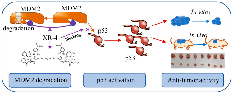
The mutation or function loss of tumour suppressor p53 plays an important role in abnormal cell proliferation and cancer generation. Murine Double Minute 2 (MDM2) is one of the key negative regulators of p53. p53 reactivation by inhibiting MDM2-p53 interaction represents a promising therapeutic option in cancer treatment. Here, to develop more effective MDM2 inhibitors with lower off-target toxicities, we synthesized a dimer, spiroindolinone pyrrolidinecarboxamide XR-4, with potent MDM2-p53 inhibition activity. Western blotting and qRT-PCR were performed to detect the impact of XR-4 on MDM2 and p53 protein levels and p53 downstream target gene levels in different cancers. Cancer cell proliferation inhibition and clonogenic activity were also investigated via the CCK8 assay and colony formation assay. A subcutaneous 22Rv1-derived xenografts mice model was used to investigate the in vivo anti-tumour activity of XR-4. The results reveal that XR-4 can induce wild-type p53 accumulation in cancer cells, upregulate the levels of the p53 target genes p21 and PUMA levels, and then inhibit cancer cell proliferation and induce cell apoptosis. XR-4 can also act as a homo-PROTAC that induces MDM2 protein degradation. Meanwhile, the in vivo study results show that XR-4 possesses potent antitumour efficacy and a favourable safety property. In summary, XR-4 is an interesting spiroindolinone pyrrolidinecarboxamide-derivative dimer with effective p53 activation activity and a cancer inhibition ability.
Keywords: MDM2 inhibitor; cancer treatment; dimer spiroindolinone pyrrolidinecarboxamide; p53 activation.
Conflict of interest statement
The authors declare no conflict of interest.
Figures


Similar articles
-
Novel MDM2 Inhibitor XR-2 Exerts Potent Anti-Tumor Efficacy and Overcomes Enzalutamide Resistance in Prostate Cancer.Front Pharmacol. 2022 Apr 25;13:871259. doi: 10.3389/fphar.2022.871259. eCollection 2022. Front Pharmacol. 2022. PMID: 35548335 Free PMC article.
-
Reactivation of p53 by novel MDM2 inhibitors: implications for pancreatic cancer therapy.Curr Cancer Drug Targets. 2010 May;10(3):319-31. doi: 10.2174/156800910791190229. Curr Cancer Drug Targets. 2010. PMID: 20370686 Free PMC article.
-
BI-907828, a novel potent MDM2 inhibitor, inhibits glioblastoma brain tumor stem cells in vitro and prolongs survival in orthotopic xenograft mouse models.Neuro Oncol. 2023 May 4;25(5):913-926. doi: 10.1093/neuonc/noac271. Neuro Oncol. 2023. PMID: 36521007 Free PMC article.
-
Reactivation of p53 gene by MDM2 inhibitors: A novel therapy for cancer treatment.Biomed Pharmacother. 2019 Jan;109:484-492. doi: 10.1016/j.biopha.2018.10.155. Epub 2018 Nov 6. Biomed Pharmacother. 2019. PMID: 30551517 Review.
-
p53-Mdm2 Interaction Inhibitors as Novel Nongenotoxic Anticancer Agents.Curr Cancer Drug Targets. 2018;18(8):749-772. doi: 10.2174/1568009617666170623111953. Curr Cancer Drug Targets. 2018. PMID: 28669344 Review.
Cited by
-
The Therapeutic Potential of Spirooxindoles in Cancer: A Focus on p53-MDM2 Modulation.Pharmaceuticals (Basel). 2025 Feb 19;18(2):274. doi: 10.3390/ph18020274. Pharmaceuticals (Basel). 2025. PMID: 40006086 Free PMC article. Review.
-
Precision oncology revolution: CRISPR-Cas9 and PROTAC technologies unleashed.Front Genet. 2024 Jul 31;15:1434002. doi: 10.3389/fgene.2024.1434002. eCollection 2024. Front Genet. 2024. PMID: 39144725 Free PMC article. Review.
References
-
- Schwaederle M., Daniels G.A., Piccioni D.E., Kesari S., Fanta P.T., Schwab R.B., Kurzrock R. Next generation sequencing demonstrates association between tumor suppressor gene aberrations and poor outcome in patients with cancer. Cell Cycle. 2015;14:1730–1737. doi: 10.1080/15384101.2015.1033596. - DOI - PMC - PubMed
MeSH terms
Substances
Grants and funding
LinkOut - more resources
Full Text Sources
Research Materials
Miscellaneous
