. 2023 Apr 22;24(9):7686.
doi: 10.3390/ijms24097686.

Cyclic Stretch-Induced Mechanical Stress Applied at 1 Hz Frequency Can Alter the Metastatic Potential Properties of SAOS-2 Osteosarcoma Cells

Affiliations

Cyclic Stretch-Induced Mechanical Stress Applied at 1 Hz Frequency Can Alter the Metastatic Potential Properties of SAOS-2 Osteosarcoma Cells

Giulia Alloisio et al. Int J Mol Sci. .

Abstract

Recently, there has been an increasing focus on cellular morphology and mechanical behavior in order to gain a better understanding of the modulation of cell malignancy. This study used uniaxial-stretching technology to select a mechanical regimen able to elevate SAOS-2 cell migration, which is crucial in osteosarcoma cell pathology. Using confocal and atomic force microscopy, we demonstrated that a 24 h 0.5% cyclic elongation applied at 1 Hz induces morphological changes in cells. Following mechanical stimulation, the cell area enlarged, developing a more elongated shape, which disrupted the initial nuclear-to-cytoplasm ratio. The peripheral cell surface also increased its roughness. Cell-based biochemical assays and real-time PCR quantification showed that these morphologically induced changes are unrelated to the osteoblastic differentiative grade. Interestingly, two essential cell-motility properties in the modulation of the metastatic process changed following the 24 h 1 Hz mechanical stimulation. These were cell adhesion and cell migration, which, in fact, were dampened and enhanced, respectively. Notably, our results showed that the stretch-induced up-regulation of cell motility occurs through a mechanism that does not depend on matrix metalloproteinase (MMP) activity, while the inhibition of ion-stretch channels could counteract it. Overall, our results suggest that further research on mechanobiology could represent an alternative approach for the identification of novel molecular targets of osteosarcoma cell malignancy.

Keywords: biomechanical response; cell migration; cell morphology; cell structural deformation; cyclic mechanical strain; malignant bone cells; mechanical stress; mechano-sensing; mechanobiology; osteosarcoma.


Conflict of interest statement

The authors declare no conflict of interest.

Figures

Figure 1
Figure 1
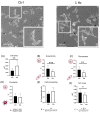
AFM analysis of the nucleus of SAOS-2 cells on a silicone plate treated or not with a 24 h 1 Hz uniaxial cyclic stretch (along the x-axis). Upper panel: cell drawing—the cell region where the measurements were taken is in black (i.e., the nucleus) (https://smart.servier.com, accessed on 1 February 2023), and there are two representative AFM pictures of untreated and stretched cells. The double-pointed arrows indicate the positions where the cell profiles were measured. Panels (A,B) report the cell profiles for untreated and stretched cells, respectively. From Panel (C) to Panel (F), the comparison of four parameters between untreated cells (black) and 24 h 1 Hz treated cells (white) (error bars represent standard error of the mean). Panel (C) displays the nuclear heights. Panel (D) shows the nuclear surface roughness. Panel (E) displays nuclear areas, and Panel (F) reports the nuclear eccentricity. AFM analyses were performed on two stretched and two unstretched samples with at least 16 cells per sample preparation. Student’s t-test was used for statistical analysis, and the results are shown as the mean ± SD. * p < 0.05; treated cells compared with control cells.
Figure 2
Figure 2
Morphological changes were induced in the whole cells by the 24 h 1 Hz cyclic stretch (along the x-axis). The upper panel shows two representative confocal microscopy pictures of treated and control SAOS-2 cells on a silicone plate. From Panels (AF), the column bar graphs report the plot means and the SD and display the difference in the mean of the cyclically stretched and unstimulated SAOS-2 cells. Panel (A) plots the cell area. Panel (B) refers to eccentricity E. Panel (C) displays the roundness index R. Panel (D) shows the circularity index C. Panel (E) displays the amount of cell processing counted per cell. Panel (F) reports the ratio between the area of the nucleus and its cytoplasm in the same cell (N/C). ImageJ 1.52 was employed for image analysis, and a paired Student’s t-test was used for statistical analysis. The analysis was performed on 3 biological replicates with at least 18 cells per condition. The results are shown as the mean ± SD. * p < 0.05, ** p < 0.01, and *** p < 0.001; treated specimens compared with control static cells.
Figure 3
Figure 3
AFM roughness changes in the peripheral cell induced by the 24 h 1 Hz cyclic stretch (along the x-axis) measured on silicone plates. Upper panel: cell drawing—the cell periphery of cells is represented in black (https://smart.servier.com, accessed on 1 February 2023), and there are two representative AFM pictures for untreated and stretched cells. The double-pointed arrows indicate the positions where the cell profiles were measured. Panels (A,B) report the cell height profiles for untreated and stretched cells, respectively, to better show the two-micron peripheral area (for further details, see Supplementary Figure S5). Roughness was measured with respect to the top surface of the cells. Panel (C): Comparison between roughness averages of the peripheral cell surfaces. Black histogram represents untreated cells and white histograms represent 1 Hz 24 h treated cells (error bars represent standard error of the mean). AFM analyses were performed on two stretched and two unstretched samples with at least 16 cells per sample. Student’s t-test was used for statistical analysis, and the results are shown as the mean ± SD. * p < 0.05; treated cells compared with the control cells.
Figure 4
Figure 4
Cell-based assays on a silicone plate of adherent SAOS-2 cells. The 1 Hz 24 h stretch stimulation impacted neither cell viability nor the osteoblastic properties of SAOS-2 cells. The comparison between measurements displays black histograms for untreated cells and white histograms for 1 Hz 24 h treated cells (error bars represent the standard error of the mean). (A) Indirect cell viability quantitation via soluble spectrometric MTS probe; (B) cell viability derived via DNA quantification based on Cy Quant fluorogenic probe; (C) on-plate detection of ALP activity using a Blue ALP kit (blue microwell substrate containing BCIP®); (D) the impact of a 24 h 1 Hz uniaxial stimulation on the gene expression of three pro-osteogenic differentiative markers (i.e., RUNX-2, COL1A1, and ALPL). Student’s t-test of the histograms of Panels (AD) showed no significant differences between the treated cells compared with the control static cells. Statistical analyses were performed on three biological replicates with at least three technical replicates per condition.
Figure 5
Figure 5
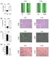
Cell motility capacity was found to be upregulated by the 24 h 1 Hz stretch stimulation (on a plastic support) (Panels (A,B)). Panel (A) shows a comparison of the 20 h cell migration between treated and untreated SAOS-2 cells using a scratch test. Panel (B) evaluated the cell transmigration comparison between the control and stretched cells using Boyden chambers. The 1 Hz cyclical stretch reduced the cell-adhesion capacity of SAOS-2 cells (Panels (C,D)). Panel (C) reports cell confluence measured for both conditions after 24 h of attachment. Panel (D) reports the cell counts measured after 24 h of attachment to precoated wells (see Materials and Methods for further details). The statistical analyses were performed on three biological replicates with six technical replicates for Panel (A,C), four technical replicates for Panel (B), and five technical replicates for Panel (D). Scale bar: 100 µm. An unpaired t-test analysis was adopted to calculate the significant difference. The results are shown as the mean ± SD. *** p < 0.001, and **** p < 0.0001; treated cells compared with control static cells.
Figure 6
Figure 6
Panel (A): The zymography displays the effect of cyclical stimulation on the gelatinolytic activity of the MMP-2 pro-enzyme (pMMP-2) found in the 24 h conditioned media of treated or untreated SAOS-2 cells. A representative image of gel zymography is reported. Panel (B,C): Western blot analysis of cell extracts treated or untreated with 24 h 1 Hz stimulation. A representative image of a Western blot for the Akt protein is reported. Filters were probed with specific antibodies for the total Akt (60 kDa), Phospo-Akt(Ser473) (60 kDa),and GAPDH (37 kDa). A densitometric analysis of the gel band areas was performed using the ImageJ free processing software and quantified using a scale of arbitrary units (error bars represent the standard error of the mean). An unpaired t-test analysis was used to calculate the significant differences. The results are shown as the mean ± SD of treated cells compared with control static cells (*** p < 0.001). The statistical analyses were performed on three biological replicates with six technical replicates: three technical replicates for Panel (A) and three technical replicates for Panel (B,C).
Figure 7
Figure 7
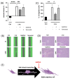
The 24 h 1 Hz stretch-induced upregulation of motile cell capacity was inhibited by stretch-activated ion-channel inhibitors (on plastic supports) (Panels (AD)). Panel (A): A comparison of cell migration between stimulated and unstimulated SAOS-2 cells, treated with GsMTx4 or ilomastat, was assessed using a scratch assay. Panel (B): Representative images of migrated cells after the 24 h 1 Hz mechanical stretch and treated with GsMTx4 or ilomastat. The area of scratches was calculated as a percentage using Tecan spark instruments (Tecan Group Ltd., Männedorf, Switzerland). Panel (C): A comparison of cell transmigration between control and stretched cells, subsequently treated with GsMTx4 or ilomastat, was made using a Boyden chamber. Panel (D): Representative images of transmigrated cells stained with crystal violet after the 24 h 1 Hz mechanical stretch and treated with GsMTx4 or ilomastat (scale bar: 100 µm). Panel (E): Sketch of the GsMTx4 inhibition of the mechanically induced, highly migratory transition (https://smart.servier.com, accessed on 1 February 2023). An unpaired t-test analysis was adopted to calculate the significant difference. The statistical analyses were performed on three biological replicates with twelve technical replicates for Panel (A) and four technical replicates for Panel (C). The results are shown as the mean ± SD. # p < 0.05 ** p < 0.01, ### p < 0.001, and **** p < 0.0001; treated cells compared with control static cells (see Materials and Methods for further details).

References

    1. Michaletti A., Gioia M., Tarantino U., Zolla L. Effects of Microgravity on Osteoblast Mitochondria: A Proteomic and Metabolomics Profile. Sci. Rep. 2017;7:15376. doi: 10.1038/s41598-017-15612-1. - DOI - PMC - PubMed
    1. Holle A.W., Engler A.J. More than a Feeling: Discovering, Understanding, and Influencing Mechanosensing Pathways. Curr. Opin. Biotechnol. 2011;22:648–654. doi: 10.1016/j.copbio.2011.04.007. - DOI - PMC - PubMed
    1. Northey J.J., Przybyla L., Weaver V.M. Tissue Force Programs Cell Fate and Tumor Aggression. Cancer Discov. 2017;7:1224–1237. doi: 10.1158/2159-8290.CD-16-0733. - DOI - PMC - PubMed
    1. Jagodzinski M., Drescher M., Zeichen J., Hankemeier S., Krettek C., Bosch U., van Griensven M. Effects of Cyclic Longitudinal Mechanical Strain and Dexamethasone on Osteogenic Differentiation of Human Bone Marrow Stromal Cells. Eur. Cell. Mater. 2004;7:35–41. doi: 10.22203/eCM.v007a04. discussion 41. - DOI - PubMed
    1. Wang L., You X., Zhang L., Zhang C., Zou W. Mechanical Regulation of Bone Remodeling. Bone Res. 2022;10:16. doi: 10.1038/s41413-022-00190-4. - DOI - PMC - PubMed