Review
. 2023 Apr 25;24(9):7800.
doi: 10.3390/ijms24097800.

Mesenchymal Stem Cells in Nerve Tissue Engineering: Bridging Nerve Gap Injuries in Large Animals

Affiliations
Review

Mesenchymal Stem Cells in Nerve Tissue Engineering: Bridging Nerve Gap Injuries in Large Animals

Mirko Lischer et al. Int J Mol Sci. .

Abstract

Cell-therapy-based nerve repair strategies hold great promise. In the field, there is an extensive amount of evidence for better regenerative outcomes when using tissue-engineered nerve grafts for bridging severe gap injuries. Although a massive number of studies have been performed using rodents, only a limited number involving nerve injury models of large animals were reported. Nerve injury models mirroring the human nerve size and injury complexity are crucial to direct the further clinical development of advanced therapeutic interventions. Thus, there is a great need for the advancement of research using large animals, which will closely reflect human nerve repair outcomes. Within this context, this review highlights various stem cell-based nerve repair strategies involving large animal models such as pigs, rabbits, dogs, and monkeys, with an emphasis on the limitations and strengths of therapeutic strategy and outcome measurements. Finally, future directions in the field of nerve repair are discussed. Thus, the present review provides valuable knowledge, as well as the current state of information and insights into nerve repair strategies using cell therapies in large animals.

Keywords: Schwann cells; cell therapy; growth factors; large animal models; nerve guidance conduit; nerve injury; nerve regeneration; stem cells.


Conflict of interest statement

The authors declare that the research was conducted in the absence of any commercial or financial relationships that could be construed as a potential conflict of interest.

Figures

Figure 1
Figure 1
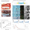
Tissue engineering of nerve guidance conduits using different MSCs and other cells. TE-NGCs were tested for bridging the nerve gap injuries in various large animal models covering rabbit, pig, dog, sheep and monkey. Therapeutic efficacy of these cell-based therapies was assessed measuring histomorphometry, electro physiological properties and behavioral recovery. Key: BM-ASC, Bone marrow-derived mesenchymal stem cells; ASC, Adipose-derived stem cells; UC-MSC, Umbilical cord-derived MSCs; AF-MSC, Amniotic fluid-derived MSCs; SK-MSCs, Skin-derived MSCs; FBC, Fibroblast cells; NSC, Neural stem cells.
Figure 2
Figure 2
Dog sciatic nerve repair and regeneration using tissue engineered nerve graft consisting of chitosan/PLGA and autologous bone marrow mesenchymal stem cells. Gross view of dog sciatic nerve repair after 60 mm gap injury: View obtained immediately (A) and 12 months (B) after a tissue-engineered nerve graft was used to bridge a 60 mm gap in dog sciatic nerve. The proximal and the distal coaptations are indicated by an arrow and an arrowhead, respectively. Minimal scale: 1 mm; (C) Hind limb functional values represent means ± standard deviations. Two-way ANOVA, in which one factor was time points and the other was grouping, and post hoc Bonferroni t test were used to analyze the data. # p < 0.05 versus autograft group, § p < 0.05 versus tissue-engineered nerve graft (TENG) group, and  p < 0.05 versus nongrafted group; (D) showing the regenerated nerve segment in different grafted groups and (E) the distal portion from which nerve sections were harvested. Meyer trichrome staining (a1e1,a2e2), immunohistochemistry with anti-NF and anti-S-100 (a3e3), transmission electron micrographs (a4e4,a5e5), obtained at 12 months postsurgery, of the sectioned regenerated nerve on the injured side in nongrafted (a1a5), scaffold (b1b5), tissue-engineered nerve graft (TENG; (c1c5)), and autograft (d1d5) groups and on the contralateral uninjured side (e1e4), respectively. The higher magnifications of the boxed areas in (a1e1) are shown in (a2e2), respectively. Scale bar: 50 µm (a1e1), 20 µm (a2e2,a3e3), 5 µm (a4e4), 0.2 µm (a5e5). Histograms showing the thickness of regenerated myelin sheath (F), the diameter of regenerated myelinated nerve fibers (G), the number of regenerated myelin lamellae (H) and the frequency distribution of myelinated nerve fiber diameters on the distal portion (I). All data are expressed as means ± standard deviations. One-way ANOVA plus post hoc Scheffé test for (FH) and Kolmogorov–Smirnov test alone for (I) were used to analyze the data. * p < 0.05 versus the contralateral uninjured side, # p < 0.05 versus autograft group, and § p< 0.05 versus TENG group. These figures were adapted from published work by Xue et al., 2011 [45].
Figure 3
Figure 3
Repair and regeneration of rhesus monkey radial nerve 25 mm gap injury using autologous bone marrow mesenchymal stem cells. Behavioral assessment of the peripheral nerve 5 months after surgery. All animals displayed a lack of wrist extension after the radial nerve surgery (A). Five months after the surgery, the monkeys implanted with the autograft (B) and BMSC-laden allografts (C) showed a remarkable restoration of wrist-extension function. However, the animals that received acellular allografts exhibited reduced wrist-extension performance, with a smaller maximum wrist extension angle (D). (E) A smaller maximum wrist extension angle was seen in the acellular group compared with the BMSC-laden and autograft groups. * p < 0.05, vs. BMSC-laden and autograft groups (n = 6 forearms). Data are expressed as mean ± SD. One-way analysis of variance with the Student-Newman-Keuls multiple comparisons method was used for statistical testing. BMSC: Bone marrow stem cell. These figures were adapted from published work by Wang et al., 2014 [60].
Figure 4
Figure 4
Functional collagen nerve guide consisting of longitudinally aligned fibers and human umbilical cord derived mesenchymal stem cells promote functional recovery after sciatic nerve 35 mm gap injury in dogs. Quantitative results of the electrophysiological evaluation made 9 months after surgery. Representative measurements of the (a) proximal, (b) middle, and (c) distal sections of the injured canine sciatic nerve. The data are shown as the mean ± standard deviation. * p < 0.05, ** p < 0.01, or *** p < 0.001, compared with the negative control (NC) group. cMAPs = compound muscle action potentials; hUC-MSCs = human umbilical cord mesenchymal stem cells; LOCC = longitudinally oriented collagen conduit. These figures were adapted from published work by Cui et al., 2018 [63].
Figure 5
Figure 5
GDNF promotes long-gap nerve regeneration after 50 mm median nerve injury of rhesus monkey. Study design: (A) Schematic depicting experimental design. (B) Photograph of the 5.2 cm PCL/GDNF nerve guide. (C) SEM of the nerve guide cross section embedded with double-walled microspheres. Mag, magnification. (D) Diagram of the PCL/GDNF nerve guide cross section. (E) SEM of a bisected double-walled PLGA/PLA microsphere. (F) SEM of microsphere adhesion to the initial PCL layer during the manufacturing process. (G) Higher magnification of a cross section of a double-walled PLGA/PLA microsphere embedded in the PCL wall [rectangle in (C)]. EHT, electron high tension; WD, working distance; Photographs of (H) exposed native nerve. (I) PCL/GDNF conduit explanted after 1 year. (J) Implanted PCL/Empty conduit. (K) PCL/Empty conduit explanted after 1 year. (L) Implanted PCL/GDNF conduit. (M) Autograft explanted after 1 year. (N) Modified Klüver board with varying well diameters used for functional training and assessment; Well 1 has a diameter of 2.5 cm, and well 2 has a diameter of 0.5 cm. (O) Photograph of the correct pinching motion. (P) Photograph of the incorrect pinching motion. (Q) Normalized functional bar graph comparing the NHPs’ 50-week functional recovery to their preoperative baselines; n.s., not significant. (R) Linear regression plot assessing functional recovery over 50 weeks for all treatment groups. n = 30 measurements per time point per NHP. Means represented with +SE/−SD. Adjusted p values presented as: ** p < 0.01; *** p < 0.001 (select comparisons shown). These figures were adapted from published work by Fadia et al., 2020 [66].
Figure 6
Figure 6
Repair and regeneration of long-peripheral nerve injuries in sheep model. (A) Following the surgery, functional tests (Fx test) were performed each month, electrophysiological tests and echography were made at 6.5 and 9 months, and samples were taken for histology at the end of the follow-up. (B) The surgical approach was performed with the animal in lateral recumbency through a lateral longitudinal skin incision. (C) Wide dissection showing the peroneal nerve location (arrow) after the sciatic nerve bifurcation into the tibial and peroneal nerve in a cadaveric sheep. (D) Resection of the common peroneal nerve under the operating microscope to create the nerve gap. (E) A 5 cm autograft was sutured again to the nerve stumps with epineural sutures (proximal suture marked with yellow arrow and distal suture marked with a yellow asterisk). (F) Detail of the 8 stitches made to join the nerve graft with the healthy nerve stump without tension. (G) After the surgery, some animals showed foot drop in the standing position. Representative immunohistochemical images of transverse sections of a control peroneal nerve (H,K,N,Q) and of an autograft of group AG5 (I,L,O,R) and of group AG7 (J,M,P,S). Sections were immunolabeled against NF200 for myelinated axons (HM), and against S100 for Schwann cells (NS). Images were taken at ×40 magnification (HJ,NP), scale bar 200 μm, and at ×400 magnification (KM,QS), scale bar 100 μm. The bottom panels show representative semithin transverse sections of the middle segment of the nerve graft stained with toluidine blue. (T) control nerve, (U) AG5, and (V) AG7 graft, at 10,000× magnification, scale bar 10 μm. These figures were adapted from published work by Contreras et al., 2023 [36].

References

    1. Böcker A., Aman M., Kneser U., Harhaus L., Siemers F., Stang F. Closing the Gap: Bridging Peripheral Sensory Nerve Defects with a Chitosan-Based Conduit a Randomized Prospective Clinical Trial. J. Pers. Med. 2022;12:900. doi: 10.3390/jpm12060900. - DOI - PMC - PubMed
    1. Hopf A., Al-Bayati L., Schaefer D.J., Kalbermatten D.F., Guzman R., Madduri S. Optimized Decellularization Protocol for Large Peripheral Nerve Segments: Towards Personalized Nerve Bioengineering. Bioengineering. 2022;9:412. doi: 10.3390/bioengineering9090412. - DOI - PMC - PubMed
    1. Bain J.R., Mackinnon S.E., Hudson A.R., Wade J., Evans P., Makino A., Hunter D. The peripheral nerve allograft in the primate immunosuppressed with Cyclosporin A: I. Histologic and electrophysiologic assessment. Plast. Reconstr. Surg. 1992;90:1036–1046. doi: 10.1097/00006534-199212000-00015. - DOI - PubMed
    1. Ding F., Wu J., Yang Y., Hu W., Zhu Q., Tang X., Liu J., Gu X. Use of tissue-engineered nerve grafts consisting of a chitosan/poly(lactic-co-glycolic acid)-based scaffold included with bone marrow mesenchymal cells for bridging 50-mm dog sciatic nerve gaps. Tissue Eng. Part A. 2010;16:3779–3790. doi: 10.1089/ten.tea.2010.0299. - DOI - PubMed
    1. Prautsch K.M., Degrugillier L., Schaefer D.J., Guzman R., Kalbermatten D.F., Madduri S. Ex-Vivo Stimulation of Adipose Stem Cells by Growth Factors and Fibrin-Hydrogel Assisted Delivery Strategies for Treating Nerve Gap-Injuries. Bioengineering. 2020;7:42. doi: 10.3390/bioengineering7020042. - DOI - PMC - PubMed