. 2023 Apr 28;24(9):7992.
doi: 10.3390/ijms24097992.

NAMPT and PARylation Are Involved in the Pathogenesis of Atopic Dermatitis

Affiliations

NAMPT and PARylation Are Involved in the Pathogenesis of Atopic Dermatitis

Ana B Arroyo et al. Int J Mol Sci. .

Abstract

Atopic dermatitis (AD) is a chronic inflammatory skin disease of very high prevalence, especially in childhood, with no specific treatment or cure. As its pathogenesis is complex, multifactorial and not fully understood, further research is needed to increase knowledge and develop new targeted therapies. We have recently demonstrated the critical role of NAD+ and poly (ADP-ribose) (PAR) metabolism in oxidative stress and skin inflammation. Specifically, we found that hyperactivation of PARP1 in response to DNA damage induced by reactive oxygen species, and fueled by NAMPT-derived NAD+, mediated inflammation through parthanatos cell death in zebrafish and human organotypic 3D skin models of psoriasis. Furthermore, the aberrant induction of NAMPT and PARP activity was observed in the lesional skin of psoriasis patients, supporting the role of these signaling pathways in psoriasis and pointing to NAMPT and PARP1 as potential novel therapeutic targets in treating skin inflammatory disorders. In the present work, we report, for the first time, altered NAD+ and PAR metabolism in the skin of AD patients and a strong correlation between NAMPT and PARP1 expression and the lesional status of AD. Furthermore, using a human 3D organotypic skin model of AD, we demonstrate that the pharmacological inhibition of NAMPT and PARP reduces pathology-associated biomarkers. These results help to understand the complexity of AD and reveal new potential treatments for AD patients.

Keywords: NAD+; NAMPT; PARP; atopic dermatitis; skin inflammation.


Conflict of interest statement

The authors declare no competing interest.

Figures

Figure 1
Figure 1
The amount of NAMPT protein and PARylation increased in lesional skin of AD patients. Representative images and analysis of biopsy sections from healthy (n = 10) and AD (n = 6) skin biopsies immunostained with anti-NAMPT (A,B) and anti-PAR (C,D). The mean ± SEM of each group is shown. p values were calculated using the nonparametric Mann–Whitney test, **** p ≤ 0.0001. Scale bar is 50 μm in all panels. CL: cornified layer; D: dermis; SL: spinous layer.
Figure 2
Figure 2
Differential expression profiles of genes encoding key NAD+ metabolic enzymes in AD. Transcriptomic data obtained from two different human AD cohorts, GSE57225 (AD) and GDS4491 (EG), from the GEO database. In cohort 1, AD was compared to healthy skin samples (AD), and in cohort 2, skin with lesional (AL) and non-lesional (ANL) AD was compared to healthy skin samples (EG). Each point represents one individual and the mean ± SEM for each group is also shown. p values were calculated using unpaired Student’s t-test, one-way ANOVA and Tukey’s multiple range, as appropriate. * p ≤ 0.05, ** p ≤ 0.01, *** p ≤ 0.001, **** p ≤ 0.0001.
Figure 3
Figure 3
Differential expression profiles of genes encoding parthanatos components in AD. Transcriptomic data obtained from two different human AD cohorts, GSE57225 (A,B) and GDS4491 (C,D), from the GEO database. In cohort 1, AD was compared to healthy skin samples (A,B), and in cohort 2, skin with lesional (AL) and non-lesional (ANL) AD was compared to healthy skin samples (C,D). Each point represents one individual and the mean ± SEM for each group is also shown. p values were calculated using unpaired Student’s t-test, one-way ANOVA and Tukey’s multiple range, as appropriate. * p ≤ 0.05, ** p ≤ 0.01, *** p ≤ 0.001, **** p ≤ 0.0001.
Figure 4
Figure 4
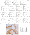
NAMPT and PARP1 levels correlated with lesional biomarkers of AD. (A) Correlation of NAMPT and PARP1 with proliferative markers. (B) Correlation of NAMPT and PARP1 with inflammatory markers. Univariate correlations were performed using transcriptomic data from GEO cohort 2 and calculated using Spearman’s correlation coefficient. (C) Representative images and analysis of biopsy sections from healthy (n = 10) and AD (n = 6) skin biopsies that have been immunostained with anti-PCNA. The mean ± SEM of each group is shown. p values were calculated using the nonparametric Mann–Whitney test, * p ≤ 0.05. Scale bar is 50 μm in all panels. CL: cornified layer; D: dermis; SL: spinous layer.
Figure 5
Figure 5
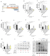
Reduction in pathology-associated biomarkers in human organotypic 3D skin model of AD. (A) Experimental design. Transcript levels of the indicated genes encoding markers of inflammation (BD,H), epidermal differentiation (E,F) and proliferation (G) were determined in human organotypic 3D skin pretreated with 10 ng/mL IL13 and IL4 in the presence of vehicle (DMSO) or the indicated inhibitors. Protein levels of interest were also determined by Western blot (I,J). The mean ± SEM of each group is shown. Results are representative of 3 independent experiments. p values were calculated by Kruskal–Wallis test and Dunn’s multiple comparisons test. * p ≤ 0.05, ** p ≤ 0.01, *** p ≤ 0.001, **** p ≤ 0.0001. M, molecular weight markers.
Figure 6
Figure 6
NAD+ and PAR metabolism is involved in AD pathogenesis. PARP1 hyperactivation in keratinocytes fueled by NAMPT-derived NAD+ exacerbates the inflammation and hyperplasia of AD. Consequently, pharmacological inhibition of NAMPT and PARP1 decreases disease-associated inflammation and proliferation, restoring skin homeostasis. Smartservier resources have been used to generate this figure.

References

    1. Ständer S. Atopic Dermatitis. N. Engl. J. Med. 2021;384:1136–1143. doi: 10.1056/NEJMra2023911. - DOI - PubMed
    1. Katsyuba E., Romani M., Hofer D., Auwerx J. NAD+ Homeostasis in Health and Disease. Nat. Metab. 2020;2:9–31. doi: 10.1038/s42255-019-0161-5. - DOI - PubMed
    1. Rajman L., Chwalek K., Sinclair D.A. Therapeutic Potential of NAD-Boosting Molecules: The In Vivo Evidence. Cell Metab. 2018;27:529–547. doi: 10.1016/j.cmet.2018.02.011. - DOI - PMC - PubMed
    1. Martínez-Morcillo F.J., Cantón-Sandoval J., Martínez-Menchón T., Corbalán-Vélez R., Mesa-Del-Castillo P., Pérez-Oliva A.B., García-Moreno D., Mulero V. Non-Canonical Roles of NAMPT and PARP in Inflammation. Dev. Comp. Immunol. 2021;115:103881. doi: 10.1016/j.dci.2020.103881. - DOI - PubMed
    1. Martínez-Morcillo F.J., Cantón-Sandoval J., Martínez-Navarro F.J., Cabas I., Martínez-Vicente I., Armistead J., Hatzold J., López-Muñoz A., Martínez-Menchón T., Corbalán-Vélez R., et al. NAMPT-Derived NAD+ Fuels PARP1 to Promote Skin Inflammation through Parthanatos Cell Death. PLoS Biol. 2021;19:e3001455. doi: 10.1371/journal.pbio.3001455. - DOI - PMC - PubMed

Substances