Review
. 2023 Apr 30;24(9):8096.
doi: 10.3390/ijms24098096.

Phafins Are More Than Phosphoinositide-Binding Proteins

Affiliations
Review

Phafins Are More Than Phosphoinositide-Binding Proteins

Tuoxian Tang et al. Int J Mol Sci. .

Abstract

Phafins are PH (Pleckstrin Homology) and FYVE (Fab1, YOTB, Vac1, and EEA1) domain-containing proteins. The Phafin protein family is classified into two groups based on their sequence homology and functional similarity: Phafin1 and Phafin2. This protein family is unique because both the PH and FYVE domains bind to phosphatidylinositol 3-phosphate [PtdIns(3)P], a phosphoinositide primarily found in endosomal and lysosomal membranes. Phafin proteins act as PtdIns(3)P effectors in apoptosis, endocytic cargo trafficking, and autophagy. Additionally, Phafin2 is recruited to macropinocytic compartments through coincidence detection of PtdIns(3)P and PtdIns(4)P. Membrane-associated Phafins serve as adaptor proteins that recruit other binding partners. In addition to the phosphoinositide-binding domains, Phafin proteins present a poly aspartic acid motif that regulates membrane binding specificity. In this review, we summarize the involvement of Phafins in several cellular pathways and their potential physiological functions while highlighting the similarities and differences between Phafin1 and Phafin2. Besides, we discuss research perspectives for Phafins.

Keywords: FYVE domain; PH domain; Phafin; PtdIns(3)P; PtdIns(4)P; autoinhibition; membrane remodeling.


Conflict of interest statement

The authors declare no conflict of interest.

Figures

Figure 1
Figure 1
Sequence alignment of Phafin proteins. Amino acid sequences corresponding to Homo sapiens Phafin1 (UniProt accession number Q96S99), Mus musculus Phafin1 (UniProt accession number Q3TB82), Drosophila melanogaster Phafin1 (UniProt accession number O76902), Danio rerio Phafin2 (UniProt accession number Q7ZUV1), Homo sapiens Phafin2 (UniProt accession number Q9H8W4), and Mus musculus Phafin2 (UniProt accession number Q91WB4) were aligned using Clustal Omega. Sequence alignment is shown and colored using MView and structural and functional elements are underlined (PH domain, black; FYVE domain, red; polyD motif, yellow; and C-terminal tail, blue). The putative PtdIns(3)P-binding sites in the PH and FYVE domains are marked by braces. Of note, three conserved sequences, the N-terminal WxxD, the central RR/KHHCR, and the C-terminal RVC motifs in the FYVE domain form a compact PtdIns(3)P-binding site.
Figure 2
Figure 2
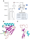
Phosphoinositides and potential PIP-binding sites in the Phafin2 PH and Phafin2 FYVE domains. (A) Molecular formulas of a glycerophospholipid and its seven derived PIPs. (B,C) Structure models of the human Phafin2 PH (B) and FYVE (C) domains generated by the SWISS-MODEL online program using templates with high sequence similarity (for PH domain, PDB accession code: 4gzu.1; for FYVE domain, PDB accession code: 3t7l.1.A). A basic sequence motif of 49-KPKAR-53 in the β1/β2 loop is shown in stick format, which corresponds to the canonical PIP-binding motif KXn(K/R)XR in PH domains.
Figure 3
Figure 3
The autophagic pathway and the role of Phafin2 in autophagy. (A) Phases of the autophagic pathway. (B) The role of Phafin2, Akt, and PIP in autophagy.
Figure 4
Figure 4
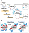
The role of Phafin2 in macropinocytosis. (A) The process of micropinocytosis. Phafin2 binds to PtdIns(3)P and PtdIns(4)P on nascent macropinosomes through a coincidence detection mechanism. Additionally, Phafin2 directly interacts with actin, shedding the dense actin cytoskeleton, and facilitating maturation of macropinosomes. (B) Phafin2 recruits JIP4 to the tubulating macropinosomes and promotes tubulation in a PIP-dependent manner.

References

    1. Conner S.D., Schmid S.L. Regulated portals of entry into the cell. Nature. 2003;422:37–44. doi: 10.1038/nature01451. - DOI - PubMed
    1. Doherty G.J., McMahon H.T. Mechanisms of Endocytosis. Annu. Rev. Biochem. 2009;78:857–902. doi: 10.1146/annurev.biochem.78.081307.110540. - DOI - PubMed
    1. Platta H.W., Stenmark H. Endocytosis and signaling. Curr. Opin. Cell Biol. 2011;23:393–403. doi: 10.1016/j.ceb.2011.03.008. - DOI - PubMed
    1. Yim W.W.-Y., Mizushima N. Lysosome biology in autophagy. Cell Discov. 2020;6:6. doi: 10.1038/s41421-020-0141-7. - DOI - PMC - PubMed
    1. Birgisdottir Å.B., Johansen T. Autophagy and endocytosis-interconnections and interdependencies. J. Cell Sci. 2020;133:jcs228114. doi: 10.1242/jcs.228114. - DOI - PubMed

MeSH terms

Substances

LinkOut - more resources