. 2023 May 2;24(9):8137.
doi: 10.3390/ijms24098137.

Polylevolysine and Fibronectin-Loaded Nano-Hydroxyapatite/PGLA/Dextran-Based Scaffolds for Improving Bone Regeneration: A Histomorphometric in Animal Study

Affiliations

Polylevolysine and Fibronectin-Loaded Nano-Hydroxyapatite/PGLA/Dextran-Based Scaffolds for Improving Bone Regeneration: A Histomorphometric in Animal Study

Elena Canciani et al. Int J Mol Sci. .

Abstract

The regeneration of large bone defects is still demanding, requiring biocompatible scaffolds, with osteoconductive and osteoinductive properties. This study aimed to assess the pre-clinical efficacy of a nano-hydroxyapatite (nano-HA)/PGLA/dextran-based scaffold loaded with Polylevolysine (PLL) and fibronectin (FN), intended for bone regeneration of a critical-size tibial defect, using an ovine model. After physicochemical characterization, the scaffolds were implanted in vivo, producing two monocortical defects on both tibiae of ten adult sheep, randomly divided into two groups to be euthanized at three and six months after surgery. The proximal left and right defects were filled, respectively, with the test scaffold (nano-HA/PGLA/dextran-based scaffold loaded with PLL and FN) and the control scaffold (nano-HA/PGLA/dextran-based scaffold not loaded with PLL and FN); the distal defects were considered negative control sites, not receiving any scaffold. Histological and histomorphometric analyses were performed to quantify the bone ingrowth and residual material 3 and 6 months after surgery. In both scaffolds, the morphological analyses, at the SEM, revealed the presence of submicrometric crystals on the surfaces and within the scaffolds, while optical microscopy showed a macroscopic 3D porous architecture. XRD confirmed the presence of nano-HA with a high level of crystallinity degree. At the histological and histomorphometric evaluation, new bone formation and residual biomaterial were detectable inside the defects 3 months after intervention, without differences between the scaffolds. At 6 months, the regenerated bone was significantly higher in the defects filled with the test scaffold (loaded with PLL and FN) than in those filled with the control scaffold, while the residual material was higher in correspondence to the control scaffold. Nano-HA/PGLA/dextran-based scaffolds loaded with PLL and FN appear promising in promoting bone regeneration in critical-size defects, showing balanced regenerative and resorbable properties to support new bone deposition.

Keywords: Polylevolysine; bone regeneration; critical-size bone defect; fibronectin; hydroxyapatite.


Conflict of interest statement

The authors declare no conflict of interest.

Figures

Figure 1
Figure 1
Structural characterization of HA. (A) XRD diffraction patterns displayed the typical diffraction maxima of a well-defined single hydroxyapatite phase, thus suggesting a high crystallinity degree; (B) TEM image showed well-defined nano-HA crystals differently oriented with respect to the image plane (indicated in squares 1 and 2, two different projections), confirming the high crystallinity degree.
Figure 2
Figure 2
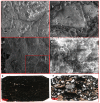
Morphological analyses of the scaffolds. The SEM analysis of the scaffold surfaces (A,B, scale bar = 10 µm and 5 µm) showed the presence of submicrometric crystals, also highlighted at the transversal section (C and insert, scale bar = 50 µm and 5 µm, respectively) and sagittal sections (D, scale bar = 5 µm). In addition, the ground sections of the control scaffold (E) and test scaffold (F) with the light microscope show the presence of porosity within both the scaffolds; the control scaffold was more compact, with less porosity, than the test scaffold (total magnification 4×).
Figure 3
Figure 3
Histological analysis of the scaffolds at 3 months after surgery. The new bone deposition was observed within the control scaffold (A,B) and test scaffold (B,D) at three months. Deposited bone (in blue/light violet) was detectable at the periphery (A,C) and inside the defect (B,D). The residual material was still present (in dark brown). Light microscope, total magnification 4× (2A, 2C) and 10× (2B, 2D); Toluidine Blue/Pyronin Yellow staining.
Figure 4
Figure 4
Histological analysis of the scaffolds at 6 months after surgery. At six months, a high amount of new bone (in blue/light violet) was found in the defects filled with the control scaffold (A,B) and test scaffold (B,D). The residual material was still present in both scaffolds (in dark brown). Light microscope, total magnification 4× (3A, 3C) and 10× (3B, 3D); Toluidine Blue/Pyronin Yellow staining.
Figure 5
Figure 5
Histological analysis of empty defects, used as the control, at 3 and 6 months after surgery. The empty defects showed a negligible amount of new bone detectable only at the periphery of the defect at both 3 and 6 months after surgical intervention (A,B, respectively). Light microscope, total magnification 4×; Toluidine Blue/Pyronin Yellow staining.
Figure 6
Figure 6
Histomorphometric analysis of new bone formation. (A) At 3 months, residual intra-defects biomaterial and bone ingrowth were comparable between the scaffolds. (B) At 6 months, scaffold B (test scaffold) was significantly more resorbed compared to scaffold A (control scaffold) and showed a significantly increased bone ingrowth (4% for control scaffold A and 7% for test scaffold B).

References

    1. Nauth A., Schemitsch E., Norris B., Nollin Z., Watson J.T. Critical-Size Bone Defects: Is There a Consensus for Diagnosis and Treatment? J. Orthop. Trauma. 2018;32((Suppl. 1)):S7–S11. doi: 10.1097/BOT.0000000000001115. - DOI - PubMed
    1. Kengelbach-Weigand A., Thielen C., Bäuerle T., Götzl R., Gerber T., Körner C., Beier J.P., Horch R.E., Boos A.M. Personalized Medicine for Reconstruction of Critical-Size Bone Defects—A Translational Approach with Customizable Vascularized Bone Tissue. Npj Regen. Med. 2021;6:49. doi: 10.1038/s41536-021-00158-8. - DOI - PMC - PubMed
    1. Velasco M.A., Narváez-Tovar C.A., Garzón-Alvarado D.A. Design, Materials, and Mechanobiology of Biodegradable Scaffolds for Bone Tissue Engineering. BioMed Res. Int. 2015;2015:e729076. doi: 10.1155/2015/729076. - DOI - PMC - PubMed
    1. Dec P., Modrzejewski A., Pawlik A. Existing and Novel Biomaterials for Bone Tissue Engineering. Int. J. Mol. Sci. 2022;24:529. doi: 10.3390/ijms24010529. - DOI - PMC - PubMed
    1. Akilbekova D., Shaimerdenova M., Adilov S., Berillo D. Biocompatible Scaffolds Based on Natural Polymers for Regenerative Medicine. Int. J. Biol. Macromol. 2018;114:324–333. doi: 10.1016/j.ijbiomac.2018.03.116. - DOI - PubMed

LinkOut - more resources