. 2023 May 6;24(9):8336.
doi: 10.3390/ijms24098336.

Clostridia and Enteroviruses as Synergistic Triggers of Type 1 Diabetes Mellitus

Affiliations

Clostridia and Enteroviruses as Synergistic Triggers of Type 1 Diabetes Mellitus

Robert Root-Bernstein et al. Int J Mol Sci. .

Abstract

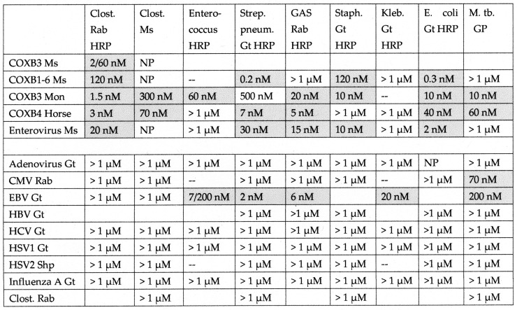
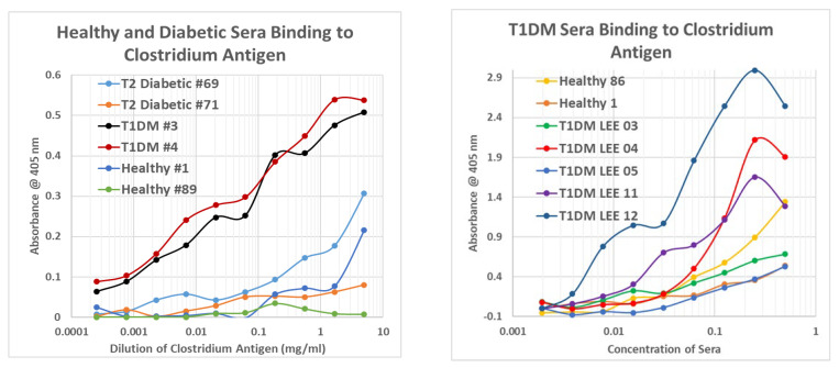
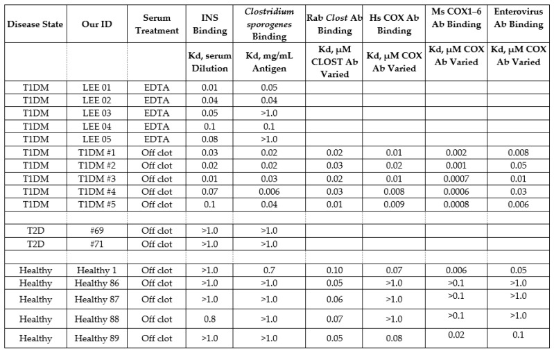
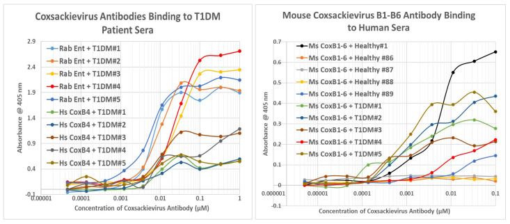
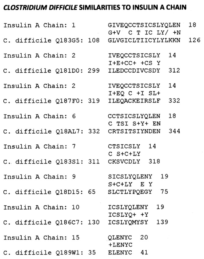
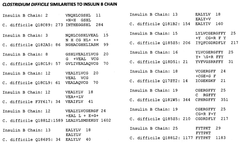
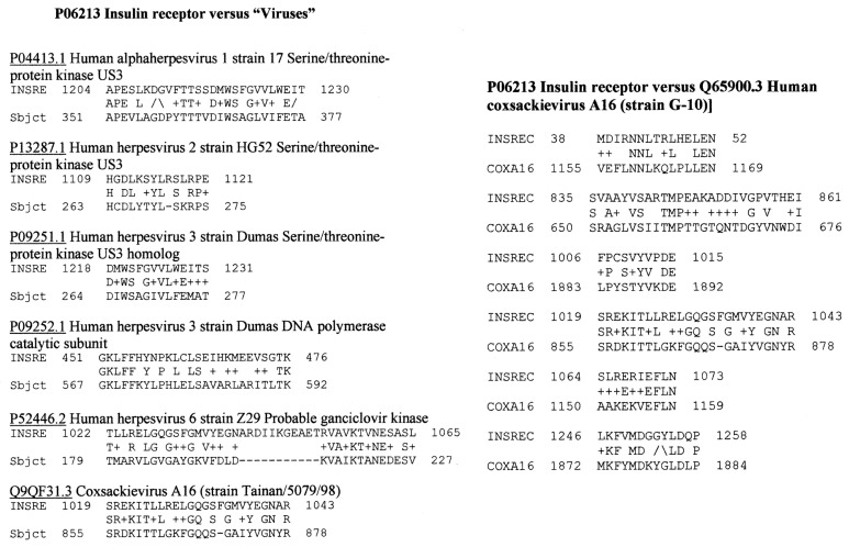
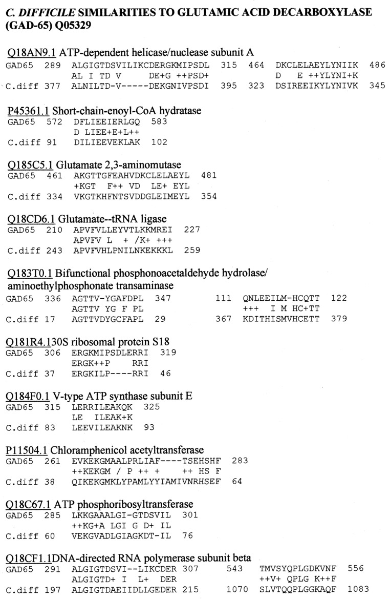
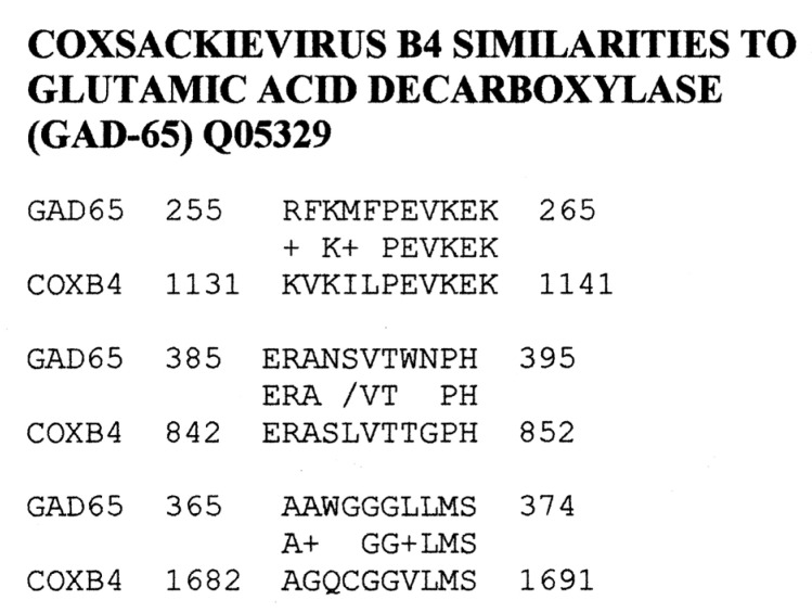
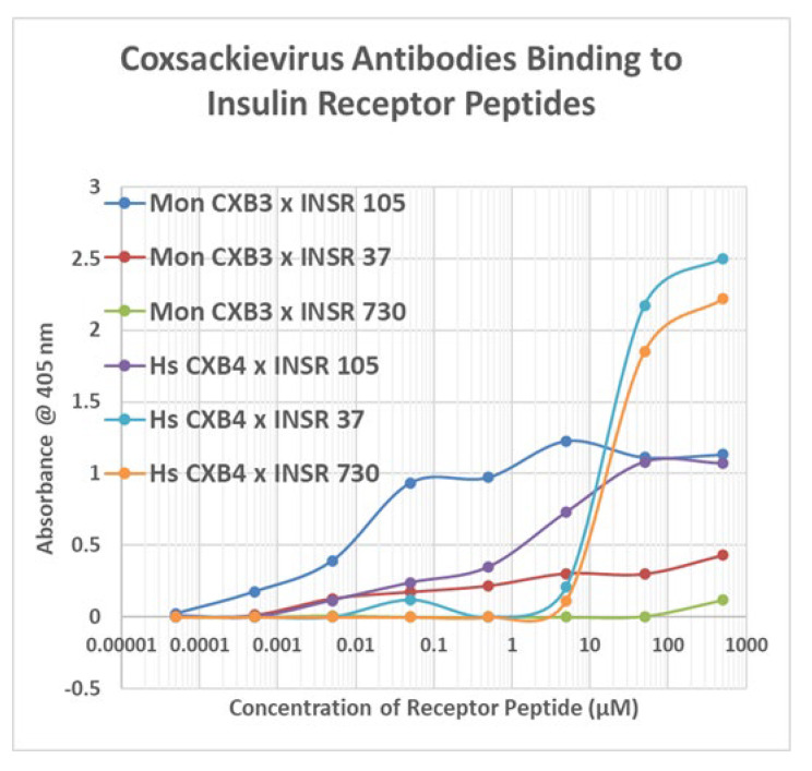
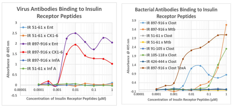
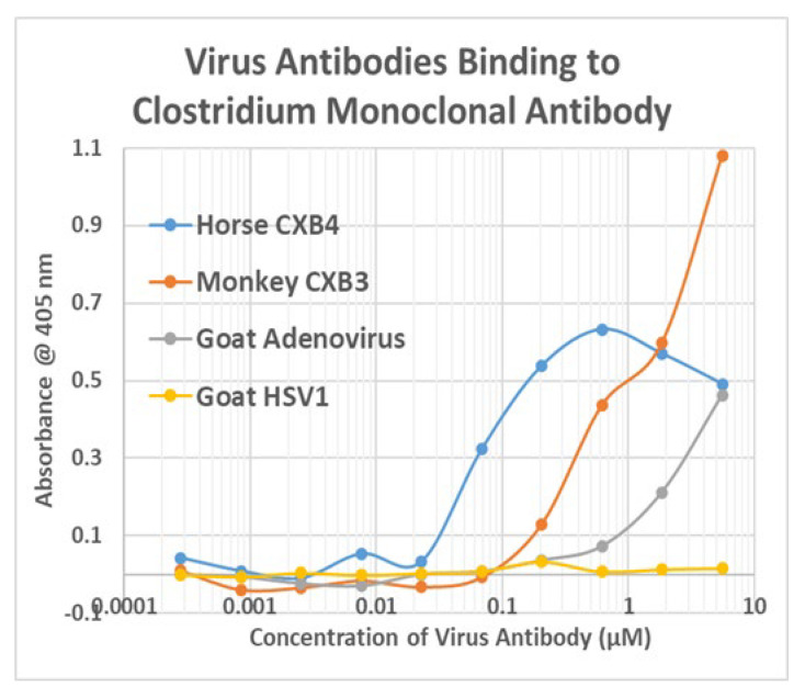
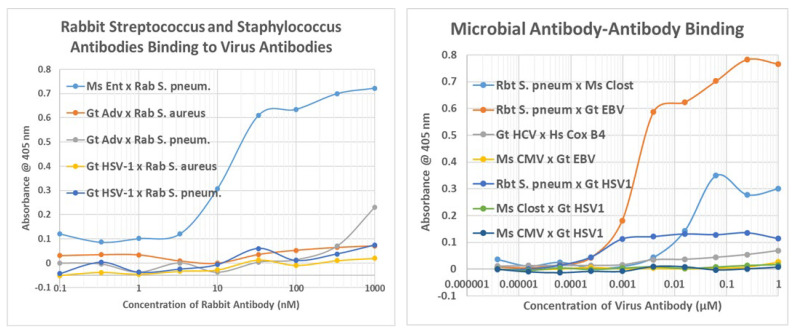
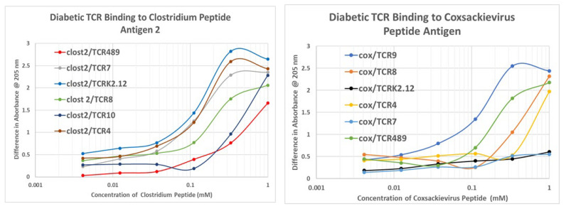
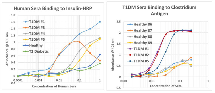
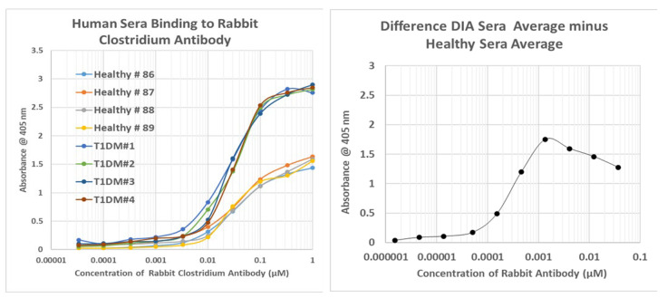
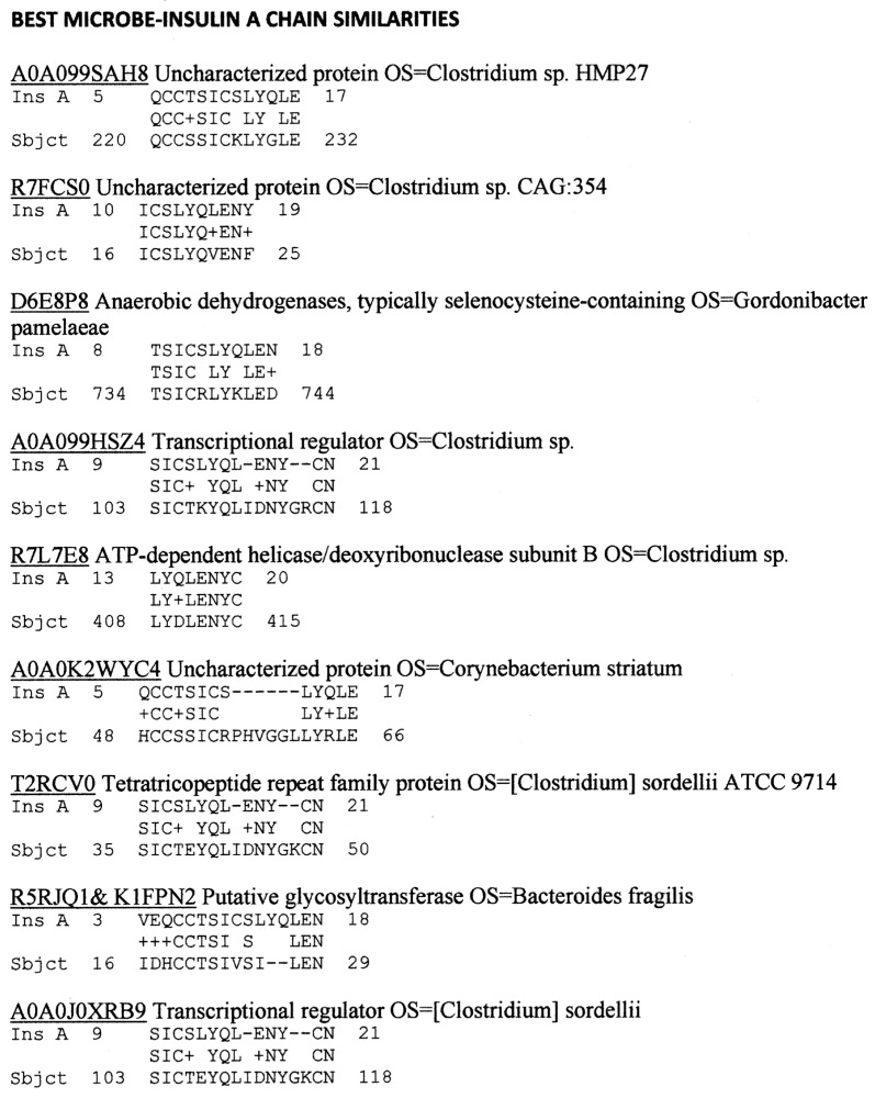
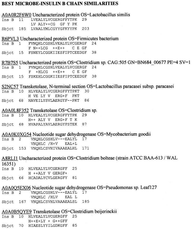
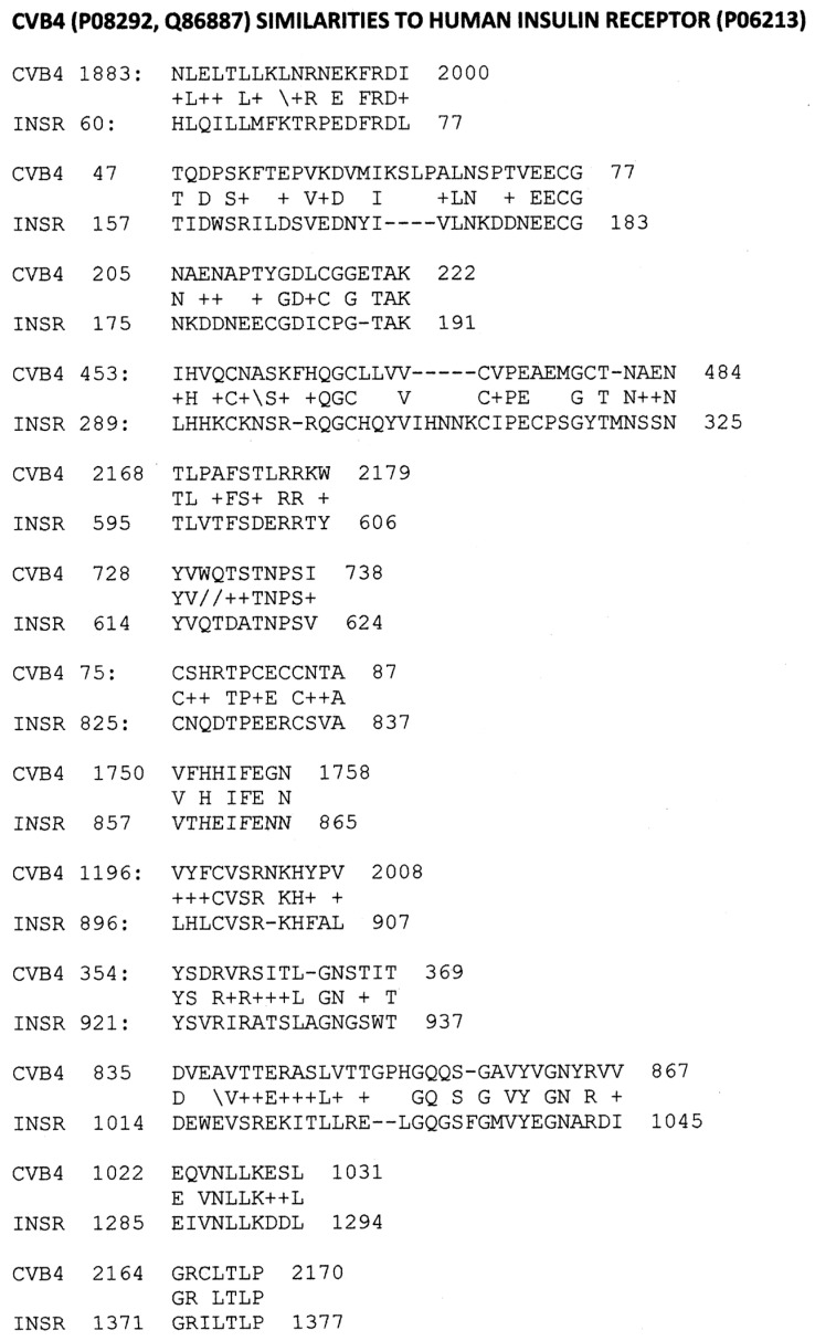
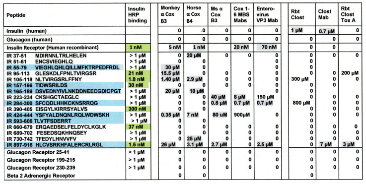
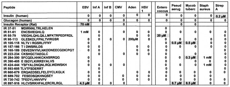
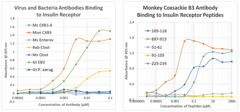
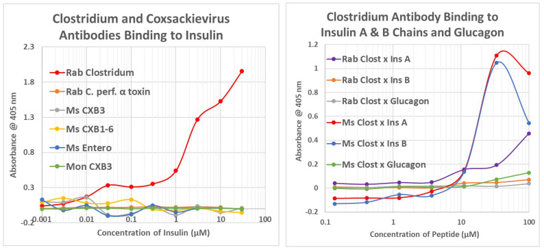
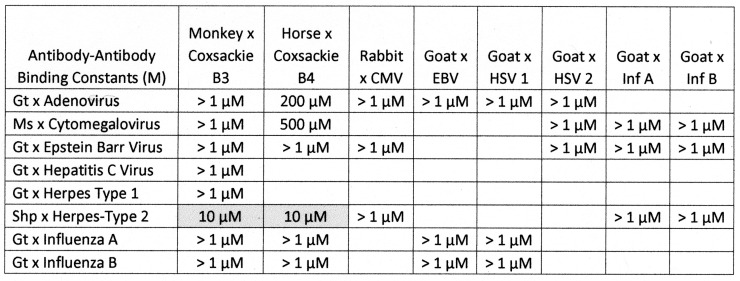
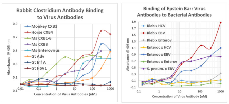
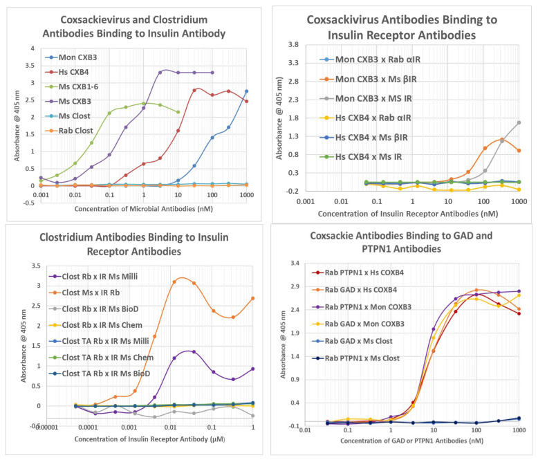
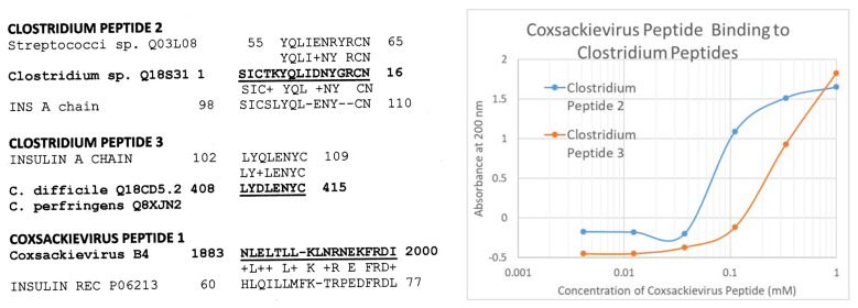
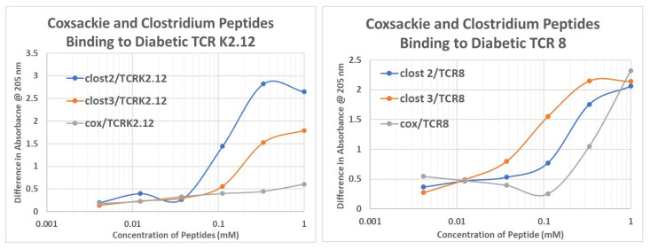
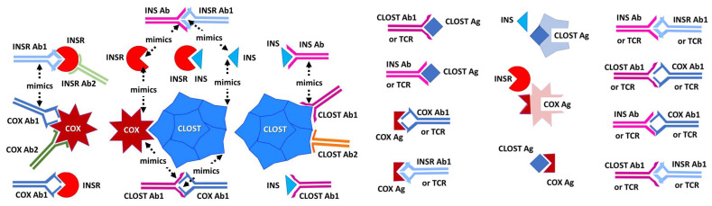
What triggers type 1 diabetes mellitus (T1DM)? One common assumption is that triggers are individual microbes that mimic autoantibody targets such as insulin (INS). However, most microbes highly associated with T1DM pathogenesis, such as coxsackieviruses (COX), lack INS mimicry and have failed to induce T1DM in animal models. Using proteomic similarity search techniques, we found that COX actually mimicked the INS receptor (INSR). Clostridia were the best mimics of INS. Clostridia antibodies cross-reacted with INS in ELISA experiments, confirming mimicry. COX antibodies cross-reacted with INSR. Clostridia antibodies further bound to COX antibodies as idiotype-anti-idiotype pairs conserving INS-INSR complementarity. Ultraviolet spectrometry studies demonstrated that INS-like Clostridia peptides bound to INSR-like COX peptides. These complementary peptides were also recognized as antigens by T cell receptor sequences derived from T1DM patients. Finally, most sera from T1DM patients bound strongly to inactivated Clostridium sporogenes, while most sera from healthy individuals did not; T1DM sera also exhibited evidence of anti-idiotype antibodies against idiotypic INS, glutamic acid decarboxylase, and protein tyrosine phosphatase non-receptor (islet antigen-2) antibodies. These results suggest that T1DM is triggered by combined enterovirus-Clostridium (and possibly combined Epstein-Barr-virus-Streptococcal) infections, and the probable rate of such co-infections approximates the rate of new T1DM diagnoses.

Keywords: COX; Clostridium; T cell receptors; circulating immune complexes; complementary antigens; diabetes; idiotype–anti-idiotype; synergism.


Conflict of interest statement

The authors declare no conflict of interest.

Figures

Figure 8
Figure 8
Summary of the binding constants (Kd) from double-antibody ELISA experiments involving a virus antibody potentially binding to a bacterial antibody. See Figure 10 and Appendix E for examples of the binding curves. The binding constants were derived from the inflection points of the curves. COXB = COX type B; CMV = cytomegalovirus; HBV = hepatitis B virus; HCV = hepatitis C virus; HSV = herpes simplex virus; Clost = Clostridium; Strep. pneum. = Streptococcus pneumoniae; GAS = group A Streptococci; Staph = Staphylococcus aureus; Kleb. = Klebsiella; E. coli = Escherichia coli; M. tb. = Mycobacterium tuberculosis; Rab = rabbit; Ms = mouse; Gt = goat; Shp = sheep. Where two numbers appear in any particular box, this indicates that the binding curve was doubly inflected, the first number providing the binding constant of the high affinity part of the curve, and the second number providing the binding constant of the low affinity part of the curve. NP = combination not possible.
Figure 14
Figure 14
Results of ELISA experiments demonstrating binding of sera from type 1 diabetic patients (T1DM), type 2 diabetic patients (T2 diabetic), and healthy controls to inactivated Clostridium sporogenes antigen. LEFT: The sera were held constant and the concentration of Closridium antigen was varied. RIGHT: the concentration of Clostridium antigen was held constant and concentration of sera was varied.
Figure 15
Figure 15
Summary of human sera binding to insulin (INS), Clostridium sporogenes antigen, rabbit (Rab) anti-Clostridium antibody (Ab), horse (Hs) and mouse (Ms) anti-coxsackievirus (COX) antibodies, and enterovirus antibodies. EDTA = etheylenediaminetetraacetic acid.
Figure 16
Figure 16
Results of double antibody ELISA experiments involving the binding of human healthy or type 1 diabetic patient (T1DM) sera to enterovirus (Ent) or COX (Cox) antibodies derived from rabbit (Rab), horse (Hs), or mouse (Ms).
Figure A1
Figure A1
BLASTP similarities between human INS A chain and C. difficile.
Figure A2
Figure A2
BLASTP similarities between human INS B chain and C. difficile.
Figure A3
Figure A3
Similarities between viruses and human insulin receptor.
Figure A4
Figure A4
A selection of BLASTP similarities between C. difficile proteins and protein tyrosine phosphatase non-receptor type 1. Many additional significant similarities exist.
Figure A5
Figure A5
A selection of BLASTP similarities between C. difficile proteins and glutamic acid decarboxylase type 65. Many additional significant similarities exist.
Figure A6
Figure A6
BLASTP similarities between glutamic acid decarboxylase type 65 and COX type B4 (which is typical of the other COX). These are all the significant similarities that were found (compared with results for GAD-65 and Clostridia above).
Figure A7
Figure A7
Comparison of monkey (Mon) coxsackievirus (CX) antibodies and horse (Hs) CX antibodies binding to IR peptides. Numbers indicate the amino acid sequence of the INS receptor from which the peptides were derived.
Figure A8
Figure A8
Results of ELISA studies concerning virus and bacteria antibodies binding to human INS receptor peptides. LEFT: Enterovirus (Ent), COX types B1-6 (CX1-6), and influenza A virus (Inf A) antibodies binding to INS receptor (IR) peptides identified by their sequence numbers. RIGHT: Clostridium (Clost), and Mycobacterium tuberculosis (Mtb) antibodies binding to INS receptor peptides identified by their sequence numbers. ToxA = toxin A.
Figure A9
Figure A9
Representative binding curves resulting from double antibody ELISA experiments involving Clostridium monoclonal antibodies binding to coxsackievirus (CX) type B3 and type B4 antibodies, adenovirus antibodies, and herpes simplex type 1 (HSV1) antibodies.
Figure A10
Figure A10
Some of the many control combinations of virus–bacterial antibody pairs tested by double antibody ELISA and summarized in Figure 12 and Figure 13. S. pneum. = Streptococcus pneumoniae; S. aureus = Staphylococcus aureus; Clost = Clostridium; Adv = adenovirus; CMV = cytomegalovirus; EBV = Epstein–Barr virus; HSV1 = herpes simplex type 1 virus; Ms = mouse; Rbt = rabbig; Gt = goat.
Figure A11
Figure A11
Ultraviolet spectrometry study of COX (cox) peptide (which mimics the INS receptor) and Clostridium peptides 2 and 3 (clost2, clost3) (which mimic INS), binding to T cell receptors (TCR) that mimic a range of peptides (see Table 1 for details). The binding constants listed in Table 1 were estimated from the inflection points of the curves.
Figure A12
Figure A12
TIDM and Control Sera binding to human insulin and Clostridium antigen. LEFT: Results of ELISA experiments demonstrating binding of sera from type 1 diabetic patients (T1DMand healthy controls to INS conjugated to horseradish peroxidase (INS–HRP). RIGHT: Results of ELISA experiments demonstrating binding of sera from type 1 diabetic patients (T1DM), and healthy controls to inactivated Clostridium sporogenes antigen. In these experiments, the concentration of Clostridium antigen was held constant, and concentration of sera was varied.
Figure A13
Figure A13
Results of double antibody ELISA experiments involving the binding of human healthy or type 1 diabetic patient (T1DM) sera to rabbit Clostridium antibodies (LEFT). Although all sera bound Clostridium antibodies, the T1DM sera bound significant more than the healthy sera (RIGHT).
Figure 1
Figure 1
Results of BLASTP similarity search comparing the human INS A chain sequence with the UNIPROTKB bacterial database curated for human pathogens and commensals.
Figure 2
Figure 2
Results of BLASTP similarity search comparing the human INS B chain sequence with the UNIPROTKB bacterial database curated for human pathogens and commensals.
Figure 3
Figure 3
BLASTP similarities between COX type B4 and the human INS receptor.
Figure 4
Figure 4
Summary of quantitative ELISA experiments regarding COX and Clostridia antibody binding to INS, glucagon, the INS receptor (IR), IR peptides, glucagon receptor peptides, and the beta 2 adrenergic receptor. Blue highlighted sequences are ones overlapping the COX-INS receptor similarities listed in Figure 3. Green highlighted figures are sequences with significant INS binding. Gray highlighted figures are sequences that displayed significant antibody binding. Results are binding constants (Kd) in micromoles determined by the inflection points of each curve. A zero (0) indicates that binding was not measurable (in practice, Kd > 1 micromolar). The IR sequences are listed by the numerical position in the IR sequence and followed by the single-letter amino acid sequence. HRP = horseradish peroxidase conjugated; α = “anti-“; Cox = coxsackivierus; Mabs = monoclonal antibodies (mouse); Rab = rabbit antibody; Clost = Clostridium; Tox A = Clostridium toxin A.
Figure 5
Figure 5
Binding of virus and bacterium antibodies to INS, glucagon, the INS receptor (INSR), and INSR peptides. EBV = Epstein–Barr virus; Inf A = Influenza A virus; Inf B = Influenza B virus; CMV = cytomegalovirus; Adeno = adenovirus; HSV1 = human herpes simplex virus type 1; Pseud aerug = Pseudomonas aeruginosa; Mycob tuber = Mycobacterium tuberculosis; Staph aureus = Staphylococcus aureus; Strep A = group A Stretpcococci. See Figure 8 for an explanation of the rest of the abbreviations and formalisms.
Figure 6
Figure 6
Results of ELISA studies concerning binding to the INS receptor (IR) and its peptides. LEFT: Monkey (Mon) anti-coxsackievirus type B3 (CXB3), Epstein–Barr virus (EBV) antibody and to a much lesser extent, Clostridium antibodies, bind to the human insulin receptor. RIGHT: Further characterization of monkey anti-CXB3 antibody binding to peptides derived from the human INSR. Ms = mouse; Rab = rabbit; CX = coxsackievirus; Enterov = enterovirus; EBV = Epstein–Barr virus; Clost = Clostridium; P. aerug. = Pseudomonas aeruginosa. Numbers indicate the amino acid sequence of the INS receptor from which the peptides were derived. Further data are provided in Appendix D.
Figure 7
Figure 7
Results of ELISA studies concerning binding to insulin (INS). LEFT: Clostridium antibody, but not coxsackievirus (X) antibodies, bind to INS. RIGHT: Clostridium antibodies bind to the INS A and B chains but not to glucagon. Rab = rabbit; Ms = mouse; C. perf toxin = Clostridium perfringens alpha toxin; Cox = COX; Clost = Clostridium; Ins = INS; A = A chain; B = B chain.
Figure 9
Figure 9
Summary of the binding constants (Kd) from double-antibody ELISA experiments involving a virus antibody potentially binding to another virus antibody. See Figure 14, Figure 15 and Figure 16 for examples. COXB = COX type B; CMV = cytomegalovirus; HBV = hepatitis B virus; HCOX = hepatitis C virus; HSV = herpes simplex virus; Ms = mouse; Gt = goat; Shp = sheep.
Figure 10
Figure 10
Representative binding curves resulting from double antibody ELISA experiments involving virus and bacterium antibodies. LEFT: Rabbit Clostridium antibodies bound to all of the COX antibodies tested as well as an enterovirus antibody but did not bind to other virus antibodies. RIGHT: Additional control combinations of virus–bacterial antibody pairs tested by double antibody ELISA and summarized in Appendix E. CXB = Coxsackievirus; Adv = adenovirus; Inf A = influenza A virus, HSV1 = herpes simplex type 1 virus; Ms = mouse; Gt = goat; Hs = horse; Mon = monkey; Enteroc = Enterococcus faecium; S. pneum. = Streptococcus pneumoniae; Adv = adenovirus; EBV = Epstein–Barr virus; HCOX = hepatitis C virus; Enterov = enterovirus. See Figure 8 and Figure 9 for species of each antibody.
Figure 11
Figure 11
Results of double antibody ELISA experiments. TOP LEFT: Coxsackievirus (CX) and Clostridium antibodies binding to INS antibody. TOP RIGHT: CX and Clostridium antibodies binding to several INS receptor antibodies. BOTTOM LEFT: Clostridia antibodies binding to INS receptor antibodies (IR-Ab). Rb IR-Ab were tested against Ms Clostridia, while Ms IR-Ab were tested against Rb Clostridia. BOTTOM RIGHT: Binding of COX type B (COXB) or Clostridium (Clost) antibody binding to glutamic acid decarboxylase (GAD-65) antibody or protein tyrosine phosphatase non-receptor type 1 (PTPN(IA2)) antibody. Clost = Clostridium; TA = toxin A; Rb = rabbit; Ms = mouse; Milli = Millipore CXB = Coxsackievirus type B; IR = INS receptor (alpha and beta indicating the chain to which the antibody is specific); Mon = monkey; Hs = horse; Rab = rabbit; Ms = mouse; Biod = Biodesign; Chem = Chemicon (see Tables of antibodies in Section 4).
Figure 12
Figure 12
Two Clostidium peptides that mimic the INS A chain (INS A) and one COX peptide that mimics the INS receptor (INS Rec) were synthesized (sequences and similarities to the LEFT) and tested for their ability to bind to each other using ultraviolet spectrometry (RIGHT). Kd was 60 µM for Clostridium peptide 2 and 200 µM for peptide 3.
Figure 13
Figure 13
Ultraviolet spectrometry study of COX (cox) peptide (which mimics the INS receptor) and Clostridium peptides 2 and 3 (clost2, clost3) (which mimic INS), binding to T cell receptors (TCR) K2.12 (which mimics the INS receptor) and TCR8 (which mimics the glucagon receptor). The binding constants listed in Table 1 were estimated from the inflection points of the curves.
Figure 17
Figure 17
Summary of the various types of complementarity and mimicry revealed by the experiments performed here. INS = insulin; INSR = INS receptor; Ab = antibody; TCR = T cell receptor; Clost = Clostridium; COX = coxsackievirus. The arrow link mimics. Steric fits represent complementarity between antibodies with their antigens as well as between pairs of antigens (e.g., COX and Clost or INS and INSR).

Similar articles

Cited by

References

    1. Mallone R., Brezar V., Boitard C. T cell recognition of autoantigens in human type 1 diabetes: Clinical perspectives. Clin. Dev. Immunol. 2011;2011:513210. doi: 10.1155/2011/513210. - DOI - PMC - PubMed
    1. Krischer J.P., Liu X., Lernmark Å., Hagopian W.A., Rewers M.J., She J.X., Toppari J., Ziegler A.G., Akolkar B., TEDDY Study Group Predictors of the Initiation of Islet Autoimmunity and Progression to Multiple Autoantibodies and Clinical Diabetes: The TEDDY Study. Diabetes Care. 2022;45:2271–2281. doi: 10.2337/dc21-2612. - DOI - PMC - PubMed
    1. Bauer W., Veijola R., Lempainen J., Kiviniemi M., Härkönen T., Toppari J., Knip M., Gyenesei A., Ilonen J. Age at Seroconversion, HLA Genotype, and Specificity of Autoantibodies in Progression of Islet Autoimmunity in Childhood. J. Clin. Endocrinol. Metab. 2019;104:4521–4530. doi: 10.1210/jc.2019-00421. - DOI - PubMed
    1. Kim S.W., Won H.K., Seok H., Lee B.W., Jung C.H., Lee W.J., Kim J.H. High prevalence of both anti-INS and anti-INS receptor antibodies in Korean patients with INS autoimmune syndrome. Diabetes Res. Clin. Pract. 2012;98:e12–e15. doi: 10.1016/j.diabres.2012.09.019. - DOI - PubMed
    1. Blackard W.G., Anderson J.H., Mullinax F. Anti-INS receptor antibodies and diabetes. Ann. Intern. Med. 1977;86:584–585. doi: 10.7326/0003-4819-86-5-584. - DOI - PubMed