

Heterogeneity of Lipopolysaccharide as Source of Variability in Bioassays and LPS-Binding Proteins as Remedy
- PMID: 37176105
- PMCID: PMC10179214
- DOI: 10.3390/ijms24098395
Heterogeneity of Lipopolysaccharide as Source of Variability in Bioassays and LPS-Binding Proteins as Remedy
Abstract
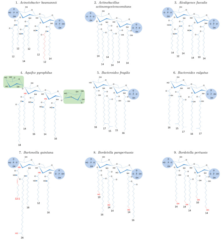
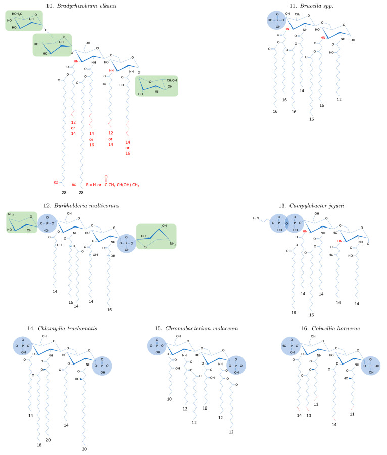
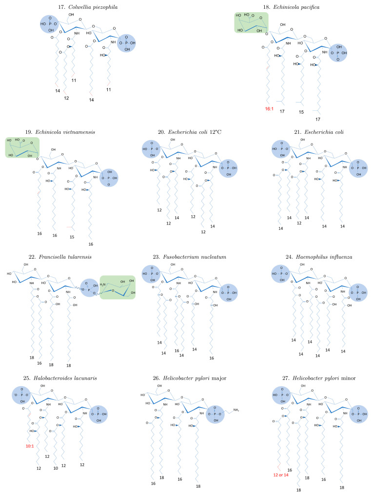
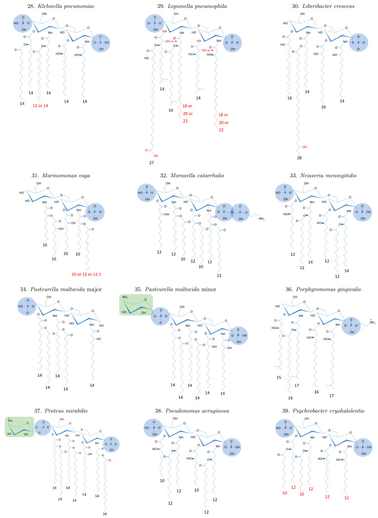
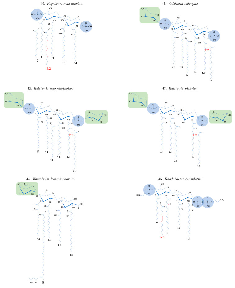
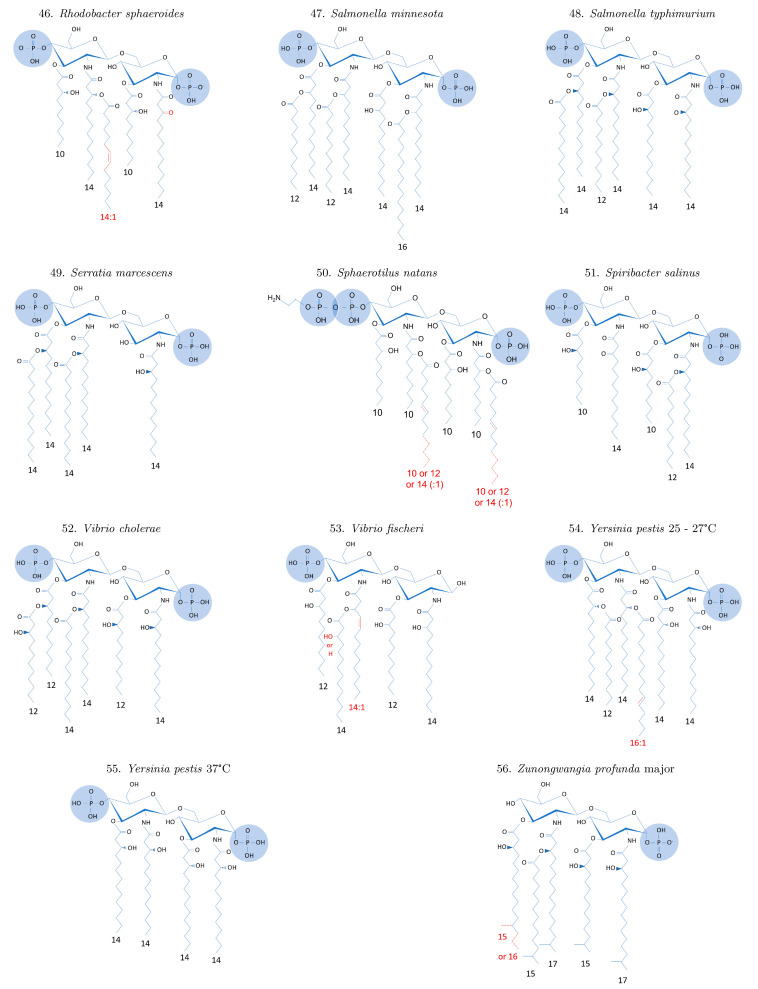
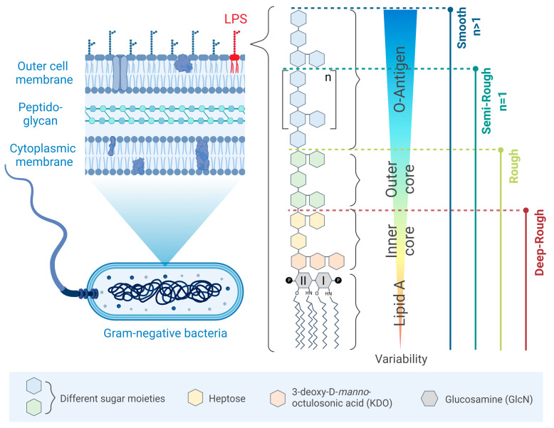
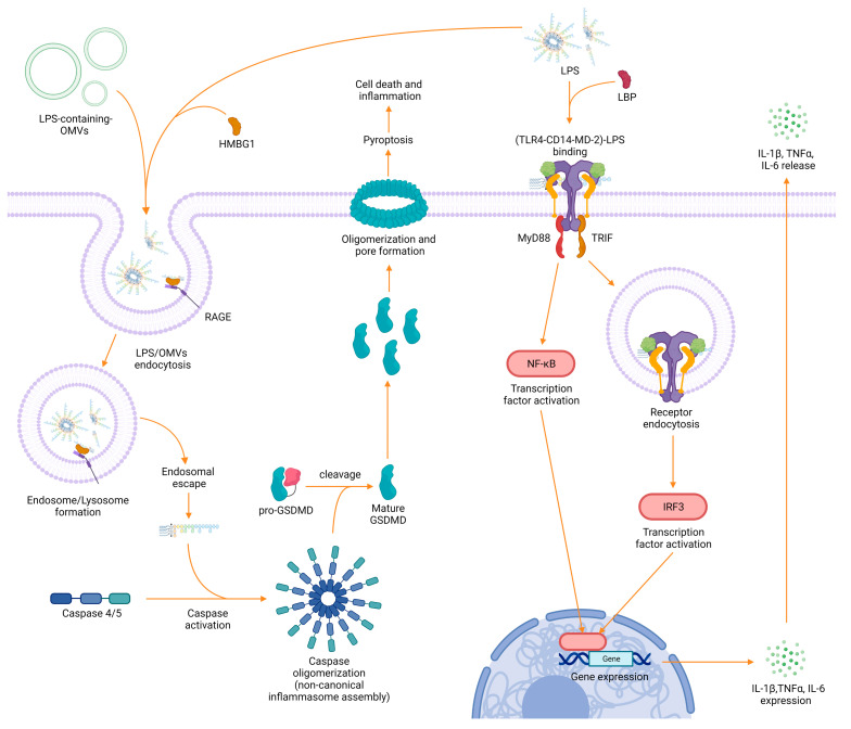
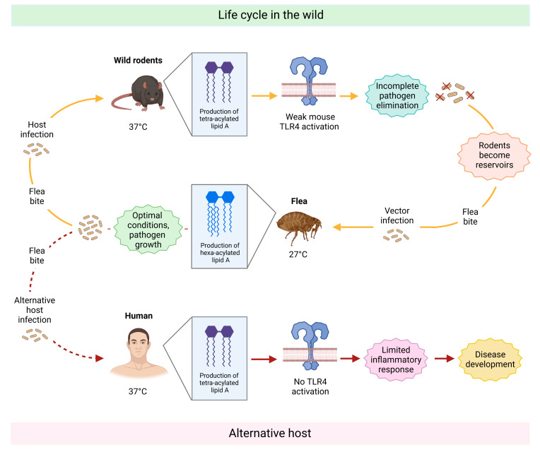
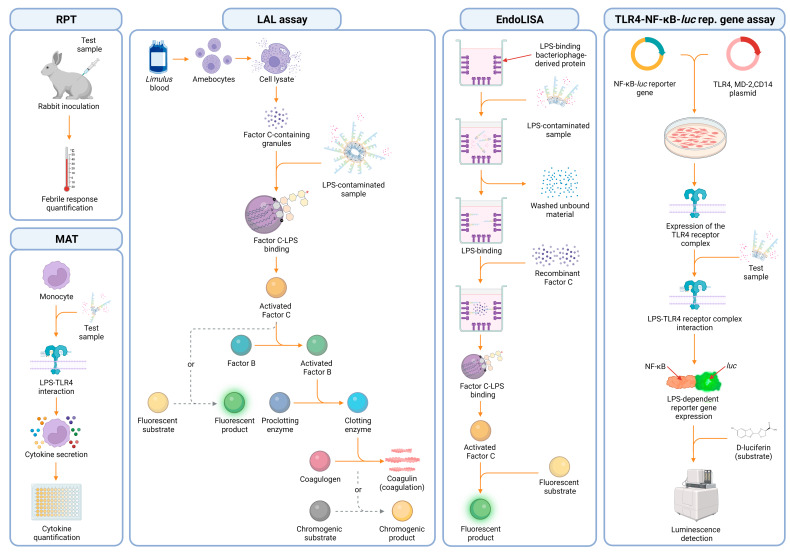
Lipopolysaccharide (LPS), also referred to as endotoxin, is the major component of Gram-negative bacteria's outer cell wall. It is one of the main types of pathogen-associated molecular patterns (PAMPs) that are known to elicit severe immune reactions in the event of a pathogen trespassing the epithelial barrier and reaching the bloodstream. Associated symptoms include fever and septic shock, which in severe cases, might even lead to death. Thus, the detection of LPS in medical devices and injectable pharmaceuticals is of utmost importance. However, the term LPS does not describe one single molecule but a diverse class of molecules sharing one common feature: their characteristic chemical structure. Each bacterial species has its own pool of LPS molecules varying in their chemical composition and enabling the aggregation into different supramolecular structures upon release from the bacterial cell wall. As this heterogeneity has consequences for bioassays, we aim to examine the great variability of LPS molecules and their potential to form various supramolecular structures. Furthermore, we describe current LPS quantification methods and the LPS-dependent inflammatory pathway and show how LPS heterogeneity can affect them. With the intent of overcoming these challenges and moving towards a universal approach for targeting LPS, we review current studies concerning LPS-specific binders. Finally, we give perspectives for LPS research and the use of LPS-binding molecules.
Keywords: LPS-binding molecules; detection; endotoxin; immunology; lipid A; low endotoxin recovery.
Conflict of interest statement
The authors declare no conflict of interest.
Figures
Similar articles
-
Lipopolysaccharide Structure and the Phenomenon of Low Endotoxin Recovery.Eur J Pharm Biopharm. 2022 Nov;180:289-307. doi: 10.1016/j.ejpb.2022.10.006. Epub 2022 Oct 19. Eur J Pharm Biopharm. 2022. PMID: 36272656 Review.
-
Host Defense Proteins and Peptides with Lipopolysaccharide-Binding Activity from Marine Invertebrates and Their Therapeutic Potential in Gram-Negative Sepsis.Mar Drugs. 2023 Nov 7;21(11):581. doi: 10.3390/md21110581. Mar Drugs. 2023. PMID: 37999405 Free PMC article. Review.
-
Abdominal Septic Shock - Endotoxin Adsorption Treatment (ASSET) - endotoxin removal in abdominal and urogenital septic shock with the Alteco® LPS Adsorber: study protocol for a double-blinded, randomized placebo-controlled trial.Trials. 2016 Dec 8;17(1):587. doi: 10.1186/s13063-016-1723-4. Trials. 2016. PMID: 27931259 Free PMC article. Clinical Trial.
-
Protective role of phospholipid oxidation products in endotoxin-induced tissue damage.Nature. 2002 Sep 5;419(6902):77-81. doi: 10.1038/nature01023. Nature. 2002. PMID: 12214235
-
Molecular mechanisms of endotoxin activity.Arch Microbiol. 1995 Dec;164(6):383-9. doi: 10.1007/BF02529735. Arch Microbiol. 1995. PMID: 8588739 Review.
Cited by
-
The interaction between oral microbiota and gut microbiota in atherosclerosis.Front Cardiovasc Med. 2024 Jun 12;11:1406220. doi: 10.3389/fcvm.2024.1406220. eCollection 2024. Front Cardiovasc Med. 2024. PMID: 38932989 Free PMC article. Review.
-
Biomass-Derived Nanoporous Carbon Honeycomb Monoliths for Environmental Lipopolysaccharide Adsorption from Aqueous Media.Int J Mol Sci. 2025 Jan 23;26(3):952. doi: 10.3390/ijms26030952. Int J Mol Sci. 2025. PMID: 39940720 Free PMC article.
-
Radiation-Detoxified Form of Endotoxin Effectively Activates Th1 Responses and Attenuates Ragweed-Induced Th2-Type Airway Inflammation in Mice.Int J Mol Sci. 2024 Jan 27;25(3):1581. doi: 10.3390/ijms25031581. Int J Mol Sci. 2024. PMID: 38338861 Free PMC article.
-
Impact on Human Health of Salmonella spp. and Their Lipopolysaccharides: Possible Therapeutic Role and Asymptomatic Presence Consequences.Int J Mol Sci. 2024 Nov 5;25(22):11868. doi: 10.3390/ijms252211868. Int J Mol Sci. 2024. PMID: 39595937 Free PMC article. Review.
-
Factors Promoting Lipopolysaccharide Uptake by Synthetic Lipid Droplets.bioRxiv [Preprint]. 2024 Oct 19:2024.10.19.619182. doi: 10.1101/2024.10.19.619182. bioRxiv. 2024. Update in: ACS Omega. 2025 Feb 10;10(6):5866-5873. doi: 10.1021/acsomega.4c09599. PMID: 39464097 Free PMC article. Updated. Preprint.
References
-
- Di Lorenzo F., Billod J.-M., Martín-Santamaría S., Silipo A., Molinaro A. Gram-Negative Extremophile Lipopolysaccharides: Promising Source of Inspiration for a New Generation of Endotoxin Antagonists. Eur. J. Org. Chem. 2017;2017:4055–4073. doi: 10.1002/ejoc.201700113. - DOI
Publication types
MeSH terms
Substances
Grants and funding
LinkOut - more resources
Full Text Sources
Research Materials
