. 2023 Apr 22;16(9):3287.
doi: 10.3390/ma16093287.

Inflammatory, Oxidative Stress and Small Cellular Particle Response in HUVEC Induced by Debris from Endoprosthesis Processing

Affiliations

Inflammatory, Oxidative Stress and Small Cellular Particle Response in HUVEC Induced by Debris from Endoprosthesis Processing

Zala Jan et al. Materials (Basel). .

Abstract

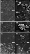
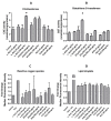
We studied inflammatory and oxidative stress-related parameters and cytotoxic response of human umbilical vein endothelial cells (HUVEC) to a 24 h treatment with milled particles simulating debris involved in sandblasting of orthopedic implants (OI). We used different abrasives (corundum-(Al2O3), used corundum retrieved from removed OI (u. Al2O3), and zirconia/silica composite (ZrO2/SiO2)). Morphological changes were observed by scanning electron microscopy (SEM). Concentration of Interleukins IL-6 and IL-1β and Tumor Necrosis Factor α (TNF)-α was assessed by enzyme-linked immunosorbent assay (ELISA). Activity of Cholinesterase (ChE) and Glutathione S-transferase (GST) was measured by spectrophotometry. Reactive oxygen species (ROS), lipid droplets (LD) and apoptosis were measured by flow cytometry (FCM). Detachment of the cells from glass and budding of the cell membrane did not differ in the treated and untreated control cells. Increased concentration of IL-1β and of IL-6 was found after treatment with all tested particle types, indicating inflammatory response of the treated cells. Increased ChE activity was found after treatment with u. Al2O3 and ZrO2/SiO2. Increased GST activity was found after treatment with ZrO2/SiO2. Increased LD quantity but not ROS quantity was found after treatment with u. Al2O3. No cytotoxicity was detected after treatment with u. Al2O3. The tested materials in concentrations added to in vitro cell lines were found non-toxic but bioactive and therefore prone to induce a response of the human body to OI.

Keywords: corundum; cytokines; endoprosthesis failure; lipid droplets; reactive oxygen species.


Conflict of interest statement

The authors declare no conflict of interest.

Figures
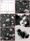
Figure 1
Figure 1
(a) XRD patterns of Al2O3, u. Al2O3 and ZrO2/SiO2 abrasives (c: corundum, u: unknown, z: monoclinic zirconia); (b,c) SEM images of Al2O3 and u. Al2O3 samples, respectively; (d) TEM image of Al2O3 sample; (e) SEM image of ZrO2/SiO2 sample; (f) TEM image of ZrO2/SiO2 sample.
Figure 2
Figure 2
Secondary Electron (SE) image of untreated cells (A1,A2) and cells treated with 100 μg/mL concentration of u. Al2O3 (B1,B2), Al2O3 (C1,C2), ZrO2/SiO2 (D1,D2) and positive control—Staurosporine (E1,E2).
Figure 3
Figure 3
Concentration of IL-6 (A), IL-1β (B) and TNF-α (C) in conditioned media of cells treated with three different types of particles (u. Al2O3, ZrO2/SiO2 and Al2O3). Particles were administered at three concentrations (10 μg/mL, 50 μg/mL and 100 μg/mL). The horizontal line denotes the average concentration of negative control samples (untreated cells). Experiments were performed in duplicate and bars represent the standard deviations. Asterisks denote statistically significant differences with respect to the control.
Figure 4
Figure 4
ChE activity (A), GST activity (B), fold change of median fluorescence intensity of ROS (CM-H2DCFA) (C) and LD (BODIPY 483/503) (D) in comparison to controls of HUVEC cells treated with either of the three different types of particles (u. Al2O3, ZrO2/SiO2 and Al2O3) at three concentrations (10 μg/mL, 50 μg/mL and 100 μg/mL), or the positive controls (5 mM H2O2 and 75 µM oleic acid). Experiments were performed in triplicate (A,B) or duplicate (C,D) and bars represent the standard deviations. Asterisks denote statistically significant differences with respect to the control.
Figure 5
Figure 5
Top: histograms showing ROS production due to treatment with u. Al2O3. Bottom: histograms showing LD production due to due to treatment with u. Al2O3. (A,B) untreated cells, (C) positive controls for ROS—cells treated with 5 mM H2O2, (D) positive controls for lipid droplets (LD)—cells treated with 75 μM oleic acid, (E) cells treated with 10 μg/mL u. Al2O3 (ROS), (F) cells treated with 10 μg/mL u. Al2O3 (LD), (G) cells treated with 50 μg/mL u. Al2O3 (ROS), (H) cells treated with 50 μg/mL u. Al2O3 (LD), (I) cells treated with 100 μg/mL u. Al2O3 (ROS), (J) cells treated with 100 μg/mL u. Al2O3 (LD). For ROS detection, cells were stained with CM-H2DCFDA; for LD detection, cells were stained with BODIPY 493/503. Gates indicate the % of ROS and LD-positive cells. Abscissa gives the fluorescence intensity of the signal release by the fluorophores, and ordinate gives the number of events (cells) detected by the flow cytometer (FCM) for a given intensity.
Figure 6
Figure 6
Cytotoxicity effect of u. Al2O3 particles on HUVEC. (A) untreated cells, (B) positive control for apoptosis (cells treated with Staurosporine), (C) HUVEC treated with 10 μg/mL of u. Al2O3, (D) HUVEC treated with 50 μg/mL of u. Al2O3, (E) HUVEC treated with 100 μg/mL of u. Al2O3. SSC-A—side scatter measurements; information on internal complexity (granularity), FSC-A—forward scatter; information on cell size. The numbers in boxes represent percent of cells in the respective gates (left) and percent of apoptotic cells (right).

References

    1. Gibon E., Amanatullah D.F., Loi F., Pajarinen J., Nabeshima A., Yao Z., Hamadouche M., Goodman S.B. The biological response to orthopaedic implants for joint replacement: Part I: Metals. J. Biomed. Mater. Res. Part B Appl. Biomater. 2017;105:2162–2173. doi: 10.1002/jbm.b.33734. - DOI - PMC - PubMed
    1. Evans E.J., Thomas I.T. The in vitro toxicity of cobalt-chrome-molybdenum alloy and its constituent metals. Biomaterials. 1986;7:25–29. doi: 10.1016/0142-9612(86)90084-0. - DOI - PubMed
    1. Mack M. Inflammation and fibrosis. Matrix Biol. 2018;68–69:106–121. doi: 10.1016/j.matbio.2017.11.010. - DOI - PubMed
    1. Mirra J.M., Marder R.A., Amstutz H.C. The pathology of failed total joint arthroplasty. Clin. Orthop. Relat. Res. 1982;170:175–183. doi: 10.1097/00003086-198210000-00023. - DOI - PubMed
    1. Kienzle A., Walter S., von Roth P., Fuchs M., Winkler T., Müller M. High Rates of Aseptic Loosening after Revision Total Knee Arthroplasty for Periprosthetic Joint Infection. JBJS OA. 2020;5:e20.00026. doi: 10.2106/JBJS.OA.20.00026. - DOI - PMC - PubMed