. 2023 Apr 29;16(9):3472.
doi: 10.3390/ma16093472.

Phytosynthesis of Silver Nanoparticles Using Leonurus cardiaca L. Extracts

Affiliations

Phytosynthesis of Silver Nanoparticles Using Leonurus cardiaca L. Extracts

Ioana Catalina Fierascu et al. Materials (Basel). .

Abstract

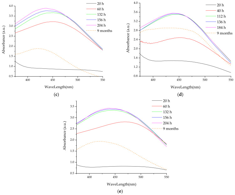
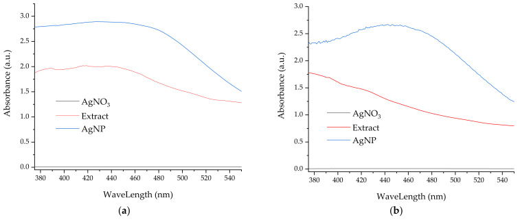
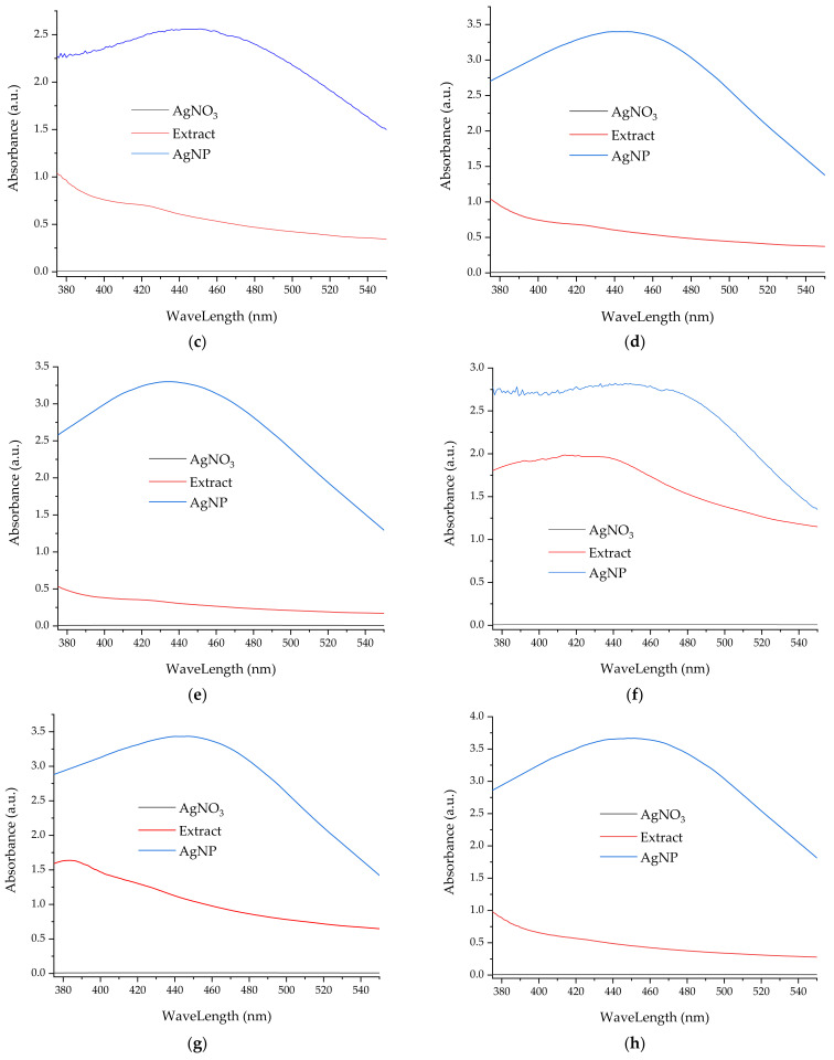
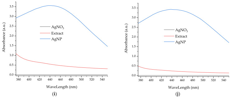
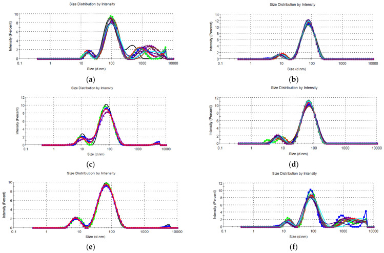
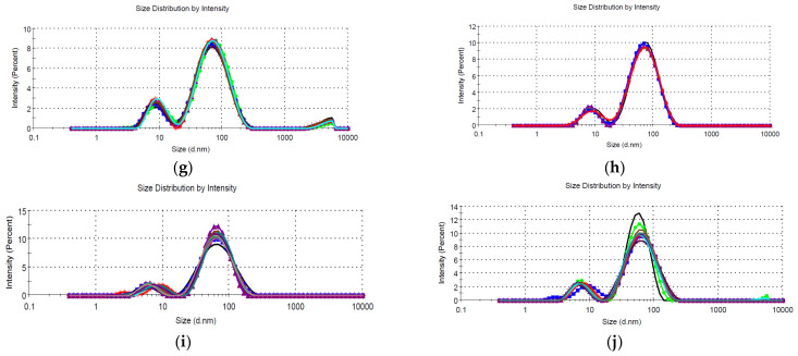
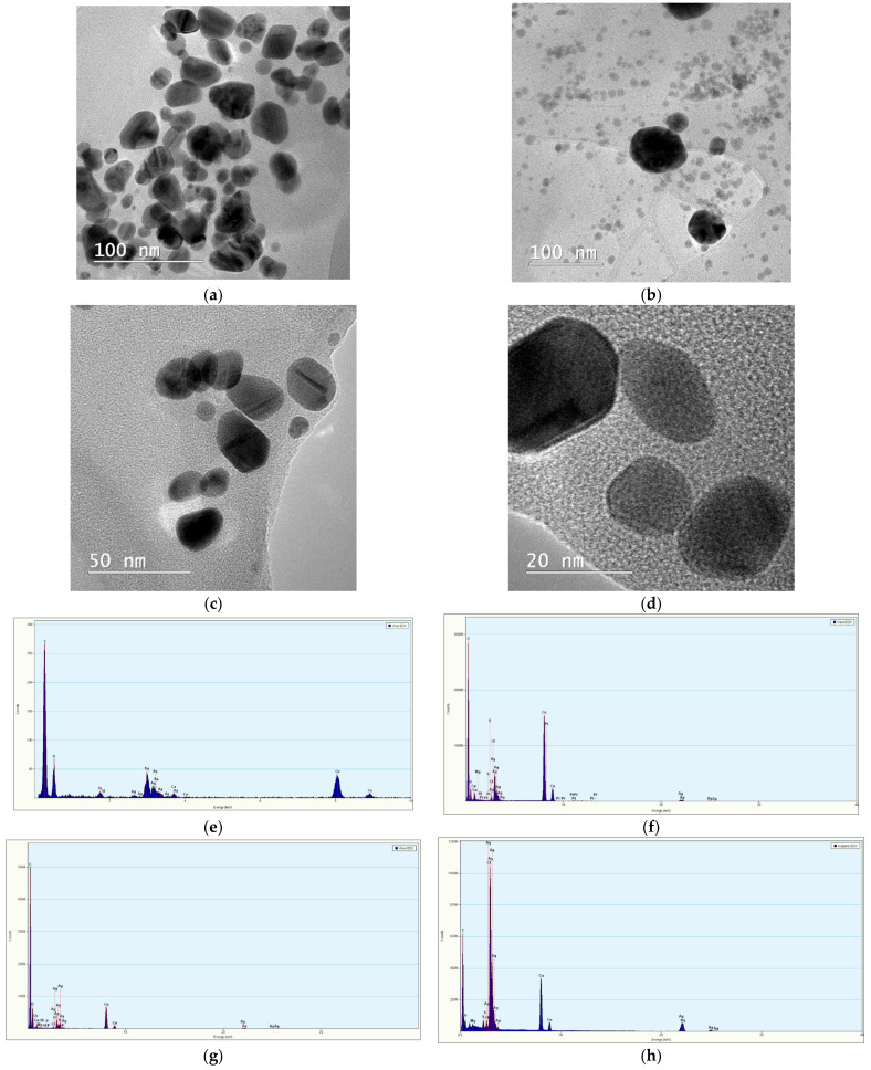
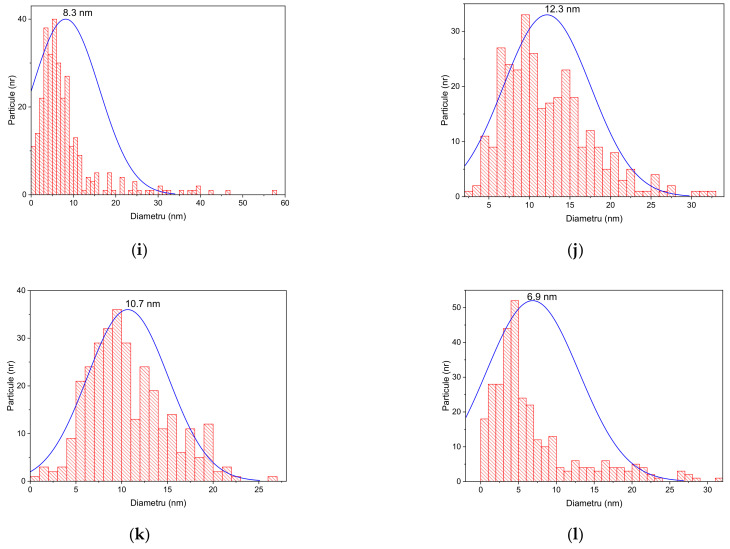
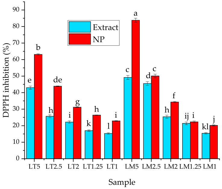
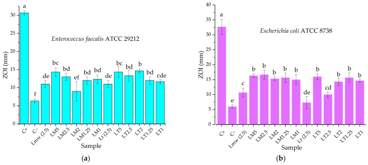
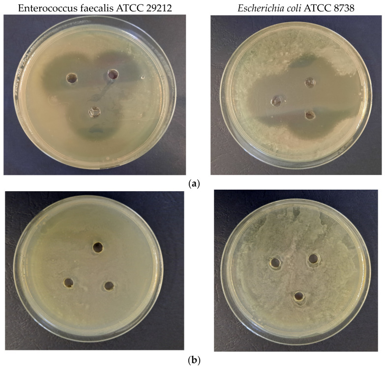
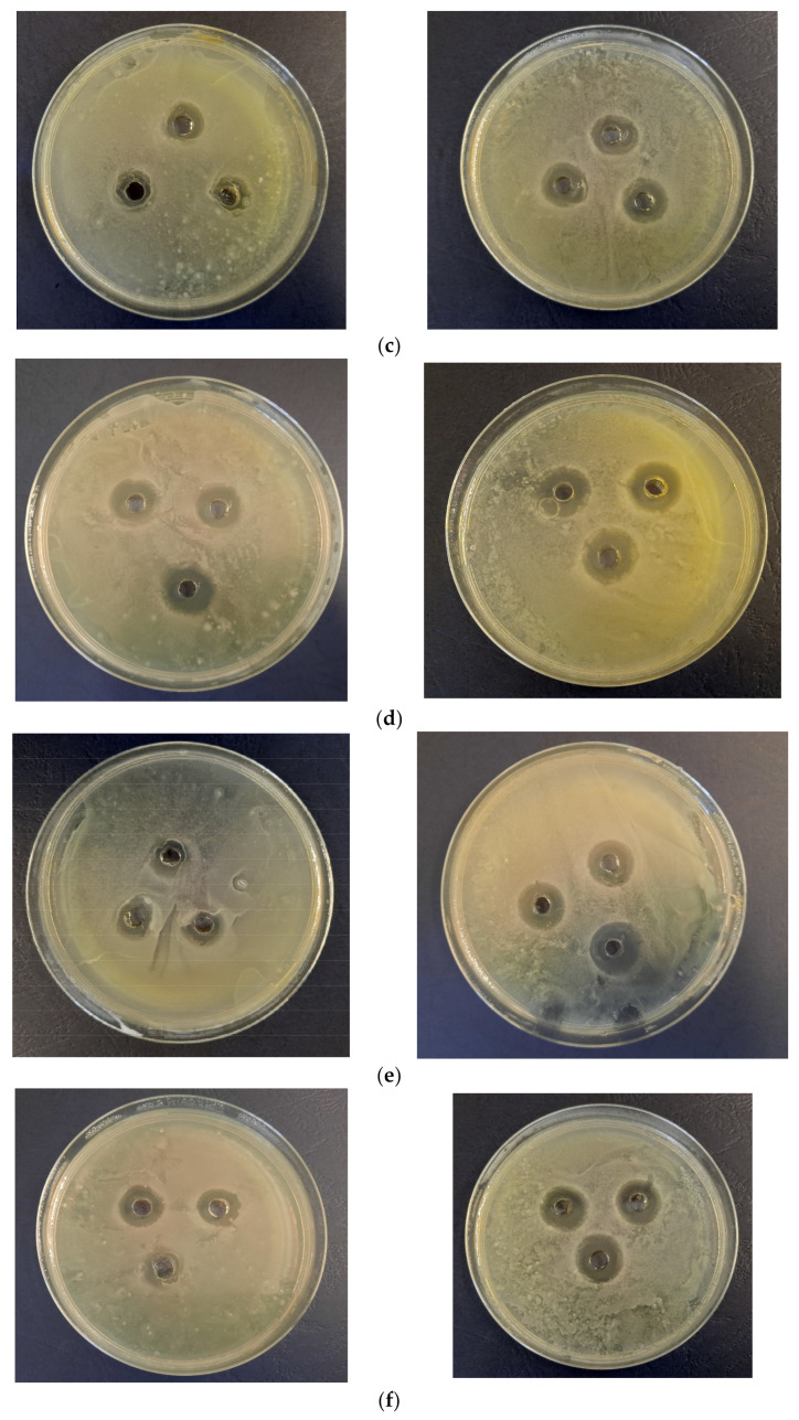
The present work describes, for the first time in the literature, the phytosynthesis of silver nanoparticles using Leonurus cardiaca L. extracts. The influence of the extraction method (classical temperature extraction and microwave extraction), as well as of the extract concentration on the characteristics of the nanoparticles, was studied using analytical methods, such as UV-Vis spectrometry, X-ray diffraction, dynamic light scattering, and transmission electron microscopy. Experimental data suggest that use of lower extract concentration leads to smaller dimensions nanoparticles, the same effect using the extract obtained by microwave-assisted extraction. The smallest recorded crystallite sizes (by X-ray diffraction) were under 3 nm. The antioxidant properties (determined by the DPPH assay) and the antimicrobial potential (determined against Gram-negative and Gram-positive strains) are enhanced by the phytosynthesis process (as demonstrated by the comparison of the nanoparticles' properties with the parent extracts). The present work could also represent an important step in obtaining nanoparticles with enhanced properties and controlled morphologies, but also offers information on the phytosynthesis of metallic nanoparticles using low extract concentrations.

Keywords: Leonurus cardiaca L.; antimicrobial; antioxidant; phytosynthesis; silver nanoparticles.


Conflict of interest statement

The authors declare no conflict of interest. The funders had no role in the design of the study; in the collection, analyses, or interpretation of data; in the writing of the manuscript; or in the decision to publish the results.

Figures

Figure 1
Figure 1
Dried and processed aerial parts of L. cardiaca used in the experiments.
Figure 2
Figure 2
UV-Vis spectra of the developed nanoparticles using L. cardiaca extracts obtained by classical temperature extraction: (a) sample LT5; (b) sample LT2.5; (c) sample LT2; (d) sample LT1.25; (e) sample LT1.
Figure 2
Figure 2
UV-Vis spectra of the developed nanoparticles using L. cardiaca extracts obtained by classical temperature extraction: (a) sample LT5; (b) sample LT2.5; (c) sample LT2; (d) sample LT1.25; (e) sample LT1.
Figure 3
Figure 3
UV-Vis spectra of the nanoparticles developed using extracts obtained by the microwave-assisted extraction: (a) sample LM5; (b) sample LM2.5; (c) sample LM2; (d) sample LM1.25; (e) sample LM1.
Figure 3
Figure 3
UV-Vis spectra of the nanoparticles developed using extracts obtained by the microwave-assisted extraction: (a) sample LM5; (b) sample LM2.5; (c) sample LM2; (d) sample LM1.25; (e) sample LM1.
Figure 4
Figure 4
Confirmation of phytosynthesis of silver nanoparticles by comparison of nanoparticles UV-Vis spectra with the corresponding extracts and AgNO3 solution’s spectra: (a) sample LT5; (b) sample LT2.5; (c) sample LT2; (d) sample LT1.25; (e) sample LT1; (f) sample LM5; (g) sample LM2.5; (h) sample LM2; (i) sample LM1.25; (j) sample LM1.
Figure 4
Figure 4
Confirmation of phytosynthesis of silver nanoparticles by comparison of nanoparticles UV-Vis spectra with the corresponding extracts and AgNO3 solution’s spectra: (a) sample LT5; (b) sample LT2.5; (c) sample LT2; (d) sample LT1.25; (e) sample LT1; (f) sample LM5; (g) sample LM2.5; (h) sample LM2; (i) sample LM1.25; (j) sample LM1.
Figure 5
Figure 5
Size distribution by intensity, determined by DLS technique, for: (a) sample LT5; (b) sample LT2.5; (c) sample LT2; (d) sample LT1.25; (e) sample LT1; (f) sample LM5; (g) sample LM2.5; (h) sample LM2; (i) sample LM1.25; (j) sample LM1.
Figure 5
Figure 5
Size distribution by intensity, determined by DLS technique, for: (a) sample LT5; (b) sample LT2.5; (c) sample LT2; (d) sample LT1.25; (e) sample LT1; (f) sample LM5; (g) sample LM2.5; (h) sample LM2; (i) sample LM1.25; (j) sample LM1.
Figure 6
Figure 6
Diffractograms of the phytosynthesized nanoparticles: (a) sample LT5; (b) sample LT2.5; (c) sample LT2; (d) sample LT1.25; (e) sample LT1; (f) sample LM5; (g) sample LM2.5; (h) sample LM2; (i) sample LM1.25; (j) sample LM1.
Figure 6
Figure 6
Diffractograms of the phytosynthesized nanoparticles: (a) sample LT5; (b) sample LT2.5; (c) sample LT2; (d) sample LT1.25; (e) sample LT1; (f) sample LM5; (g) sample LM2.5; (h) sample LM2; (i) sample LM1.25; (j) sample LM1.
Figure 7
Figure 7
TEM images of phytosynthesized nanoparticles: (a) sample LT5; (b) sample LT2; (c) sample LM5; (d) sample LM2; EDX spectra obtained for the NPs solutions: (e) sample LT5; (f) sample LT2; (g) sample LM5; (h) sample LM2; size distribution of the NPs determined from the measurement of over 150 particles: (i) sample LT5; (j) sample LT2; (k) sample LM5; (l) sample LM2.
Figure 7
Figure 7
TEM images of phytosynthesized nanoparticles: (a) sample LT5; (b) sample LT2; (c) sample LM5; (d) sample LM2; EDX spectra obtained for the NPs solutions: (e) sample LT5; (f) sample LT2; (g) sample LM5; (h) sample LM2; size distribution of the NPs determined from the measurement of over 150 particles: (i) sample LT5; (j) sample LT2; (k) sample LM5; (l) sample LM2.
Figure 8
Figure 8
Antioxidant activity (expressed as DPPH inhibition—%) for the phytosynthesized nanoparticles and corresponding extracts. Values represent the mean ± SE, n = 5 per treatment group; values without a common superscript letter differ statistically (p < 0.05) as analyzed by one-way ANOVA and the TUKEY test.
Figure 9
Figure 9
Antimicrobial activity of the tested samples against: (a) Enterococcus faecalis ATCC 29212; (b) Escherichia coli ATCC 8738. C+—positive control (as described in the Methods section); C-—negative control (water); Lmw (2.5)—microwave-assisted extract at 2.5 mg/mL; Lt (2.5)—classical temperature extract at 2.5 mg/mL. Values represent the mean ± SE, n = 3 per treatment group; values without a common superscript letter differ statistically (p < 0.05) as analyzed by one-way ANOVA and the TUKEY test.
Figure 10
Figure 10
Aspect of the antimicrobial experiments (left—Enterococcus faecalis ATCC 29212, right—Escherichia coli ATCC 8738): (a) positive control; (b) negative control (as described in the Methods section); (c) LT1; (d) LT5; (e) LM1; (f) LM5.
Figure 10
Figure 10
Aspect of the antimicrobial experiments (left—Enterococcus faecalis ATCC 29212, right—Escherichia coli ATCC 8738): (a) positive control; (b) negative control (as described in the Methods section); (c) LT1; (d) LT5; (e) LM1; (f) LM5.

References

    1. Paiva-Santos A.C., Herdade A.M., Guerra C., Peixoto D., Pereira-Silva M., Zeinali M., Mascarenhas-Melo F., Paranhos A., Veiga F. Plant-mediated green synthesis of metal-based nanoparticles for dermopharmaceutical and cosmetic applications. Int. J. Pharm. 2021;597:120311. doi: 10.1016/j.ijpharm.2021.120311. - DOI - PubMed
    1. Saha J., Begum A., Mukherjee A., Kumar S. A novel green synthesis of silver nanoparticles and their catalytic action in reduction of Methylene Blue dye. Sustain. Environ. Res. 2017;27:245–250. doi: 10.1016/j.serj.2017.04.003. - DOI
    1. Ungureanu C., Tihan G.T., Zgârian R.G., Fierascu I., Baroi A.M., Răileanu S., Fierăscu R.C. Metallic and Metal Oxides Nanoparticles for Sensing Food Pathogens—An Overview of Recent Findings and Future Prospects. Materials. 2022;15:5374. doi: 10.3390/ma15155374. - DOI - PMC - PubMed
    1. Baroi A.M., Sieniawska E., Świątek Ł., Fierascu I. Grape Waste Materials—An Attractive Source for Developing Nanomaterials with Versatile Applications. Nanomaterials. 2023;13:836. doi: 10.3390/nano13050836. - DOI - PMC - PubMed
    1. Korde P., Ghotekar S., Pagar T., Pansambal S., Oza R., Mane D. Plant extract assisted eco-benevolent synthesis of selenium nanoparticles-a review on plant parts involved, characterization and their recent applications. J. Chem. Rev. 2020;2:157–168.

LinkOut - more resources