

Hypertensive Heart Disease-The Imaging Perspective
- PMID: 37176563
- PMCID: PMC10179093
- DOI: 10.3390/jcm12093122
Hypertensive Heart Disease-The Imaging Perspective
Abstract
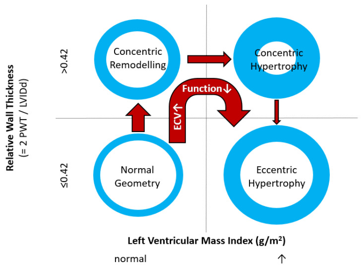
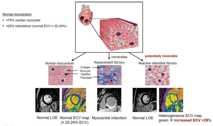
Hypertensive heart disease (HHD) develops in response to the chronic exposure of the left ventricle and left atrium to elevated systemic blood pressure. Left ventricular structural changes include hypertrophy and interstitial fibrosis that in turn lead to functional changes including diastolic dysfunction and impaired left atrial and LV mechanical function. Ultimately, these changes can lead to heart failure with a preserved (HFpEF) or reduced (HFrEF) ejection fraction. This review will outline the clinical evaluation of a patient with hypertension and/or suspected HHD, with a particular emphasis on the role and recent advances of multimodality imaging in both diagnosis and differential diagnosis.
Keywords: Fabry’s disease; PET; cardiac amyloidosis; cardiovascular magnetic resonance; computed tomography; echocardiography; hypertensive heart disease; hypertrophic cardiomyopathy; myocardial perfusion scintigraphy.
Conflict of interest statement
The authors declare no conflict of interest.
Figures
References
-
- NCD Risk Factor Collaboration (NCD-RisC) Worldwide trends in hypertension prevalence and progress in treatment and control from 1990 to 2019: A pooled analysis of 1201 population-representative studies with 104 million participants. Lancet. 2021;398:957–980. doi: 10.1016/S0140-6736(21)01330-1. - DOI - PMC - PubMed
Publication types
LinkOut - more resources
Full Text Sources
