Review
. 2023 Dec;26(4):799-807.
doi: 10.1007/s40477-023-00779-3. Epub 2023 May 13.

Imaging of ovarian lymphoma

Affiliations
Review

Imaging of ovarian lymphoma

Diana Donatello et al. J Ultrasound. 2023 Dec.

Erratum in

  • Correction to: Imaging of ovarian lymphoma.
    Donatello D, Battista G, Sassi C. Donatello D, et al. J Ultrasound. 2023 Dec;26(4):959. doi: 10.1007/s40477-023-00799-z. J Ultrasound. 2023. PMID: 37269426 Free PMC article. No abstract available.

Abstract

Objective: The aim of the study is to describe the radiological spectrum of appearances of ovarian lymphoma (OL). The manuscript describes the radiological aspects of OL to assist the radiologist in achieving correct orientation of the diagnosis.

Methods: We conducted a retrospective evaluation of imaging studies of 98 cases of non-Hodgkin's lymphoma, with extra-nodal localisation (ovaries) in three cases (1 primary, 2 secondary). A literature review was also performed.

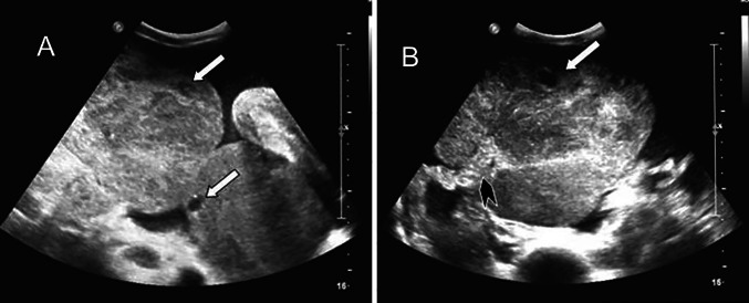
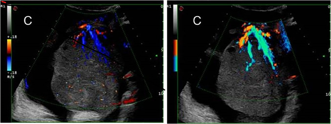
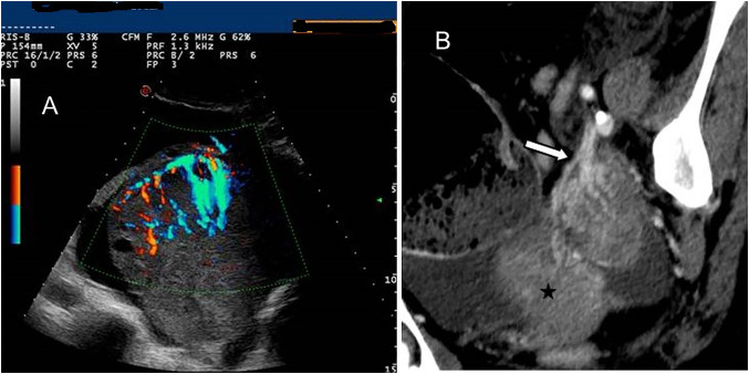
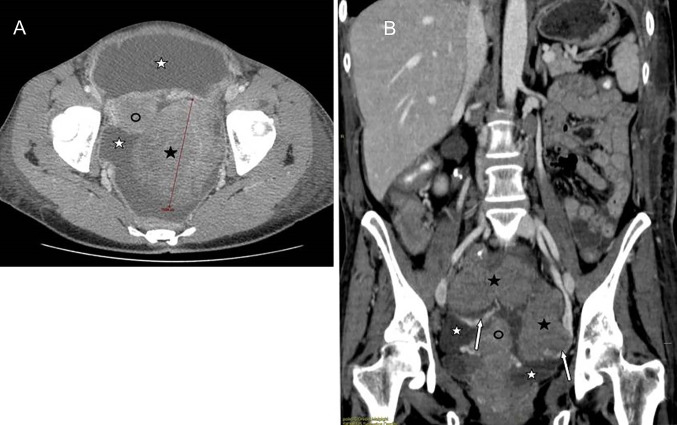
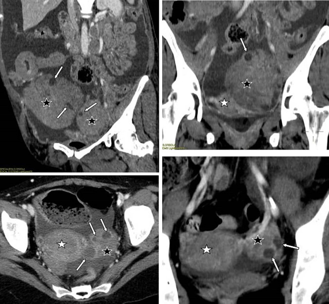
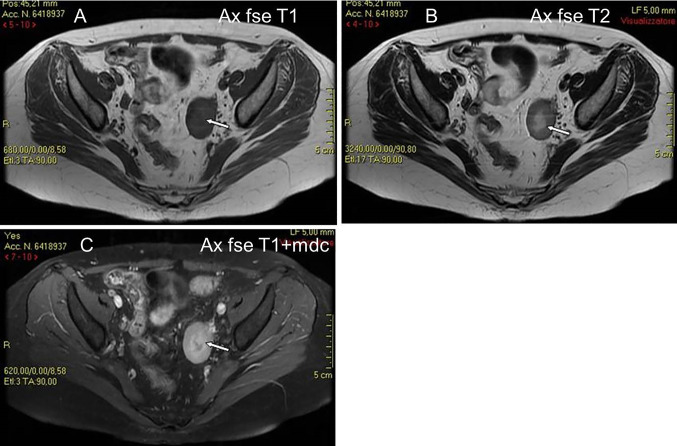
Results: Of the three evaluated women, one had a primary ovarian involvement and two had a secondary ovarian involvement. The most common lesion characteristics were a well-defined, solid homogeneous and hypoechoic mass at US. CT depicts OL as a well-defined, non-infiltrating, homogeneous hypodense solid mass, with mild contrast enhancement. On T1-weight MRI, OL appears as a homogeneous mass of low signal intensity, which enhances avidly following intravenous gadolinium.

Conclusion: Clinical and serological presentation of OL can be similar to that of primary ovarian cancer. As imaging plays a central role in the diagnosis of OL, the radiologist should be familiar with US, CT and MRI appearances of this condition to correctly orient the diagnosis and so avoid unnecessary adnexectomy.

Keywords: Lead vessel; Lymphoma; Ovarian lymphoma; Ovarian neoplasms.


Conflict of interest statement

The authors have not disclosed any competing interests.

Figures

Fig. 1
Fig. 1
A 44 years old woman with occult NHL (Burkitt type) presenting with abdominal pain and distension, and bilateral solid ovarian masses (same patient as Fig. 1). Sovrapubic US shows small anechoic areas of cystic appearance lining at the periphery of the ovarian solid masses, indicative of preserved ovarian follicles (A, B). The vascular pedicle is clearly visible entering from the border to the core of the ovarian mass (B). (White arrow: ovarian follicle. Black arrowhead: vascular pedicle)
Fig. 2
Fig. 2
TV Colour and Power Doppler TVS clearly depict the main vessel (the “lead vessel”) entering from the periphery to the centre of the ovarian mass, with many branching vessels of thinner width. (White arrow: ovarian follicle)
Fig. 3
Fig. 3
Colour Doppler TV US shows a left ovarian mass with the “lead vessel” sign: a main vessel with many thinner branching vessels entering from the periphery to the core of the mass; two an-hecoic follicles are visible at the periphery of the mass (A). Contrast enhanced oblique reformatted CT image (B) depicts the left ovarian mass and his vascular pedicle at the edge (white arrow): a tree-shaped main vessel with thinner branching vessels goes from the edge through the centre of the mass (the “lead vessel”). (Black star: uterus)
Fig. 4
Fig. 4
A 45 years old woman with peritoneal and ovarian high grade NHL presenting with ascites and bilateral ovarian masses. Contrast enhanced CT, axial (A) and coronal (B) images: the ovarian masses (black star) appear as well-defined lobulated hypodense solid lesions, with a homogeneous structure, non-infiltrating, with mild contrast enhancement. Coronal reformatted image (B) depicts the vascular pedicles of both masses (white arrows). Ascites (white star): hypodense fluid circumscribing ovarian masses and uterus (black circle)
Fig. 5
Fig. 5
Lymphoma involves ovaries but preserves their normal structure (images from different patients having ovarian lymphomatous involvement from NHL): contrast enhanced CT shows monolateral or bilateral solid omogeneous ovarian masses (black star) with some centimetric cystic areas in a linear arrangement in the periphery, corresponding to anechogenic cysts at US, and referred to preserved ovarian follicles in the cortex. (White star: uterus)
Fig. 6
Fig. 6
A 32 years old woman with NHL (DLBC type) relapsed to the left ovary (same patient as Fig. 6). MR shows a solid ovarian mass of low signal intensity on T1W images (A), mildly low signal intensity on T2W images (B), and with mildly high signal intensity on gadolinium enhanced T1W images (C). The mass has a central area of low signal intensity on T1W images (A) and mildly high signal intensity on T2W images (B), and without enhancement on gadolinium T1W images (C) (white arrows), suggestive of necrotic tissue
Fig. 7
Fig. 7
A 33 years old woman with high grade NHL relapse presenting with abdominal pain. 18-FDG-PET CT shows high uptake (SUV: 26) within the right ovarian mass referred to lymphomatous involvement (white arrows) (A). After chemotherapy, both degree and extension of right ovarian mass FDG uptake decrease to standard values (B)

References

    1. Ferry JA, Young RH. Malignant lymphoma, pseudolymphoma and hemopoeitic disorders of the female genital tract. Pathol Annu. 1991;26:227–263. - PubMed
    1. Crasta JA, Vallikad E. Ovarian lymphoma. Indian J Med Pediatric Oncol. 2009;30:28–30. doi: 10.4103/0971-5851.56333. - DOI - PMC - PubMed
    1. Yun J, Kim SJ, Won JH, Choi CW, Eom HS, Kim JS, Kim MK, Kwak J, Suh WSK. Clinical features and prognostic relevance of ovarian involvement in non-Hodgkin's lymphoma: a consortium for improving survival of lymphoma (CISL) report. Leuk Res. 2010;34:1175–1179. doi: 10.1016/j.leukres.2010.02.010. - DOI - PubMed
    1. Dimopoulos MA, Daliani D, Pugh W, Gershenson D, Cabanillas F, Sarris AH. Primary ovarian non-Hodgkin’s lymphoma: outcome after treatment with combination chemotherapy. Gynecol Oncol. 1997;64:446–450. doi: 10.1006/gyno.1996.4583. - DOI - PubMed
    1. Crawshaw J, Sohaib SA, Wotherspoon A, Shepherd J. A primary non-Hodgkin's lymphoma of the ovaries: imaging findings. Br J Radiol. 2007;80:155–158. doi: 10.1259/bjr/35049074. - DOI - PubMed

MeSH terms