. 2023 Sep 21;30(9):1090-1103.e7.
doi: 10.1016/j.chembiol.2023.04.007. Epub 2023 May 12.

Identification of structurally diverse FSP1 inhibitors that sensitize cancer cells to ferroptosis

Affiliations

Identification of structurally diverse FSP1 inhibitors that sensitize cancer cells to ferroptosis

Joseph M Hendricks et al. Cell Chem Biol. .

Abstract

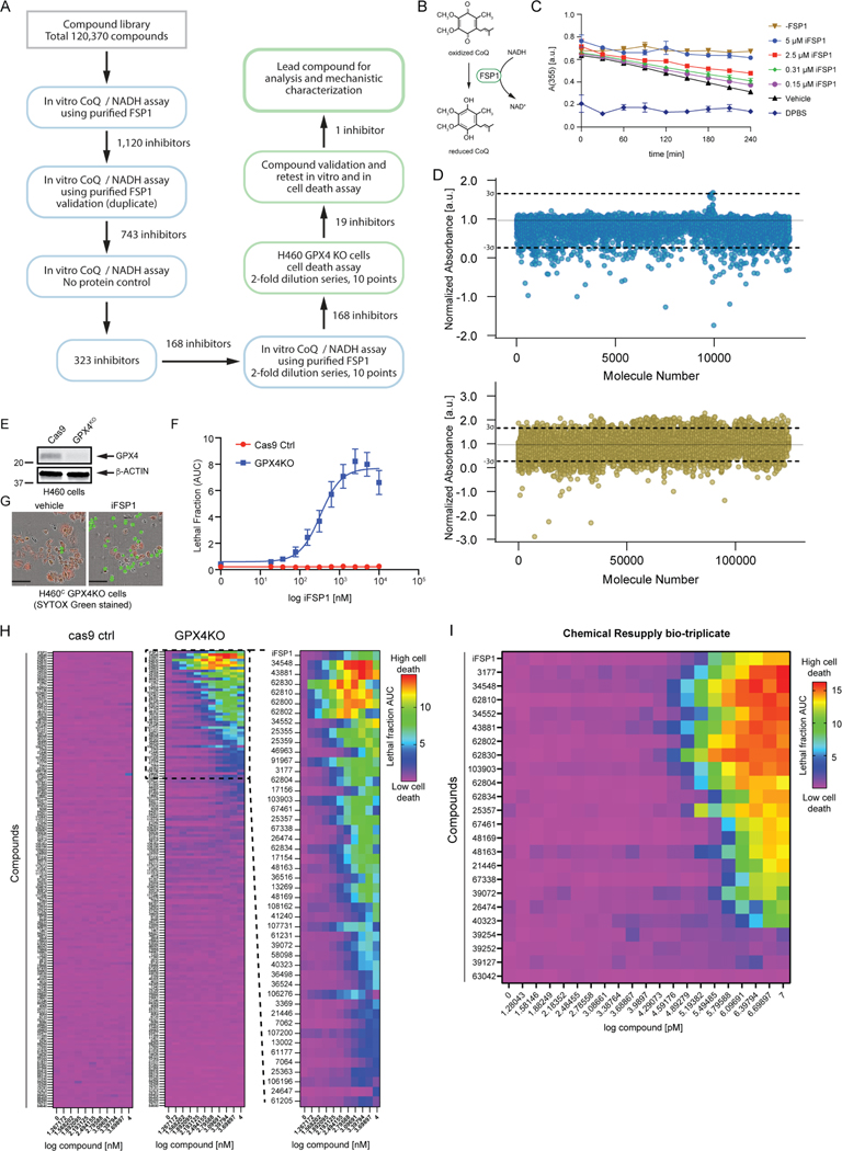
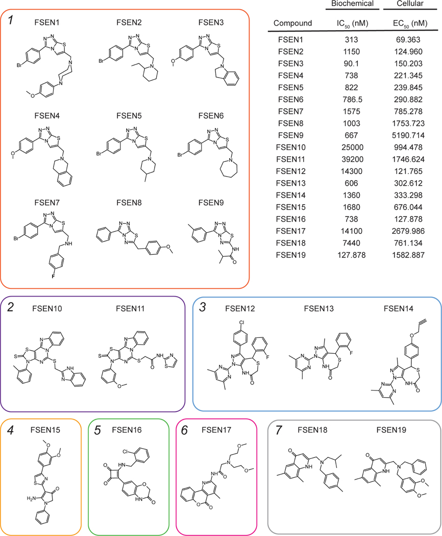
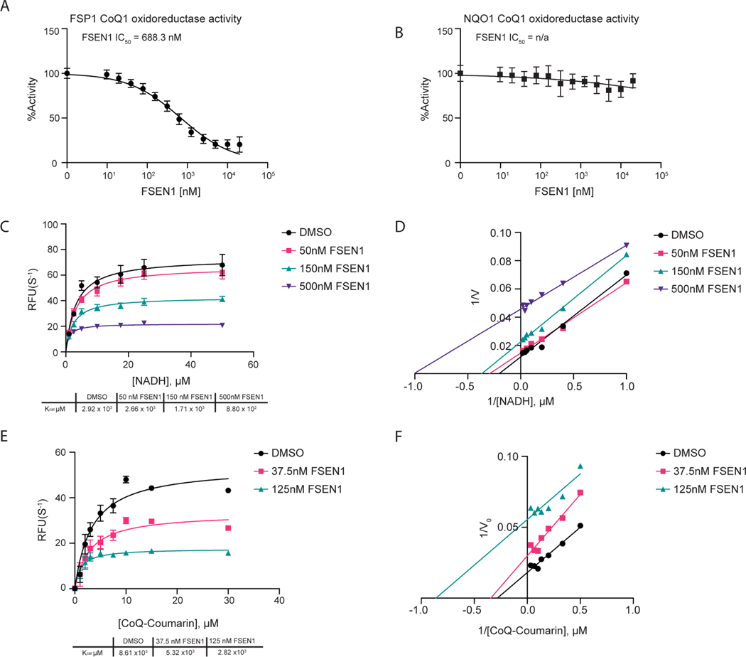
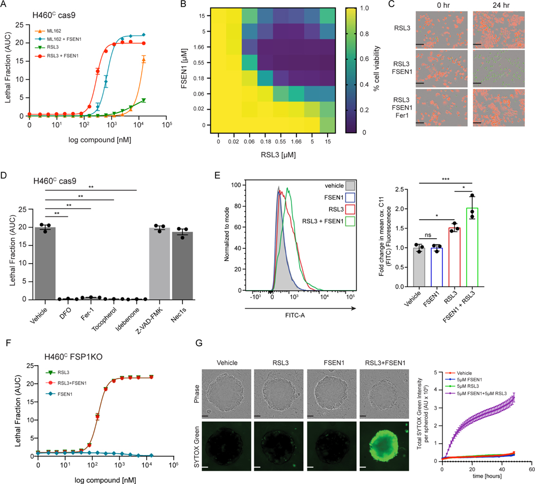
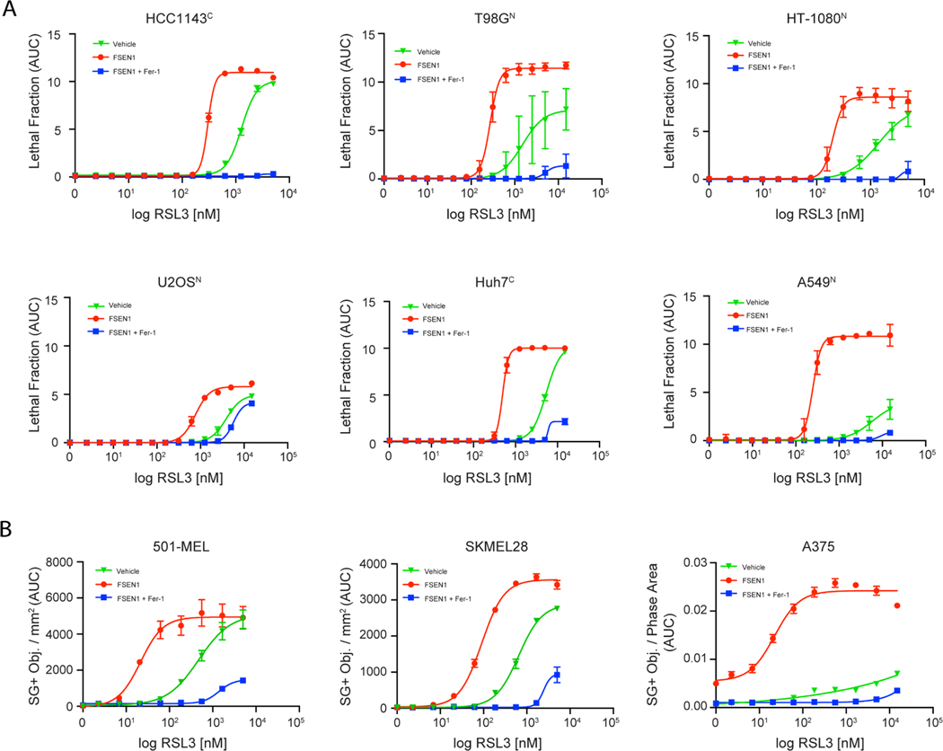
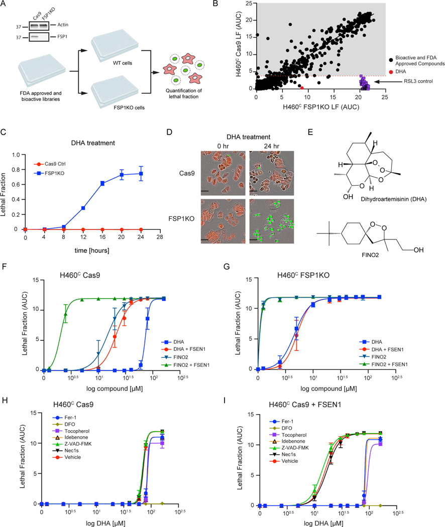
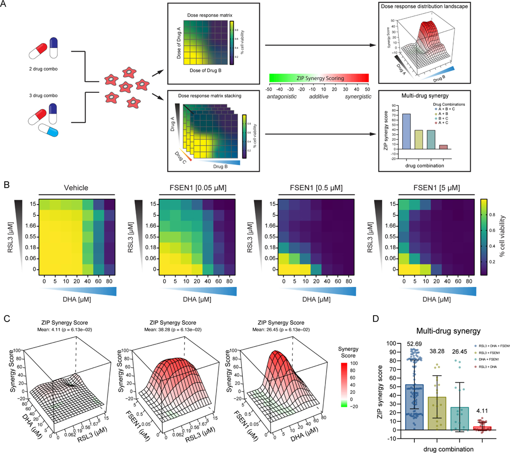
Ferroptosis is a regulated form of cell death associated with the iron-dependent accumulation of phospholipid hydroperoxides. Inducing ferroptosis is a promising approach to treat therapy-resistant cancer. Ferroptosis suppressor protein 1 (FSP1) promotes ferroptosis resistance in cancer by generating the antioxidant form of coenzyme Q10 (CoQ). Despite the important role of FSP1, few molecular tools exist that target the CoQ-FSP1 pathway. Through a series of chemical screens, we identify several structurally diverse FSP1 inhibitors. The most potent of these compounds, ferroptosis sensitizer 1 (FSEN1), is an uncompetitive inhibitor that acts selectively through on-target inhibition of FSP1 to sensitize cancer cells to ferroptosis. Furthermore, a synthetic lethality screen reveals that FSEN1 synergizes with endoperoxide-containing ferroptosis inducers, including dihydroartemisinin, to trigger ferroptosis. These results provide new tools that catalyze the exploration of FSP1 as a therapeutic target and highlight the value of combinatorial therapeutic regimes targeting FSP1 and additional ferroptosis defense pathways.

Keywords: FSP1; GPX4; cancer; cell death; coenzyme Q10; endoperoxide; ferroptosis; glutathione; lipid peroxidation; small molecule screen.


Conflict of interest statement

Declaration of interests J.A.O. is a member of the scientific advisory board for Vicinitas Therapeutics. S.J.D. is a member of the scientific advisory board for Ferro Therapeutics and Hillstream BioPharma, Inc. J.S. is a member of the board of directors for Zenith Therapeutics and a scientific advisor to Lyterian Biosciences and Organos. S.J.D., J.A.O., J.M.H., E.W., J.S., C.E.D., and K.B. have ferroptosis-related patent applications.

Figures

Figure 1.
Figure 1.. Small molecule screens identify FSP1 inhibitors
A) Flow chart of the chemical screens and experimental validation employed to identify FSP1 inhibitors. B) Schematic of the in vitro activity assay using purified recombinant FSP1. C) FSP1 activity was measured using NADH absorbance in the presence of increase amounts of iFSP1. Mean ± SEM of three technical replicates. D) Scatter plots of 15,000 antibacterial compounds and 100,000 diverse + 5,370 FDA and Bioactive compounds assayed with in vitro absorbance-based assay of FSP1 activity. “Hits” are defined by a normalized absorbance value of <0.267. Dashed lines represent SD of 3-sigma cutoff defined by activity range of vehicle and No Protein control. E) Western blot analysis of H460C Cas9 and GPX4KO cells. F) Representative images of H460 GPX4KO cells treated with vehicle and iFSP1. Dead cells are marked with SYTOX Green. Scale bar represents 50 μm. Images from one of three independent experiments are shown. G) Dose response of iFSP1 induced cell-death in H460C Cas9 and GPX4KO cells. Lethal fraction was calculated by IncuCyte quantification of the ratio of dead cells over the total amount of cells over 24 hr. The mean ± SEM bars represent three biological replicates. H) Lethal fraction (AUC) was calculated in H460C Cas9 and GPX4KO cells incubated with increasing doses of 168 small molecules that inhibited FSP1 in vitro. The heat map reflects n=3 cell biological replicates. I) Lethal fraction was calculated H460C GPX4KO cells incubated with increasing doses of the most potent 19 FSP1 inhibitors and iFSP1. Resupplied, validated small molecules were used. The heat map reflects n=3 cell biological replicates.
Figure 2.
Figure 2.. Multiple structurally distinct FSP1 inhibitor scaffolds
The structures of FSEN1–19 are shown with their calculated IC50 values from end-point assays measuring inhibition of FSP1 activity in vitro (n=2 independent biological replicates) and their calculated EC50 values from lethal fraction (AUC) quantification in H460C GPX4KO cells (n=3 cell biological replicates).
Figure 3.
Figure 3.. Mechanisms of inhibition of FSP1 by FSEN1
A,B) Purified recombinant FSP1 (A) and NQO1 (B) CoQ1 oxidoreductase activities were measured in the presence of increasing concentrations of FSEN1. Data was normalized to the slopes calculated from DMSO and No Protein controls. IC50 values displayed in nM were calculated from a non-linear regression curve fit. Data are mean ± SEM bars (n=3 independent biological replicates). C,D) Michaelis-Menten and Line Weaver-Burk plots of FSP1 treated with increasing concentrations of NADH in the presence of vehicle or FSEN1. 10 μM CoQ-Coumarin was used as the co-substrate for FSP1, and reduced CoQ-Coumarin fluorescence was measured as a read-out of enzymatic product formation. Error bars represent linear regression standard error of initial rates taken from three biological replicates. Vmax and Km were calculated from the non-linear regression curve fit. E,F) Michaelis-Menten and Line Weaver-Burk plots of FSP1 treated with increasing concentrations of CoQ-Coumarin in the presence of vehicle or FSEN1. 200 μM NADH was included as a co-substrate for FSP1, and reduced CoQ-Coumarin fluorescence was measured as the read-out of enzymatic product formation. Error bars represent linear regression standard error of initial rates (n=3 independent biological replicates). Vmax and Km were calculated from the non-linear regression curve fit.
Figure 4.
Figure 4.. FSEN1 is synthetic lethal with GPX4 inhibitors and sensitizes cancer cells to ferroptosis through inhibition of FSP1
A) Dose response of RSL3 and ML162-induced cell death in H460C Cas9 cells co-treated with 1 μM FSEN1 and 2 μM Fer-1 where indicated. Data are mean ± SEM (n=3 cell biological replicates). B) Heat map represents the fraction of viable H460C Cas9 cells co-treated with increasing doses of FSEN1 and RSL3 (n=3 cell biological replicates). C) Representative images of H460C Cas9 cells co-treated with 1.6 μM RSL3, 1 μM FSEN1 and 2 μM Fer-1 as indicated. Scale bar = 200 μm. Images from one of three independent experiments are shown. D) Lethal Fraction (AUC) of 5 μM RSL3-induced cell death in H460C Cas9 cells co-treated with 1 μM FSEN1 together with the indicated inhibitors of ferroptosis (Fer-1 [2 μM], DFO [100 μM], idebenone [10 μM], tocopherol [10 μM]), apoptosis (Z-VAD [10 μM]), and necroptosis (Nec1s [1 μM]). Data are mean ± SEM (n=3 cell biological replicates). **p<0.01 by one-way ANOVA with Dunnett’s multiple comparisons test. E) Representative flow cytometry histogram (left panel) and quantification (right panel) of H460C Cas9 cells treated with 200 nM RSL3 and/or 1 μM FSEN1 and labeled with the lipid peroxidation sensor BODIPY 581/591 C11. Green fluorescence intensity was analyzed by flow cytometry with the FITC channel. Data are mean ± SD (n=3 cell biological replicates). *p<0.05, ***p=0.0003 by one-way ANOVA with Tukey’s multiple comparisons test. F) Dose response of RSL3-induced cell death in H460C FSP1KO cells co-treated with 1 μM FSEN1. Data are mean ± SEM (n=3 cell biological replicates). G) H460C spheroids were treated with vehicle, 5 μM FSEN1, and 5 μM RSL3 as indicated. Representative images from one of 20 independent experiments are shown. Scale bar represents 100 μm. The total intensity of the SYTOX green signal for each spheroid was quantified and the mean ± SEM plotted (n=20 independent spheroids).
Figure 5.
Figure 5.. FSEN1 sensitizes cancer cells from different origins to ferroptosis
A) Dose response of RSL3-induced cell death in the indicated cancer cell lines treated in the presence and absence of 1 μM FSEN1 and 2 μM Fer-1 as indicated. Data are mean ± SEM (n=3 cell biological replicates). B) Quantification of cell death in melanoma cell lines treated as in (A), calculated as SYTOX green positive object per mm2. Data are mean ± SEM (n=2 cell biological replicates).
Figure 6.
Figure 6.. FDA library screens identify DHA as inducer of ferroptosis in FSP1KO cells
A) Western blot analysis of H460C Cas9 and FSP1KO cell lysates and screen schematic. H460C Cas9 and FSP1KO cells were incubated with 40 μM compounds and lethal fraction was quantified. B) Scatter plot of the lethal fraction (LF) for each compound in H460C Cas9 and H460C FSP1KO cells (n=1 cell biological replicate). Red dotted line represents the cutoff defined by the RSL3 positive control. C) Dose response validation of lethal fraction over time comparing H460C Cas9 and FSP1KO cells treated with 40 μM DHA. D) Representative IncuCyte images of H460C Cas9 and FSP1KO cells treated with DHA. Scale bar represents 50 μm. Images from one of two independent experiments are shown. E) The chemical structures of DHA and FINO2. F,G) Dose response of DHA and FINO2-induced cell death in H460C Cas9 (F) and FSP1KO (G) cells co-treated with vehicle or 1 μM FSEN1. Data are mean ± SEM bars (n=2 biological replicates). H,I) Dose response of DHA-induced cell death in H460C cells co-treated with vehicle (H) or 1 μM FSEN1 (I) together with the indicated inhibitors of ferroptosis (Fer-1 [2 μM], DFO [100 μM], idebenone [10 μM], tocopherol [10 μM]), apoptosis (Z-VAD [10 μM]), and necroptosis (Nec1s [1 μM]). Data are mean ± SEM (n=2 cell biological replicates).
Figure 7.
Figure 7.. FSEN1 is synergistic with DHA
A) Overview of the experimental design and analysis of synergy for two and three drug combinations. B) Heat maps of the fraction of viable cells in H460C Cas9 cells co-treated with vehicle and varying doses of FSEN1 (0.05, 0.5, 5 μM) in the presence of increasing concentrations of DHA and RSL3. Data are mean (n=3 cell biological replicates). C) 3D-dose response landscape plots illustrating the distribution and depth of synergy between the different drug combinations and doses tested in H460C Cas9 cells with the corresponding average ZIP synergy scores and p-values. D) Multi-drug synergy bar plots of H460C Cas9 cells illustrating mean ZIP synergy scores for all possible drug combinations tested. Data are mean ± SEM (n=3 cell biological replicates). P-values represents the significance of the difference between the estimated average synergy score over the whole dose-response matrix and 0% inhibition under the null hypothesis of non-response.

Comment in

Similar articles

Cited by

References

    1. Dixon SJ, Lemberg KM, Lamprecht MR, Skouta R, Zaitsev EM, Gleason CE, Patel DN, Bauer AJ, Cantley AM, Yang WS, et al. (2012). Ferroptosis: an iron-dependent form of nonapoptotic cell death. Cell 149, 1060–1072. 10.1016/j.cell.2012.03.042. - DOI - PMC - PubMed
    1. Jiang X, Stockwell BR, and Conrad M. (2021). Ferroptosis: mechanisms, biology and role in disease. Nat Rev Mol Cell Biol 22, 266–282. 10.1038/s41580-020-00324-8. - DOI - PMC - PubMed
    1. Yang WS, SriRamaratnam R, Welsch ME, Shimada K, Skouta R, Viswanathan VS, Cheah JH, Clemons PA, Shamji AF, Clish CB, et al. (2014). Regulation of ferroptotic cancer cell death by GPX4. Cell 156, 317–331. 10.1016/j.cell.2013.12.010. - DOI - PMC - PubMed
    1. Bersuker K, Hendricks JM, Li Z, Magtanong L, Ford B, Tang PH, Roberts MA, Tong B, Maimone TJ, Zoncu R, et al. (2019). The CoQ oxidoreductase FSP1 acts parallel to GPX4 to inhibit ferroptosis. Nature 575, 688–692. 10.1038/s41586-019-1705-2. - DOI - PMC - PubMed
    1. Doll S., Freitas FP., Shah R., Aldrovandi M., da Silva MC., Ingold I., Goya Grocin A., Xavier da Silva TN., Panzilius E., Scheel CH., et al.. (2019). FSP1 is a glutathione-independent ferroptosis suppressor. Nature 575, 693–698. 10.1038/s41586-019-1707-0. - DOI - PubMed

Publication types