

Extramedullary hematopoiesis (EMH) in the liver allograft presenting with a mass-like lesion
- PMID: 37179804
- PMCID: PMC10173390
- DOI: 10.1016/j.radcr.2023.02.026
Extramedullary hematopoiesis (EMH) in the liver allograft presenting with a mass-like lesion
Abstract
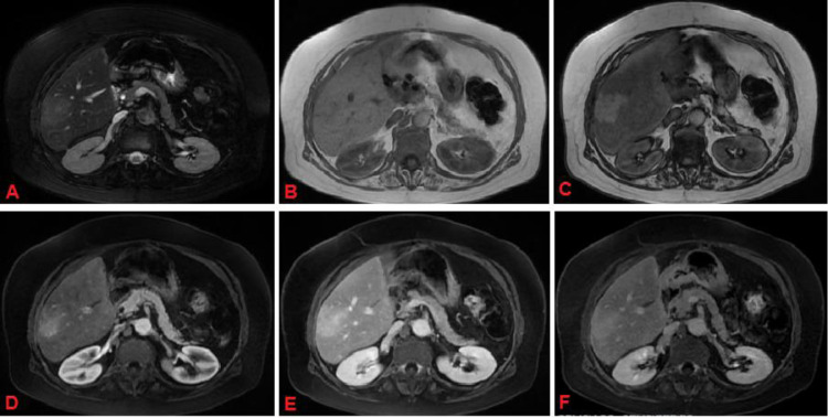
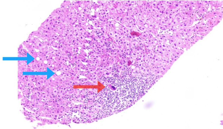
This is a rare case of extramedullary hematopoiesis (EMH) presenting as a mass-like lesion in liver allograft. Our patient was a 57-year-old woman who had undergone liver transplantation due to hepatic epithelioid hemangioendothelioma. She presented with an ill-defined hypoechoic lesion on ultrasound which showed features of focal EMH on pathologic examinations. While transient intrahepatic hematopoiesis has been reported in liver transplant patients, focal EMH mass lesion is a rarely encountered phenomenon. Therefore, focal EMH may need to be considered as a differential diagnosis when encountering a mass in post liver transplant patients.
Keywords: Extramedullary hematopoiesis (EMH); Hepatobiliary imaging; Hepatobiliary pathology; Liver transplant.
© 2023 Published by Elsevier Inc. on behalf of University of Washington.
Figures

References
-
- Golub R, Cumano A. Embryonic hematopoiesis. Blood Cells Mol Dis J. 2013;51(4):226–231. - PubMed
-
- Wong Y, Chen F, Tai K, Yip L, Tsang K, Chan F, et al. Imaging features of focal intrahepatic extramedullary haematopoiesis. Brit J Radiol. 1999;72(861):906–910. - PubMed
-
- Tsamandas AC, Jain AB, Raikow RB, Demetris AJ, Nalesnik MA, Randhawa PS. Extramedullary hematopoiesis in the allograft liver. Mod Pathol J. 1995;8(6):671–674. - PubMed
-
- Schlitt HJ, Schäfers S, Deiwick A, Eckardt KU, Pietsch T, Ebell W, et al. Extramedullary erythropoiesis in human liver grafts. Hepatol J. 1995;21(3):689–696. - PubMed
Publication types
LinkOut - more resources
Full Text Sources
