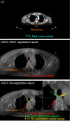
Improving the registration stability of cone-beam computed tomography with the Sphere-Mask Optical Positioning System: a feasibility study
- PMID: 37179916
- PMCID: PMC10167422
- DOI: 10.21037/qims-22-989
Improving the registration stability of cone-beam computed tomography with the Sphere-Mask Optical Positioning System: a feasibility study
Abstract
Background: Cone-beam computed tomography (CBCT) is an important tool for patient positioning in radiotherapy due to its outstanding advantages. However, the CBCT registration shows errors due to the limitations of the automatic registration algorithm and the nonuniqueness of manual verification results. The purpose of this study was to verify the feasibility of using the Sphere-Mask Optical Positioning System (S-M_OPS) to improve the registration stability of CBCT through clinical trials.
Methods: From November 2021 to February 2022, 28 patients who received intensity-modulated radiotherapy and site verification with CBCT were included in this study. S-M_OPS was used as an independent third-party system to supervise the CBCT registration result in real time. The supervision error was calculated based on the CBCT registration result and using the S-M_OPS registration result as the standard. For the head and neck, patients with a supervision error ≥3 or ≤-3 mm in 1 direction were selected. For the thorax, abdomen, pelvis, or other body parts, patients with a supervision error ≥5 or ≤-5 mm in 1 direction were selected. Then, re-registration was performed for all patients (selected and unselected). The registration errors of CBCT and S-M_OPS were calculated based on the re-registration results as the standard.
Results: For selected patients with large supervision errors, CBCT registration errors (mean ± standard deviation) in the latitudinal (LAT; left/right), vertical (VRT; superior/inferior), and longitudinal (LNG; anterior/posterior) directions were 0.90±3.20, -1.70±0.98, and 7.30±2.14 mm, respectively. The S-M_OPS registration errors were 0.40±0.14, 0.32±0.66, and 0.24±1.12 mm in the LAT, VRT, and LNG directions, respectively. For all patients, CBCT registration errors in the LAT, VRT, and LNG directions were 0.39±2.69, -0.82±1.47, and 2.39±2.93 mm, respectively. The S-M_OPS registration errors were -0.25±1.33, 0.55±1.27, and 0.36±1.34 mm for all patients in the LAT, VRT, and LNG directions, respectively.
Conclusions: This study shows that S-M_OPS registration offers comparable accuracy to CBCT for daily registration. S-M_OPS, as an independent third-party tool, can prevent large errors in CBCT registration, thereby improving the accuracy and stability of CBCT registration.
Keywords: Cone-beam computed tomography (CBCT); Sphere-Mask Optical Positioning System (S-M_OPS); registration; stability.
2023 Quantitative Imaging in Medicine and Surgery. All rights reserved.
Conflict of interest statement
Conflicts of Interest: All authors have completed the ICMJE uniform disclosure form (available at https://qims.amegroups.com/article/view/10.21037/qims-22-989/coif). The authors have no conflicts of interest to declare.
Figures


Similar articles
-
Setup error assessment based on "Sphere-Mask" Optical Positioning System: Results from a multicenter study.Front Oncol. 2022 Oct 4;12:918296. doi: 10.3389/fonc.2022.918296. eCollection 2022. Front Oncol. 2022. PMID: 36267985 Free PMC article.
-
Innovative integration of augmented reality and optical surface imaging: A coarse-to-precise system for radiotherapy positioning.Med Phys. 2023 Jul;50(7):4505-4520. doi: 10.1002/mp.16417. Epub 2023 Apr 15. Med Phys. 2023. PMID: 37060328
-
Analysis of precision in tumor tracking based on optical positioning system during radiotherapy.J Xray Sci Technol. 2016 Mar 19;24(3):443-55. doi: 10.3233/XST-160562. J Xray Sci Technol. 2016. PMID: 27257880
-
Surface guided frameless positioning for lung stereotactic body radiation therapy.J Appl Clin Med Phys. 2021 Sep;22(9):215-226. doi: 10.1002/acm2.13370. Epub 2021 Aug 18. J Appl Clin Med Phys. 2021. PMID: 34406710 Free PMC article.
-
A review of setup error in supine breast radiotherapy using cone-beam computed tomography.Med Dosim. 2016 Autumn;41(3):225-9. doi: 10.1016/j.meddos.2016.05.001. Epub 2016 Jun 14. Med Dosim. 2016. PMID: 27311516 Review.
References
-
- Sarkar B, Munshi A, Ganesh T, Manikandan A, Krishnankutty S, Chitral L, Pradhan A, Kalyan Mohanti B. Technical Note: Rotational positional error corrected intrafraction set-up margins in stereotactic radiotherapy: A spatial assessment for coplanar and noncoplanar geometry. Med Phys 2019;46:4749-54. 10.1002/mp.13810 - DOI - PubMed
-
- Ai XQ, Tang CQ, Wu H, Garbo T, Wang X, Liu JP, Cao YF, Jin H. Comparison of positioning accuracy of different registration methods and dosimetric analysis of adaptive radiotherapy for breast cancer after breast conserving surgery. Transl Cancer Res 2020;9:3274-81. 10.21037/tcr.2020.04.18 - DOI - PMC - PubMed
LinkOut - more resources
Full Text Sources
Research Materials
