

doi: 10.21037/qims-22-922.
Epub 2023 Mar 17.
Surgical application of an implantable biliary access device in the treatment of refractory recurrent cholangiolithiasis
Affiliations
- PMID: 37179917
- PMCID: PMC10167432
- DOI: 10.21037/qims-22-922
Item in Clipboard
Surgical application of an implantable biliary access device in the treatment of refractory recurrent cholangiolithiasis
Quant Imaging Med Surg.
.
No abstract available
Conflict of interest statement
Conflicts of Interest: All the authors have completed the ICMJE uniform disclosure form (available at https://qims.amegroups.com/article/view/10.21037/qims-22-922/coif). The authors report that this work was supported by the Nanchang Science and Technology Support Plan Program (No. HONGKEZI2021129). The authors have no other conflicts of interest to declare.
Figures

Preoperative imaging data of the patient. (A) Multiple previous surgical scars and T-tube can be seen in the abdomen. (B) The plain abdominal image. (C) The image of a common bile duct and duodenum after the injection of T-tube contrast agents. (D) The image of the intrahepatic bile duct. No stones were found.

The view from the choledochoscope. (A) Choledochoscopy in the duodenum showing duodenal mucosal folds. (B) The guidewire inserted through the duodenal papilla. (C) The guidewire in the duodenum. (D) The guidewire in the duodenum.
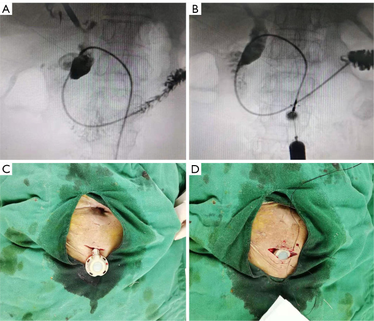
The view in DSA. (A) The IBAD catheter in the duodenum. (B) The IBAD base is connected, and the duodenum can be seen after the injection of the contrast agent through the IBAD base. (C) A small incision close to the T-tube sinus tract. (D) The IBAD implanted in the subcutaneous layer of the abdominal wall. DSA, digital subtraction angiography; IBAD, implantable biliary access device.
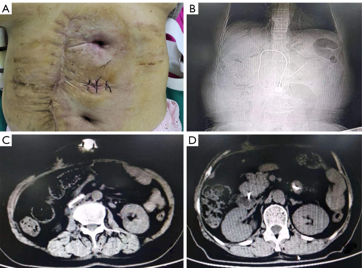
Placement of the IBAD. (A) The implantation of the IBAD was completed, the T-tube was removed, and the small incision was sutured. (B) The X-ray view after IBAD implantation. (C,D) After IBAD implantation, a CT scan showed that the IBAD base was located subcutaneously in the abdominal wall, and the IBAD catheter was located in the duodenum. IBAD, implantable biliary access device; CT, computed tomography.
Similar articles
-
Efficiency of percutaneous transhepatic cholangioscopy in the treatment of biliary complications after liver transplantation.HPB (Oxford). 2023 Apr;25(4):463-471. doi: 10.1016/j.hpb.2023.01.010. Epub 2023 Jan 25. HPB (Oxford). 2023. PMID: 36746707
-
[The diagnosis and treatment of cholangiolithiasis after cholecystectomy].Khirurgiia (Mosk). 2002;(4):4-10. Khirurgiia (Mosk). 2002. PMID: 12001681 Russian.
-
Minimally invasive treatment of intrahepatic cholangiolithiasis after stricture of hepaticojejunal anastomosis.Wideochir Inne Tech Maloinwazyjne. 2018 Mar;13(1):111-115. doi: 10.5114/wiitm.2018.72667. Epub 2018 Jan 10. Wideochir Inne Tech Maloinwazyjne. 2018. PMID: 29643967 Free PMC article.
-
Infection of totally implantable venous access devices: A review of the literature.J Vasc Access. 2018 May;19(3):230-242. doi: 10.1177/1129729818758999. Epub 2018 Mar 7. J Vasc Access. 2018. PMID: 29512430 Review.
-
Cardiac implantable electronic device and vascular access: Strategies to overcome problems.J Vasc Access. 2018 Nov;19(6):521-527. doi: 10.1177/1129729818762981. Epub 2018 Mar 19. J Vasc Access. 2018. PMID: 29552930 Review.
References
-
- Imaizumi H, Kida M, Takezawa M, Kikuchi H, Saigenji K. Early complications of endoscopic sphincterotomy for common bile duct stones. Dig Endoscop 2007;19:S57-9. 10.1111/j.1443-1661.2007.00710.x - DOI
LinkOut - more resources
Full Text Sources