. 2023 May 1;13(5):3088-3103.
doi: 10.21037/qims-22-1008. Epub 2023 Mar 13.

Deep learning for fully automated segmentation and volumetry of Couinaud liver segments and future liver remnants shown with CT before major hepatectomy: a validation study of a predictive model

Affiliations

Deep learning for fully automated segmentation and volumetry of Couinaud liver segments and future liver remnants shown with CT before major hepatectomy: a validation study of a predictive model

Tingting Xie et al. Quant Imaging Med Surg. .

Abstract

Background: Recent reports have shown the potential for deep learning (DL) models to automatically segment of Couinaud liver segments and future liver remnant (FLR) for liver resections. However, these studies have mainly focused on the development of the models. Existing reports lack adequate validation of these models in diverse liver conditions and thorough evaluation using clinical cases. This study thus aimed to develop and perform a spatial external validation of a DL model for the automated segmentation of Couinaud liver segments and FLR using computed tomography (CT) in various liver conditions and to apply the model prior to major hepatectomy.

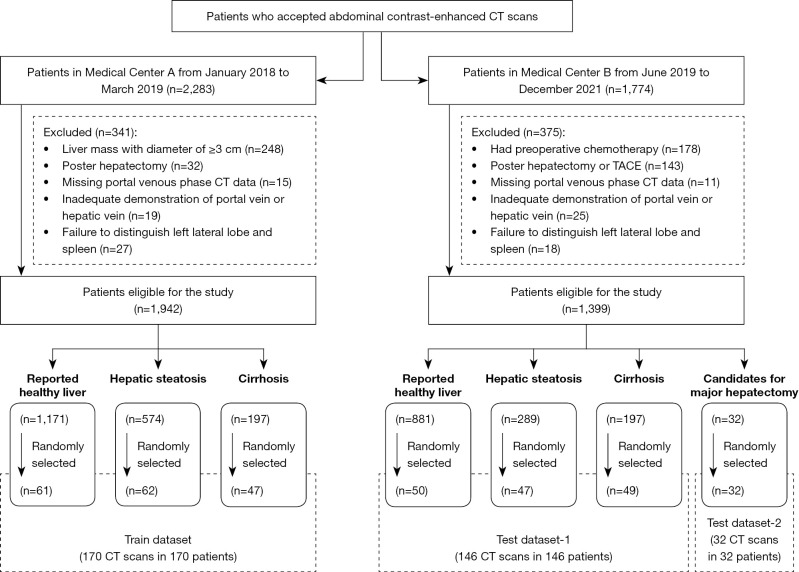
Methods: This retrospective study developed a 3-dimensional (3D) U-Net model for the automated segmentation of Couinaud liver segments and FLR on contrast-enhanced portovenous phase (PVP) CT scans. Images were obtained from 170 patients from January 2018 to March 2019. First, radiologists annotated the Couinaud segmentations. Then, a 3D U-Net model was trained in Peking University First Hospital (n=170) and tested in Peking University Shenzhen Hospital (n=178) in cases with various liver conditions (n=146) and in candidates for major hepatectomy (n=32). The segmentation accuracy was evaluated using the dice similarity coefficient (DSC). Quantitative volumetry to evaluate the resectability was compared between manual and automated segmentation.

Results: The DSC in the test data sets 1 and 2 for segments I to VIII was 0.93±0.01, 0.94±0.01, 0.93±0.01, 0.93±0.01, 0.94±0.00, 0.95±0.00, 0.95±0.00, and 0.95±0.00, respectively. The mean automated FLR and FLR% assessments were 493.51±284.77 mL and 38.53%±19.38%, respectively. The mean manual FLR and FLR% assessments were 500.92±284.38 mL and 38.35%±19.14%, respectively, in test data sets 1 and 2. For test data set 1, when automated segmentation of the FLR% was used, 106, 23, 146, and 57 cases were categorized as candidates for a virtual major hepatectomy of types 1, 2, 3, and 4, respectively; however, when manual segmentation of the FLR% was used, 107, 23, 146, and 57 cases were categorized as candidates for a virtual major hepatectomy of types 1, 2, 3, and 4, respectively. For test data set 2, all cases were categorized as candidates for major hepatectomy when automated and manual segmentation of the FLR% was used. No significant differences in FLR assessment (P=0.50; U=185,545), FLR% assessment (P=0.82; U=188,337), or the indications for major hepatectomy were noted between automated and manual segmentation (McNemar test statistic 0.00; P>0.99).

Conclusions: The DL model could be used to fully automate the segmentation of Couinaud liver segments and FLR with CT prior to major hepatectomy in an accurate and clinically practicable manner.

Keywords: Couinaud liver segments; Liver; deep learning (DL); future liver remnant (FLR); segmentation.


Conflict of interest statement

Conflicts of Interest: All authors have completed the ICMJE uniform disclosure form (available at https://qims.amegroups.com/article/view/10.21037/qims-22-1008/coif). YZ and DZ are employees of Beijing Smart Tree Medical Technology Co., Ltd. The other authors have no conflicts of interest to declare.

Figures

Figure 1
Figure 1
The flowchart showing the inclusion criteria, exclusion criteria, and distribution of CT scans in the data sets used in this study. CT, computed tomography; TACE, transcatheter arterial chemo-embolization.
Figure 2
Figure 2
Key steps in predicting the FLR. FLR, future liver remnant.
Figure 3
Figure 3
Successful automated segmentation results of 8 functional Couinaud liver segments and future liver remnant in various liver conditions of test data set 1 and test data set 2.
Figure 4
Figure 4
The differences of the segmentation results between automated and manual segmentation in test data set 2. (A) A case of hepatocellular carcinoma. For the segmentation of Couinaud liver segments, misidentification of segment V and VI occurred around the hepatic lesion. The automated segmentation underestimated the manual segmentation of the hepatic lesion. (B) A case of hepatocellular carcinoma. For the segmentation of Couinaud liver segments, misidentification of segment II occurred manly due to the relatively rare and irregular shape. The automated segmentation underestimated the manual segmentation of the hepatic lesion.
Figure 5
Figure 5
A bar plot of the average DSC of the segmentation model of the 8 Couinaud segments. Similar segmentation performances were obtained among the 8 Couinaud segments. The whiskers indicate the standard deviation of the average DSC values. DSC, dice similarity coefficient.
Figure 6
Figure 6
A bar plot of the average DSC of the segmentation model in various liver conditions. The DSC differences between healthy livers and those hepatic steatoses and between those with hepatic steatosis and candidates for major hepatectomy were statistically significant (***P<0.001). DSC, dice similarity coefficient.
Figure 7
Figure 7
Bland-Altman plots of the segmentation model in future liver remnant assessment. The segmentation model slightly underestimated manual segmentations in healthy livers, hepatic steatosis and cirrhosis but slightly overestimated manual segmentation in candidates for major hepatectomy.
Figure 8
Figure 8
Bland-Altman plots of the segmentation model in the FLR% assessment. The segmentation model slightly overestimated the segmentation compared to the manual segmentation in test data set 1 but slightly underestimated the segmentation compared to manual segmentation in test data set 2. FLR, future liver remnant.
Figure 9
Figure 9
Comparisons of the volumetry at the lobe level. Our results were similar to those obtained by Huang et al. (15), Ruskó et al. (16), Butdee et al. (17), and Le et al. (6), with a difference of less than 5%.
Figure 10
Figure 10
Boxplots showing the absolute difference of the FLR% obtained by manual and automated segmentation in test data set 1 and test data set 2. All values were within 5%, and the evaluations of resectability between the evaluations made by the model and human doctors were considered consistent, except in 3.73% of cases. FLR, future liver remnant.
Figure 11
Figure 11
Examples of successful automated segmentations in candidates for major hepatectomy. (A) A case of cholangiocarcinoma occupying 4 Couinaud liver segments (VIII, VII, V, and VI). (B) A case of hepatic hemangioma occupying 3 Couinaud liver segments (II, III, and IV). (C) A case of hepatocellular carcinoma occupying 4 Couinaud liver segments (VIII, VII, V, and VI).

References

    1. Lodewick TM, Arnoldussen CW, Lahaye MJ, van Mierlo KM, Neumann UP, Beets-Tan RG, Dejong CH, van Dam RM. Fast and accurate liver volumetry prior to hepatectomy. HPB (Oxford) 2016;18:764-72. 10.1016/j.hpb.2016.06.009 - DOI - PMC - PubMed
    1. Zhang T, Li Q, Wei Y, Yao S, Yuan Y, Deng L, Wu D, Nie L, Wei X, Tang H, Song B. Preoperative evaluation of liver regeneration following hepatectomy in hepatocellular carcinoma using magnetic resonance elastography. Quant Imaging Med Surg 2022;12:5433-51. 10.21037/qims-22-306 - DOI - PMC - PubMed
    1. Gotra A, Sivakumaran L, Chartrand G, Vu KN, Vandenbroucke-Menu F, Kauffmann C, Kadoury S, Gallix B, de Guise JA, Tang A. Liver segmentation: indications, techniques and future directions. Insights Imaging 2017;8:377-92. 10.1007/s13244-017-0558-1 - DOI - PMC - PubMed
    1. Dello SA, Stoot JH, van Stiphout RS, Bloemen JG, Wigmore SJ, Dejong CH, van Dam RM. Prospective volumetric assessment of the liver on a personal computer by nonradiologists prior to partial hepatectomy. World J Surg 2011;35:386-92. 10.1007/s00268-010-0877-6 - DOI - PMC - PubMed
    1. Jia X, Qian C, Yang Z, Xu H, Han X, Ren H, Wu X, Ma B, Yang D, Min H. Boundary-Aware Dual Attention Guided Liver Segment Segmentation Model. KSII Transactions on Internet and Information Systems 2022;16:16-37.