. 2023 May 10;13(21):14224-14235.
doi: 10.1039/d3ra01665j. eCollection 2023 May 9.

Electroactive nanofibrous membrane with temperature monitoring for wound healing

Affiliations

Electroactive nanofibrous membrane with temperature monitoring for wound healing

Liguo Yang et al. RSC Adv. .

Abstract

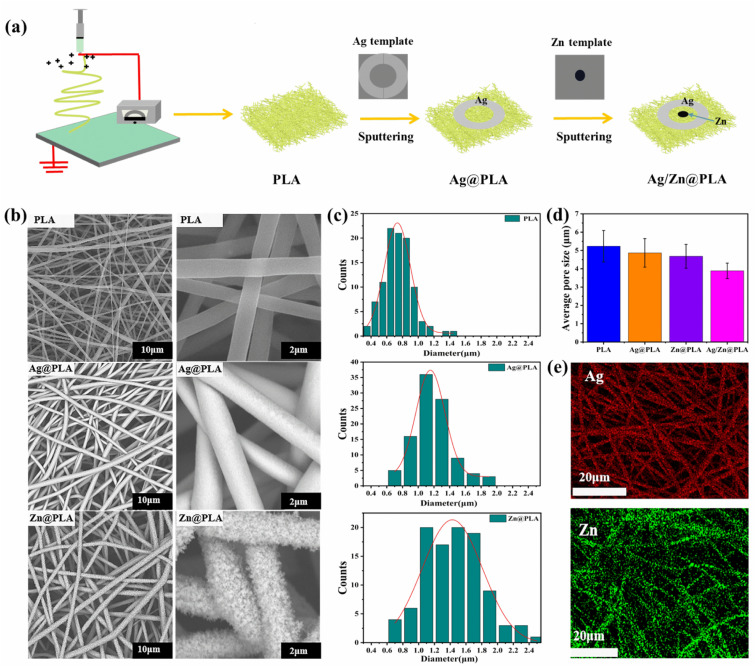
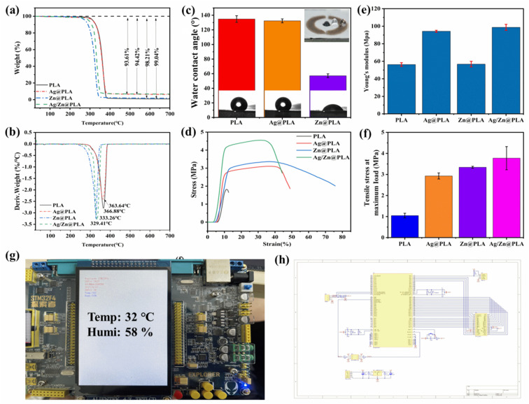
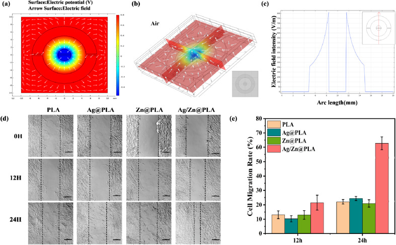
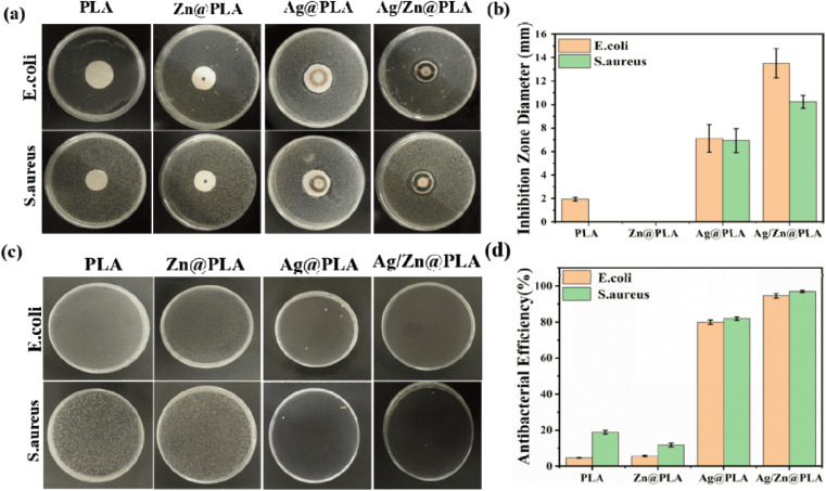
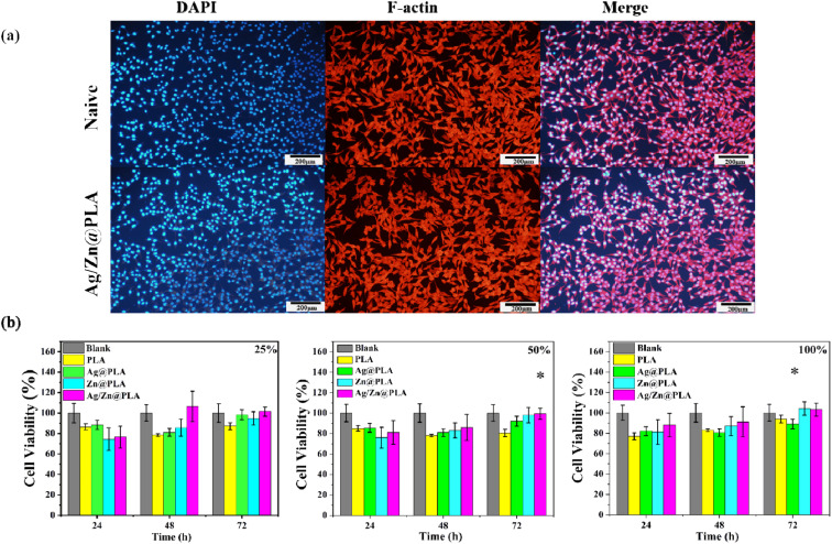
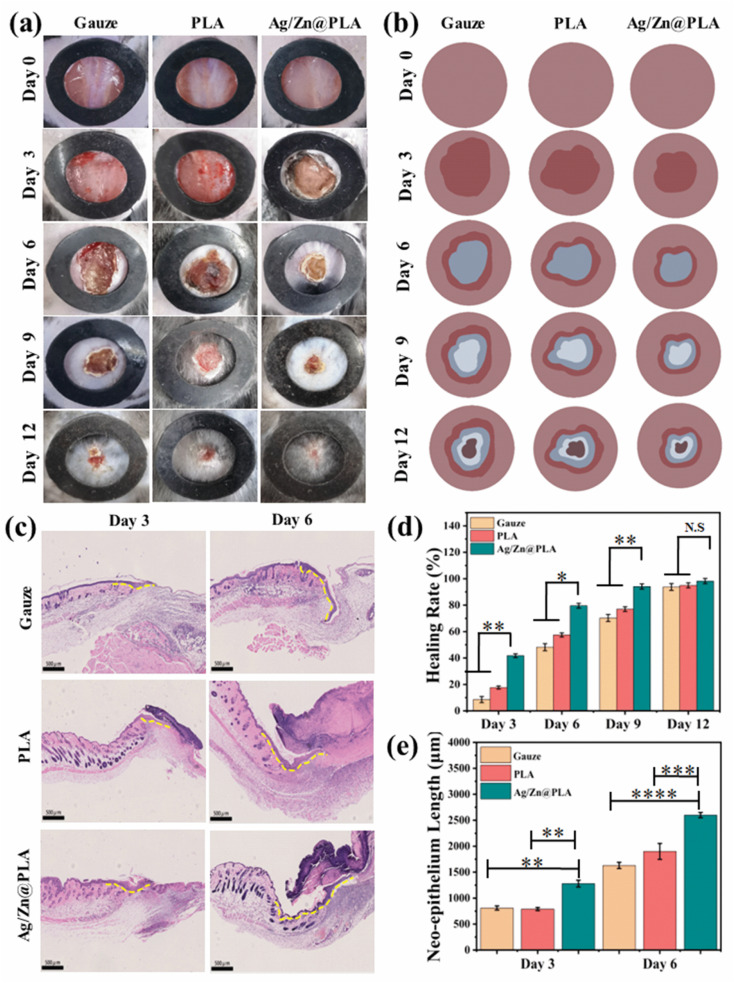
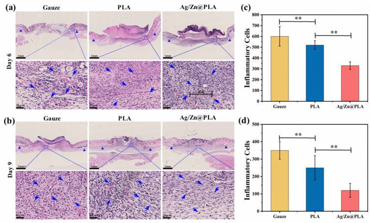
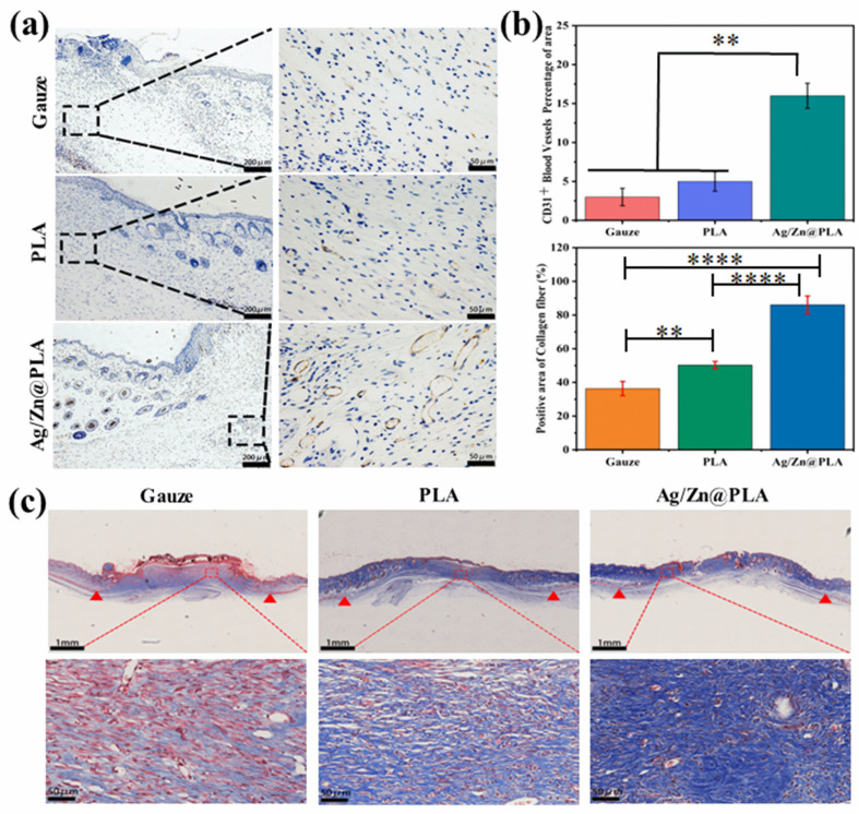
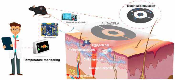
Developing functional dressings for promoting cellular activities and monitoring the healing progress is receiving increasingly widespread attention. In this study, Ag/Zn electrodes were deposited on the surface of a polylactic acid (PLA) nanofibrous membrane which can mimic the extracellular matrix. When wetted by wound exudate, the Ag/Zn electrodes could generate an electric stimulation (ES), promoting the migration of fibroblasts that heal wounds. Moreover, the Ag/Zn@PLA dressing showed excellent antibacterial activity against E. coli (95%) and S. aureus (97%). The study found that the electrostatic (ES) effect and the release of metal ions mainly contribute to the wound healing properties of Ag/Zn@PLA. In vivo mouse models demonstrated that Ag/Zn@PLA could promote wound healing by improving re-epithelialization, collagen deposition, and neovascularization. Additionally, the integrated sensor within the Ag/Zn@PLA dressing can monitor the wound site's temperature in real-time, providing timely information on wound inflammatory reactions. Overall, this work suggests that combining electroactive therapy and wound temperature monitoring may provide a new strategy for designing functional wound dressings.


Conflict of interest statement

There are no conflicts to declare.

Figures

Fig. 1
Fig. 1. (a) Schematic illustration of the fabrication process of the Ag/Zn@PLA dressing. (b) SEM images of the PLA, Ag@PLA, and Zn@PLA. (c) Diameter distribution of the PLA, Ag@PLA, and Zn@PLA. (d) The average pore size of the PLA, Ag@PLA, and Zn@PLA. (e) EDX spectra of the Ag deposited PLA (red, Ag element) and Zn deposited PLA (green, Zn element).
Fig. 2
Fig. 2. (a) Thermogram and (b) DTG curves of PLA, Ag@PLA, Zn@PLA, and Ag/Zn@PLA. (c) The contact angle of PLA, Ag@PLA, and Zn@PLA. (d) The stress–strain curves of PLA, Ag@PLA, and Zn@PLA. (e) Young's modulus and (f) tensile strength of PLA, Ag@PLA, and Zn@PLA. (g) Single-chip monitor displays temperature obtained by a temperature sensor. (h) Schematic circuit diagram of the dressing device.
Fig. 3
Fig. 3. (a) The electrical field of Ag/Zn@PLA was simulated by finite element software COMSOL in a two-dimensional plane. (b) The electrical field of Ag/Zn@PLA was modeled and simulated by finite element software COMSOL in a three-dimensional plane. (c) Detailed electric field intensity. Truncation started from x = 0, y = −10, and ended at x = 0, y = 10. (d) Representative images show cell migration at 12 and 24 h after scratching. (e) The cell migration rate of different samples treated fibroblasts.
Fig. 4
Fig. 4. Antimicrobial activity analysis. (a) Photograph of the agar plate in the disk diffusion method. (b) Inhibition zone diameters of different samples. (c) Photographs of colonies in the bacterial growth inhibition test. (d) Antimicrobial efficiency of different samples.
Fig. 5
Fig. 5. (a) Fluorescence images of fibroblast cells co-cultured with various samples for 48 h. (b) Cell viability was determined by CCK-8 assay for different sample extracts after 24, 48, and 72 h incubation.
Fig. 6
Fig. 6. In situ evaluation of Ag/Zn@PLA in promoting wound healing. (a) Representative photographs of rat wounds in different treatment groups on postoperative days 0, 3, 6, 9, and 12. (b) Illustrations of the mice wound areas on postoperative days 0, 3, 6, 9, and 12. (c) Representative H&E staining images and the yellow dashed line indicated the length of the new wound (the scale bar is 500 μm). (d) Healing rate in the different groups on postoperative days 3, 6, 9, and 12. (e) Statistics of new epithelium length in the wound on postoperative days 3 and 6. N.S, p > 0.05, *p < 0.05, **p < 0.01, ***p < 0.001, ****p < 0.0001. n = 3.
Fig. 7
Fig. 7. (a) HE staining of tissue sections from mice healed for 6 days, blue triangles indicate the junction between the wound and normal skin, and blue arrows indicate inflammatory cells. (b) Number of inflammatory cells after healing for 6 days. (c) HE staining of tissue sections from mice healed for 9 days, with blue dashed lines indicating the junction between the wound and normal skin and blue arrows indicating inflammatory cells. (d) Number of inflammatory cells at 9 days of healing.
Fig. 8
Fig. 8. (a) Immunohistochemical staining for CD31 in tissue sections from mice healed for 12 days. (b) Number and percentage of CD31 + cells. (c) Masson staining of tissue sections from mice healed for 12 days, with red triangles indicating the junction between the wound and normal skin (**p < 0.01, ***p < 0.001. n = 3.)
Fig. 9
Fig. 9. Illustration of the possible wound healing promoting mechanism of Ag/Zn@PLA with miniature temperature sensor.

Similar articles

Cited by

References

    1. Yang Y. Du Y. Zhang J. Zhang H. Guo B. Adv. Fiber Mater. 2022;4:1027–1057.
    1. Wang C. Liang Y. Huang Y. Li M. Guo B. J. Mater. Sci. Technol. 2022;121:207–219.
    1. Wu K. Yang Q. Zhang L. Xu P. Wu X. Yang H. Zhou H. Lin X. Yang L. J. Mater. Sci. Technol. 2023;133:123–134.
    1. Yu L. Zhang H. Xiao L. Fan J. Li T. ACS Appl. Mater. Interfaces. 2022;14:21814–21821. - PubMed
    1. Liang J. Zeng H. Qiao L. Jiang H. Ye Q. Wang Z. Liu B. Fan Z. ACS Appl. Mater. Interfaces. 2022;14:30507–30522. - PubMed