

A scalable corneal xenograft platform: simultaneous opportunities for tissue engineering and circular economic sustainability by repurposing slaughterhouse waste
- PMID: 37180037
- PMCID: PMC10168539
- DOI: 10.3389/fbioe.2023.1133122
A scalable corneal xenograft platform: simultaneous opportunities for tissue engineering and circular economic sustainability by repurposing slaughterhouse waste
Abstract
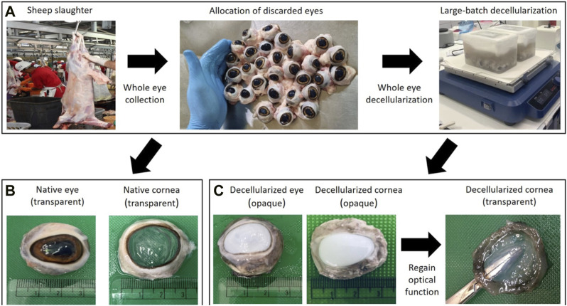
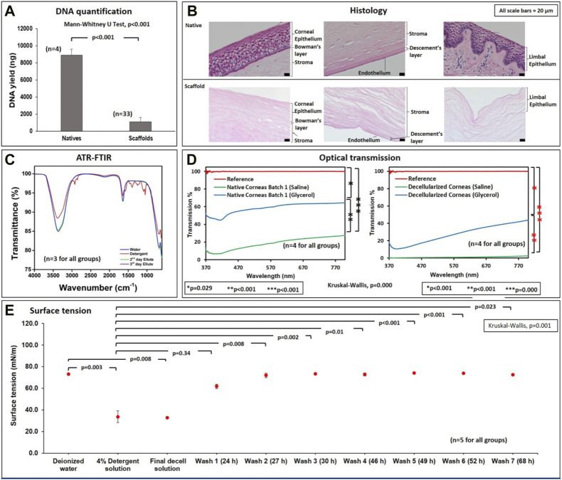
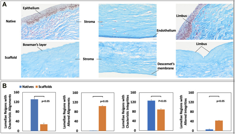
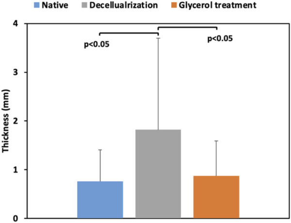
Introduction: Corneal disease is a leading cause of blindness globally that stems from various etiologies. High-throughput platforms that can generate substantial quantities of corneal grafts will be invaluable in addressing the existing global demand for keratoplasty. Slaughterhouses generate substantial quantities of underutilized biological waste that can be repurposed to reduce current environmentally unfriendly practices. Such efforts to support sustainability can simultaneously drive the development of bioartificial keratoprostheses. Methods: Scores of discarded eyes from the prominent Arabian sheep breeds in our surrounding region of the United Arab Emirates (UAE) were repurposed to generate native and acellular corneal keratoprostheses. Acellular corneal scaffolds were created using a whole-eye immersion/agitation-based decellularization technique with a widely available, eco-friendly, and inexpensive 4% zwitterionic biosurfactant solution (Ecover, Malle, Belgium). Conventional approaches like DNA quantification, ECM fibril organization, scaffold dimensions, ocular transparency and transmittance, surface tension measurements, and Fourier-transform infrared (FTIR) spectroscopy were used to examine corneal scaffold composition. Results: Using this high-throughput system, we effectively removed over 95% of the native DNA from native corneas while retaining the innate microarchitecture that supported substantial light transmission (over 70%) after reversing opacity, a well-established hallmark of decellularization and long-term native corneal storage, with glycerol. FTIR data revealed the absence of spectral peaks in the frequency range 2849 cm-1 to 3075 cm-1, indicating the effective removal of the residual biosurfactant post-decellularization. Surface tension studies confirmed the FTIR data by capturing the surfactant's progressive and effectual removal through tension measurements ranging from approximately 35 mN/m for the 4% decellularizing agent to 70 mN/m for elutes highlighting the effective removal of the detergent. Discussion: To our knowledge, this is the first dataset to be generated outlining a platform that can produce dozens of ovine acellular corneal scaffolds that effectively preserve ocular transparency, transmittance, and ECM components using an eco-friendly surfactant. Analogously, decellularization technologies can support corneal regeneration with attributes comparable to native xenografts. Thus, this study presents a simplified, inexpensive, and scalable high-throughput corneal xenograft platform to support tissue engineering, regenerative medicine, and circular economic sustainability.
Keywords: circular economy; corneas; decellularization; keratoprosthesis; tissue engineering; xenograft.
Copyright © 2023 Wang, Shakeel, Salih, Vurivi, Daoud, Desidery, Khan, Shibru, Ali, Butt, Chan and Corridon.
Conflict of interest statement
The authors declare that the research was conducted in the absence of any commercial or financial relationships that could be construed as a potential conflict of interest.
Figures
References
-
- Ahearne M. (2020). “Chapter 5 - corneal extracellular matrix decellularization,” in Methods in cell biology. Editors Caballero D., Kundu S. C., Reis R. L. (Academic Press; ), 157, 81–95. - PubMed
-
- Ahearne M., Fernández-Pérez J., Masterton S., Madden P. W., Bhattacharjee P. (2020). Designing scaffolds for corneal regeneration. Adv. Funct. Mater. 30 (44), 1908996. 10.1002/adfm.201908996 - DOI
LinkOut - more resources
Full Text Sources