Case Reports
. 2023 May 3:2023:7976780.
doi: 10.1155/2023/7976780. eCollection 2023.

Tracheal Resection for Critical Airway Obstruction in Morquio A Syndrome

Affiliations
Case Reports

Tracheal Resection for Critical Airway Obstruction in Morquio A Syndrome

Claire Frauenfelder et al. Case Rep Pediatr. .

Abstract

Introduction: The primary cause of death in Morquio A syndrome (mucopolysaccharidosis (MPS) IVA) is airway obstruction, brought about by an inexorable and pathognomonic multilevel airway tortuosity, buckling, and obstruction. The relative pathophysiological contributions of an inherent cartilage processing defect versus a mismatch in longitudinal growth between the trachea and the thoracic cage are currently a subject of debate. Enzyme replacement therapy (ERT) and multidisciplinary management continue to improve life expectancy for Morquio A patients by slowing many of the multisystem pathological consequences of the disease but are not as effective at reversing established pathology. An urgent need has developed to consider alternatives to palliation of progressive tracheal obstruction to preserve and maintain these patients' hard-won good quality of life, as well as to facilitate spinal and other required surgery. Case Report. Following multidisciplinary discussion, transcervical tracheal resection with limited manubriectomy was successfully performed, without the need for cardiopulmonary bypass, in an adolescent male on ERT with the severe airway manifestations of Morquio A syndrome. His trachea was found to be under significant compressive forces at surgery. On histology, chondrocyte lacunae appeared enlarged, but intracellular lysosomal staining and extracellular glycosaminoglycan staining was comparable to control trachea. At 12 months, this has resulted in a significant improvement in respiratory and functional status, with corresponding enhancement to his quality of life.

Conclusion: This addressing of tracheal/thoracic cage dimension mismatch represents a novel surgical treatment approach to an existing clinical paradigm and may be useful for other carefully selected individuals with MPS IVA. Further work is needed to better understand the role and optimal timing of tracheal resection within this patient cohort so as to individually balance considerable surgical and anaesthetic risks against the potential symptomatic and life expectancy benefits.


Conflict of interest statement

The authors declare that have no conflicts of interest.

Figures
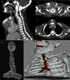
Figure 1
Figure 1
CT angiogram sagittal and (b) axial views immediately above and (c) below the manubrial head demonstrate multifactorial airway obstruction. Abnormal manubrium orientation and interposed brachiocephalic vasculature compressing the midtrachea into the c-spine, tracheal tortuosity, and tracheal ring deformity; shortened thorax with upper mediastinal compression; and cervico-thoracic kyphoscoliosis. (d-e) 3D-reconstructed images clearly demonstrate a tight thoracic inlet narrowing ().
Figure 2
Figure 2
Clinical photographs at cricotracheal resection: (a) tracheal exposure via limited manubriectomy showed a first bend 36 mm distal to the glottis, leading into a 16 mm tortuosity terminating 28 mm above the carina. (b) The deformed tracheal segment, which had concertinaed leading to omega-shaped tracheal ring buckling, was resected without (c) cardiopulmonary bypass,
Figure 3
Figure 3
Histopathological characterisation of resected trachea: periodic acid Schiff staining showed comparable GAG staining to controls in (a) mucosa, (b) cartilage, and (c) pericartilagenous junctions. Lysosomal LAMP1 staining showed no additional staining intensity in (d) epithelium or (e) chondrocytes, however, (f) chondrocyte lacunae were markedly larger in diameter.
Figure 4
Figure 4
Narrative account by parents articulating the changes in quality of life after the procedure. We note that whilst the account is presented verbatim in the parents' own words, all identifying information has been removed.

References

    1. Northover H., Cowie R. A., Wraith J. E. Mucopolysaccharidosis type IVA (Morquio syndrome): a clinical review. Journal of Inherited Metabolic Disease . 1996;19(3):357–365. doi: 10.1007/bf01799267. - DOI - PubMed
    1. Sawamoto K., Suzuki Y., Mackenzie W. G., et al. Current therapies for Morquio A syndrome and their clinical outcomes. Expert Opinion on Orphan Drugs . 2016;4(9):941–951. doi: 10.1080/21678707.2016.1214572. - DOI - PMC - PubMed
    1. Frigeni M., Rodriguez-Buritica D. F., Saavedra H., et al. The youngest pair of siblings with Mucopolysaccharidosis type IVA to receive enzyme replacement therapy to date: a case report. American Journal of Medical Genetics, Part A . 2021;185(11):3510–3516. doi: 10.1002/ajmg.a.62469. - DOI - PubMed
    1. Nakamura-Utsunomiya A., Nakamae T., Kagawa R., et al. A case report of a Japanese boy with morquio A syndrome: effects of enzyme replacement therapy initiated at the age of 24 months. International Journal of Molecular Sciences . 2020;21(3):p. 989. doi: 10.3390/ijms21030989. - DOI - PMC - PubMed
    1. Lavery C., Hendriksz C. Mortality in Patients with Morquio Syndrome A. JIMD Reports . Berlin, Germany: Springer; 2014. - PMC - PubMed

Publication types

LinkOut - more resources