. 2023 Apr 6;8(15):14208-14218.
doi: 10.1021/acsomega.3c01124. eCollection 2023 Apr 18.

Polyethylene Glycol 20k. Does It Fluoresce?

Affiliations

Polyethylene Glycol 20k. Does It Fluoresce?

Bethany F Laatsch et al. ACS Omega. .

Abstract

Polyethylene glycol (PEG) is a polyether compound commonly used in biological research and medicine because it is biologically inert. This simple polymer exists in variable chain lengths (and molecular weights). As they are devoid of any contiguous π-system, PEGs are expected to lack fluorescence properties. However, recent studies suggested the occurrence of fluorescence properties in non-traditional fluorophores like PEGs. Herein, a thorough investigation has been conducted to explore if PEG 20k fluoresces. Results of this combined experimental and computational study suggested that although PEG 20k could exhibit "through-space" delocalization of lone pairs of electrons in aggregates/clusters, formed via intermolecular and intramolecular interactions, the actual contributor of fluorescence between 300 and 400 nm is the stabilizer molecule, i.e., 3-tert-butyl-4-hydroxyanisole present in the commercially available PEG 20k. Therefore, the reported fluorescence properties of PEG should be taken with a grain of salt, warranting further investigation.


Conflict of interest statement

The authors declare no competing financial interest.

Figures

Figure 1
Figure 1
100 mg/mL PEG 20k was excited at wavelengths of 280–300 nm in increments of 5 nm to determine the maximum emissions. Each fluorescence spectrum represents an average of three trials.
Figure 2
Figure 2
Solutions containing 100 mg/mL PEG, 100 mM NaCl, and 30 mM phosphate buffer of pH 7.4 were excited at 295 nm for molecular weights of PEG 600, PEG 2k, PEG 8k, and PEG 20k. Each fluorescence spectrum represents an average of three trials. No significant fluorescence was observed for any other PEG molecules except for PEG 20k.
Figure 3
Figure 3
(a) GC–MS spectrum of 3-tert-butyl-4-hydroxyanisole (3-BHA), which is a 96% match to the spectrum of 3-BHA from the NIST Chemistry Webbook. (b) Chromatogram of the 3-BHA peak under three different washing conditions of 400 mg/mL PEG 20k. A full 30 min GC–MS analysis was run, and the 3-BHA peak is seen at approximately 18 min. Chromatogram only shows 15–20 min for clarity. (c) Peak intensity for the three trials of PEG 20k with different levels of purity. Data were taken at the apex of each 3-BHA peak, all within 0.02 s of each other and showing a match to 3-BHA in the NIST database. (d) Fluorescence spectra of 100 mg/mL PEG 20k before and after washing with diethyl ether.
Figure 4
Figure 4
AFM images of the PEG 20 k on the HOPG substrate. (a) Height-retraced and (b) amplitude-retraced images of PEG 20k on the HOPG substrate. Each AFM image size is 5 μm × 5 μm. The key in each image indicates a length of 1 μm. The image of the free HOPG substrate is shown in Figure S3.
Figure 5
Figure 5
(a) DLS autocorrelation functions of PEG 20k of 100, 200, and 300 mg/mL dissolved in 100 mM NaCl and 30 mM phosphate buffer of pH 7.4. (b) Regularization fit for 100 mg/mL PEG 20k dissolved in 100 mM NaCl and 30 mM phosphate buffer of pH 7.4.
Figure 6
Figure 6
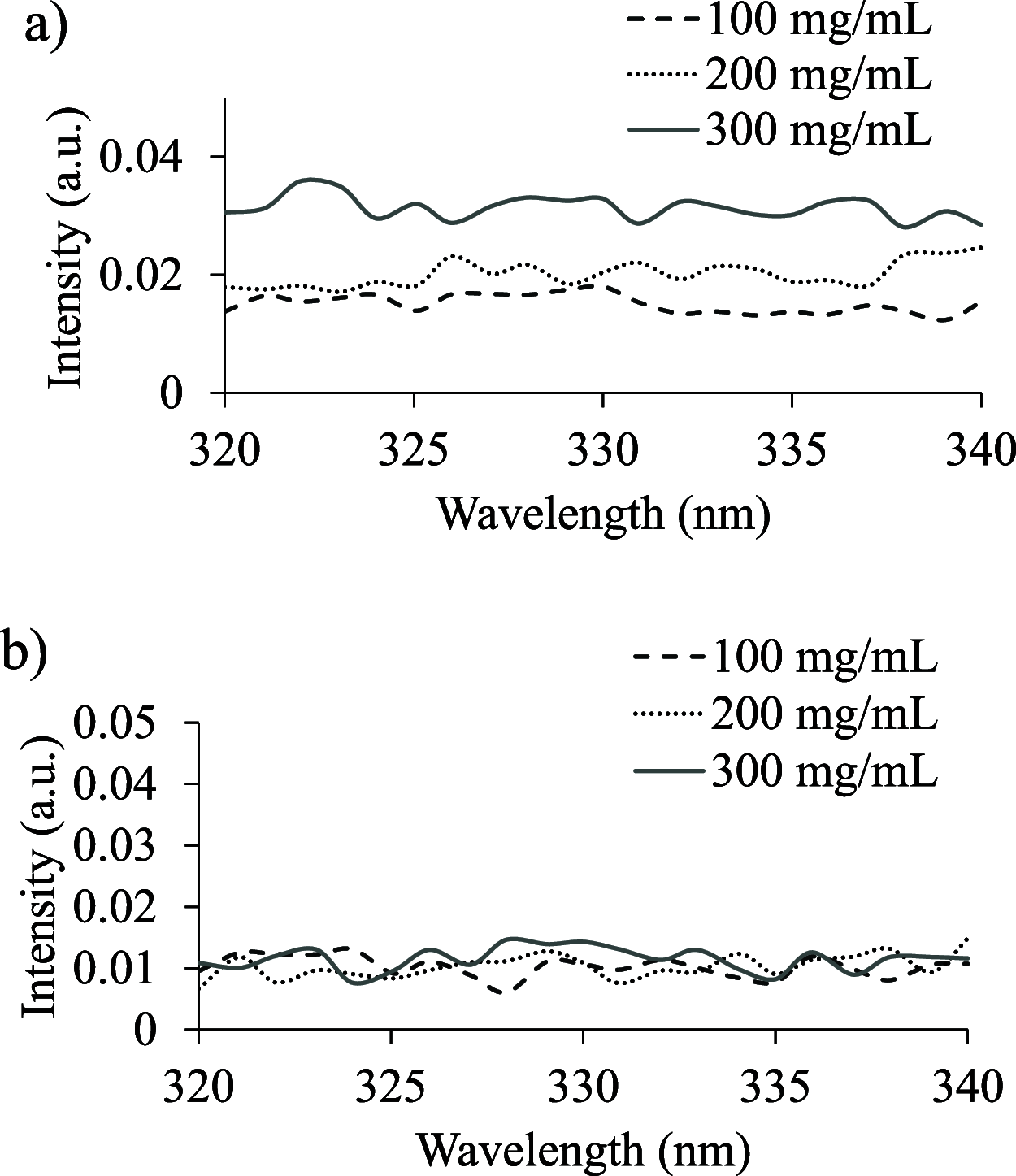
Fluorescence anisotropy of PEG 20k of 100, 200, and 300 mg/mL dissolved in 100 mM NaCl and 30 mM phosphate buffer of pH 7.4. Samples were excited at 288 nm, and emissions were read from 320 to 340 nm. (a) Anisotropy at 25 °C and (b) anisotropy at 50 °C.
Figure 7
Figure 7
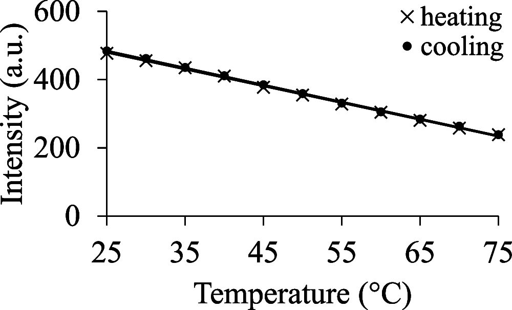
Solution containing 100 mg/mL PEG 20k, 100 mM NaCl, and 30 mM phosphate buffer of pH 7.4 was excited at 295 nm. The emission was recorded at 320 nm. The temperature was increased at 5° increments from 25 to 75 °C (marked with X). Similarly, cooling was done at 5° increments from 75 to 25 °C (marked with filled circles).
Figure 8
Figure 8
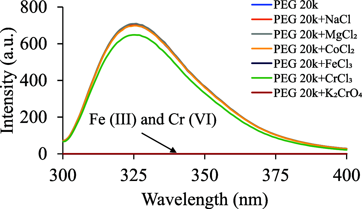
Solutions of 100 mg/mL PEG 20k in the presence of metal ions of 10 mM concentration were excited at 295 nm. Each fluorescence spectrum represents an average of three trials. Na+, Mg2+, and Co2+ had no effect on the fluorescence of PEG 20k. Cr3+ displayed minimal quenching, whereas Fe3+ and Cr6+ quenched the fluorescence of PEG 20k completely.
Figure 9
Figure 9
1H NMR spectra of PEG 20k in D2O at different concentrations.
Figure 10
Figure 10
Folded structure of PEG 20k, which was obtained after the gas-phase folded structure was immersed in water and then simulated for 100 ns. The following structures, displayed through VMD, represent planes of the folded PEG 20k—(a) the side view, (b) the top view, and (c) the zoomed-in top view of the planes.
Figure 11
Figure 11
Simulated partially folded PEG 20k molecule in the water sphere.
Figure 12
Figure 12
Organized structures of heptameric PEG 20k after 200 ns of gaseous simulation (a) followed by 200 ns of an aqueous simulation (b). These structures are displayed and rendered through VMD. The varying colors represent individual PEG 20k molecules.
Figure 13
Figure 13
Delocalization of the electron cloud as observed through electronic structure calculation of clusters of variable sizes, extracted from a folded PEG 20k polymer. The HOMO in each cluster is shown; the purple and green colors represent the positive and negative amplitudes, respectively, of the molecular orbital’s wavefunction. Hydrogen atoms are omitted for clarity.
Figure 14
Figure 14
Simulated electronic spectra of EG (monomer) and PEG 20k clusters computed using time-dependent density functional theory.

References

    1. Bruusgaard-Mouritsen M. A.; Johansen J. D.; Garvey L. H. Clinical Manifestations and Impact on Daily Life of Allergy to Polyethylene Glycol (PEG) in Ten Patients. Clin. Exp. Allergy 2021, 51, 463–470. 10.1111/cea.13822. - DOI - PubMed
    1. Turner P. J.; Ansotegui I. J.; Campbell D. E.; Cardona V.; Ebisawa M.; El-Gamal Y.; Fineman S.; Geller M.; Gonzalez-Estrada A.; Greenberger P. A.; Leung A. S. Y.; Levin M. E.; Muraro A.; Sánchez Borges M.; Senna G.; Tanno L. K.; Yu-Hor Thong B.; Worm M. COVID-19 Vaccine-Associated Anaphylaxis: A Statement of the World Allergy Organization Anaphylaxis Committee. World Allergy Organ. J. 2021, 14, 100517.10.1016/j.waojou.2021.100517. - DOI - PMC - PubMed
    1. Klimek L.; Novak N.; Cabanillas B.; Jutel M.; Bousquet J.; Akdis C. A. Allergenic Components of the MRNA-1273 Vaccine for COVID-19: Possible Involvement of Polyethylene Glycol and IgG-Mediated Complement Activation. Allergy 2021, 76, 3307–3313. 10.1111/all.14794. - DOI - PMC - PubMed
    1. Paik S. P.; Ghatak S. K.; Dey D.; Sen K. Poly(Ethylene Glycol) Vesicles: Self-Assembled Site for Luminescence Generation. Anal. Chem. 2012, 84, 7555–7561. 10.1021/ac301731x. - DOI - PubMed
    1. Alper A.; Pashankar D. S. Polyethylene Glycol: A Game-Changer Laxative for Children. J. Pediatr. Gastroenterol. Nutr. 2013, 57, 134–140. 10.1097/mpg.0b013e318296404a. - DOI - PubMed