. 2023 May 9;2(5):pgad125.
doi: 10.1093/pnasnexus/pgad125. eCollection 2023 May.

First gene-edited calf with reduced susceptibility to a major viral pathogen

Affiliations

First gene-edited calf with reduced susceptibility to a major viral pathogen

Aspen M Workman et al. PNAS Nexus. .

Abstract

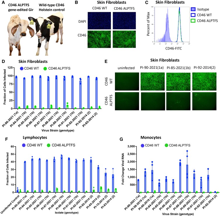
Bovine viral diarrhea virus (BVDV) is one of the most important viruses affecting the health and well-being of bovine species throughout the world. Here, we used CRISPR-mediated homology-directed repair and somatic cell nuclear transfer to produce a live calf with a six amino acid substitution in the BVDV binding domain of bovine CD46. The result was a gene-edited calf with dramatically reduced susceptibility to infection as measured by reduced clinical signs and the lack of viral infection in white blood cells. The edited calf has no off-target edits and appears normal and healthy at 20 months of age without obvious adverse effects from the on-target edit. This precision bred, proof-of-concept animal provides the first evidence that intentional genome alterations in the CD46 gene may reduce the burden of BVDV-associated diseases in cattle and is consistent with our stepwise, in vitro and ex vivo experiments with cell lines and matched fetal clones.

Keywords: BVDV; CD46; CRISPR; bovine viral diarrhea virus; gene editing.


Figures

Fig. 1.
Fig. 1.
Comparing the CD46 A82LPTFS87 substitution with the CD46 gene deletion in MDBK cells. A) Physical map of CD46 protein domains. RLI, receptor–ligand interaction domain; CCP, complement control protein domain; TM, transmembrane domain. B) Genomic DNA sequence of CD46 in the region of the G82QVLAL87 motif showing the amino acid translation, the gRNA heteroduplex, and the alignment of the synthetic ssODN used for replacing the G82QVLAL87 residues with A82LPTFS87. gRNA, synthetic guide RNA; ssODN, single-stranded oligonucleotide donor template. C) Ribbon representations of AlphaFold2 predictions of the extracellular domain of wild-type bovine CD46 (G82QVLAL87 strand) aligned with that containing the A82LPTFS87 substitution. The inset shows a closeup of the BVDV binding platform rotated to visualize the predicted structure differences in residues 82–84. D) Immunofluorescence staining of CD46 (FITC) and nuclei (DAPI; 10× magnification). E) Flow cytometric quantification of CD46 protein expression levels (n = 3). MFI, median fluorescence intensity. F) CD46-edited cells were infected with cytopathic or non-cytopathic BVDV isolates at an MOI of 2, and infection efficiency was determined at 20 hpi by flow cytometry using a monoclonal anti-BVDV E2 antibody. G) Serum from BVDV-PI calves was inoculated on cells. Infection efficiency was quantified at 72 hpi by flow cytometry. In panels F and G, the results represent the mean±standard deviation of n ≥ 3 independent experiments.
Fig. 2.
Fig. 2.
Reproductive cloning and BVDV susceptibility testing of primary cells from 100-day fetal tissues. A) Schematic representation of reproductive cloning. Primary skin fibroblasts were edited and subsequently fused to enucleated oocytes (somatic cell nuclear transfer) and the resultant embryos implanted into synchronized recipient cows. B) Flow cytometric quantification of CD46 surface expression. C) Cells were inoculated with serum (genotypes 1a and 1b) or low-passage virus isolates (genotype 2) from BVDV-PI calves. Infection efficiency was determined at 48 hpi using an anti-BVDV monoclonal antibody and FITC labeled secondary antibody. Nuclei were stained with DAPI to ensure images were taken in regions with complete cell monolayers (not shown). Cells imaged at 10× magnification. Sm. Int, small intestine; Esoph, esophageal.
Fig. 3.
Fig. 3.
Ex vivo challenge of fibroblasts, monocytes, and lymphocytes from CD46 A82LPTFS87-edited calf. A) Picture of study participants. B) Immunofluorescence staining of CD46 (FITC) and nuclei were stained with DAPI . C) Flow cytometric quantification of CD46 protein expression levels. D) Cells were inoculated with serum from BVDV-PI calves, and infection efficiency was quantified at 72 hpi by flow cytometry using an anti-BVDV E2 antibody. Results represent the mean±standard deviation of n = 3 independent experiments. E) BVDV infection was visualized for a representative set of samples from panel D by IF using an anti-BVDV antibody and FITC labeled secondary antibody (10× magnification). F) Flow cytometric quantification of BVDV-infected cells at 24 hpi. Results represent the mean±SD of n = 3 independent experiments. G) Monocytes were inoculated with serum from BVDV-PI calves (genotypes 1a and 1b) or a low-passage BVDV-2 isolate at an MOI of 0.01 (PI-92-2014). Viral RNA was detected by RT-qPCR at 48 hpi, and fold change in viral RNA relative to the input sample (0 hpi) was calculated. Results represent the mean±SD of n ≥ 3 independent experiments. WT, wild-type.
Fig. 4.
Fig. 4.
Natural exposure challenge study of CD46 A82LPTFS87-edited calf. A) Timeline of challenge study. The CD46-edited Gir calf and the unedited wild-type (WT) control Holstein calf were challenged with BVDV by cohabitation with a BVDV-PI calf for 7 days (exposure window). Nasal and fecal samples were collected on days −5 to 30. Blood was collected on days −5 to 44. B) BVDV RT-qPCR. Samples were extracted in duplicate and run on the same PCR plate. Samples that tested negative for BVDV RNA are plotted having a cycle threshold (Ct) equal to 40. C) Rectal temperatures taken daily through day 21. D) BVDV-1 serum neutralization (SN) titers.

References

    1. Baker JC. 1995. The clinical manifestations of bovine viral diarrhea infection. Vet Clin North Am Food Anim Pract. 11:425–445. - PubMed
    1. Bolin SR. 2002. Bovine viral diarrhea virus in mixed infections. In: Brogden KA, Guthmiller JM, editors. Polymicrobial diseases. Washington (DC): ASM Press. - PubMed
    1. Bruschke CJ, Weerdmeester K, Van Oirschot JT, Van Rijn PA. 1998. Distribution of bovine virus diarrhoea virus in tissues and white blood cells of cattle during acute infection. Vet Microbiol. 64:23–32. - PubMed
    1. Liess B, Moennig V, Pohlenz J, Trautwein G. 2012. Ruminant pestivirus infections: virology, pathogenesis, and perspectives of prophylaxis. Berlin (Germany): Springer Science & Business Media.
    1. McClurkin AW, et al. 1984. Production of cattle immunotolerant to bovine viral diarrhea virus. Can J Comp Med. 48:156–161. - PMC - PubMed