Review
. 2023 Mar 21;3(2):185-197.
doi: 10.1016/j.jacasi.2022.11.016. eCollection 2023 Apr.

Can Most Calcified Coronary Stenosis Be Optimized With Coronary Intravascular Lithotripsy?

Affiliations
Review

Can Most Calcified Coronary Stenosis Be Optimized With Coronary Intravascular Lithotripsy?

Rohit Mody et al. JACC Asia. .

Abstract

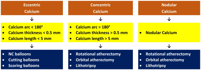
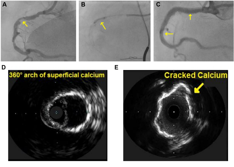
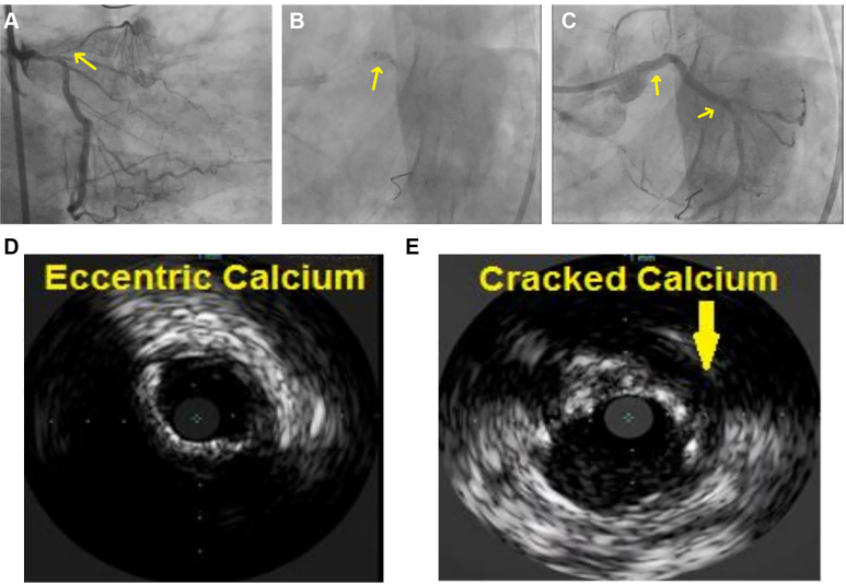
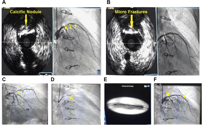
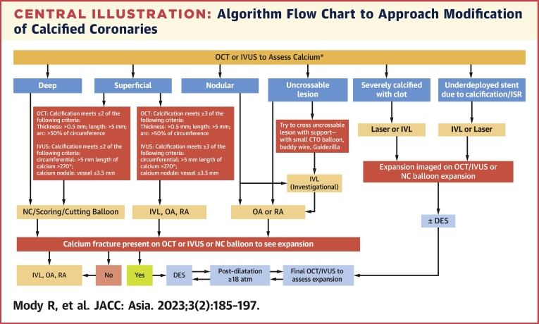
Intravascular lithotripsy can be used as an effective therapy for lesion preparation in severely calcified lesions. The mechanism, as shown by optical coherence tomography, is calcium fractures. The aforementioned modification is performed with minimal risk of perforation, no-reflow and a low incidence of flow-limiting dissection and myocardial infarctions. Other techniques, such as cutting or scoring balloons and rotational atherectomy have also been shown to increase luminal diameter, but other complications, such as distal embolization, induced by these treatment modalities, are a source of concern. This review describes a single-center study of all-comer patients, including those with complex characteristics. This therapy is very effective, with a very low risk of complications. In this article, we characterize the mechanism of action of the intravascular lithotripsy catheter, its optical coherence tomography validation, clinical applications, and comparison with other calcium-modifying technologies, as well as future directions, which can be used to improve the technology.

Keywords: coronary arteriosclerosis; percutaneous atherectomy; percutaneous transluminal angioplasty.


Figures

None
Graphical abstract
Figure 1
Figure 1
Types and Extent of Calcium and Their Modification by IVL (A) Types of calcium on OCT. (a) Deep calcific plaque characterized by signal-poor region with sharply delineated borders located deeply (asterisk). (b) Superficial calcific plaque characterized by signal-poor region with sharply delineated borders located superficial (asterisk). (c) Nodular calcific plaque characterized by signal-poor region with sharply delineated borders in nodular morphology (arrow). (B) Superficial calcified plaque determining the extent of calcified lesion. (a) Angle of superficial calcified plaque (round arrow showing the angle, which is 360°). (b) Calcified plaque showing thickness (arrow). (c) Longitudinal view of calcified plaque showing length (arrow). (C) Calcified plaque after modification. (a) Dissections in calcified plaque after modification (arrow). (b) Fissures in calcified plaque after modification (arrow). (c) Fractures in calcified plaque after modification (arrow). (D) OCT showing post modification by IVL device with calcified plaque showing fractures. IVL = intravascular lithotripsy; OCT = optical coherence tomography.
Figure 2
Figure 2
Effects on Calcified Plaque Induced by RA and OA (A) In the shape of a polished groove, calcium ablation (yellow arrows) is demonstrated by OCT after RA. (B) Similar in shape to RA, calcium ablation (yellow arrows) is demonstrated by OCT method after OA. Ablation is highlighted by both cross sections by either device at the wire bias site, with the modification of plaque occurred in the segment adjacent to the OCT catheter and the guidewire. OA = orbital atherectomy; RA = rotational atherectomy; other abbreviations as in Figure 1.
Figure 3
Figure 3
De Novo Calcium Modified by IVL (A) severe circumferential calcification in coronary artery is demonstrated by OCT cross-section method. (B) At the artery circumference (yellow arrows), deep fractures are modified by IVL. Abbreviations as in Figure 1.
Figure 4
Figure 4
Underexpanded Stent in a Severely Calcified Lesion Modified by ELCA (A) In a severely calcified lesion, there is an underexpanded stent. (B) Luminal gain and calcium fracture (yellow arrow) is demonstrated by OCT after ELCA. ELCA = excimer laser coronary atherectomy. Abbreviation as in Figure 1.
Figure 5
Figure 5
Modification of Various Subtypes of Calcium Defined by Imaging The different calcium plaque morphologies by imaging and also the extent and degree of calcification and Arrow heads show the flow of the calcium modification procedure. NC = noncompliant; other abbreviations as in Figure 1.
Figure 6
Figure 6
A Large RCA With Concentric Calcium Treated With IVL (A) Pre-PCI CAG shows significant calcified plaques in proximal and distal RCA (arrow). (B) Inflated NC balloon shows dog boning indicating unyielding calcified lesion (arrow). (C) Post-PCI CAG shows fully expanded stent deployed after IVL modification of the lesion (arrows). (D) Pre-IVL IVUS imaging shows a superficial 360° arch of superficial calcium. (E) IVUS post-IVL shows calcified plaque after IVL, which shows fractures (arrow). CAG = coronary angiogram; IVUS = intravascular ultrasound; PCI = percutaneous coronary intervention; RCA = right coronary artery; other abbreviations as in Figures 1 and 5.
Figure 7
Figure 7
A Case of LM to LCX With Eccentric Calcium Treated With IVL (A) Pre-PCI CAG shows significant calcified plaques in LM to LCx (arrow). (B) Inflated NC balloon shows dog boning, indicating an unyielding calcified lesion (arrow). (C) Post-PCI CAG shows the fully expanded stent deployed after IVL modification of the lesion (arrows). (D) Pre-IVL IVUS imaging shows the eccentric calcified plaque. (E) IVUS imaging after IVL shows calcified plaque after IVL, which shows fractures (arrow). LCx = left circumflex; LM = left main; other abbreviations as in Figures 1, 5, and 6.
Figure 8
Figure 8
A Case of a Calcified Nodule Treated With IVL (A) IVUS examination before IVL shows the calcified nodule (arrow). (B) IVUS examination after IVL shows the microfractures in calcified nodule (arrow). (C) Pre-PCI CAG shows the significant calcified plaques in proximal LAD (arrow). (D) Inflated NC balloon shows dog boning indicating unyielding calcified lesion (arrow). (E) Syncvision image shows fully expanded balloon after IVL, indicating good modification. (F) Post-IVL CAG shows fully expanded stent deployed after IVL modification. LAD = left anterior descending artery; other abbreviations as in Figures 1 and 6.
Figure 9
Figure 9
A Case of an Underdeployed Stent Owing to Calcification Treated With IVL (A) Pre-PCI CAG showing diffuse ISR in RCA from proximal to distal. (B) Pre-IVL IVUS examination showing the underdeployed stent and calcified ISR. (C) Inflated NC shows underexpansion, indicating an unyielding calcified lesion. (D) Post-IVL IVUS examination shows some expansion of the stent in the lesion. (E) The fully expanded stent after IVL after stenting. (F) Post-PCI CAG showing the fully expanded stents from proximal to distal RCA. ISR = in-stent restenosis; other abbreviations as in Figures 1 and 6.
Central Illustration
Algorithm Flow Chart to Approach Modification of Calcified Coronaries Imaging with OCT or IVUS assesses the calcified plaque and divides it into various imagining subclasses. The role of various modalities to modify calcium subtypes identified by imaging like NC, scoring, cutting (balloons), IVL, OA, RA, or laser is described in the flow chart. Arrowheads show the flow of the calcium modification procedure. CTO = chronic total occlusion; DES = drug-eluting stent(s); ISR = in-stent restenosis; IVL = intravascular lithotripsy; IVUS = intravascular ultrasound; OCT = optical coherence tomography; OA = orbital atherectomy; RA = rotational atherectomy.

References

    1. Yamamoto M.H., Maehara A., Karimi Galougahi K., et al. Mechanisms of orbital versus rotational atherectomy plaque modification in severely calcified lesions assessed by optical coherence tomography. J Am Coll Cardiol Intv. 2017;10:2584–2586. - PubMed
    1. Wiemer M., Butz T., Schmidt W., Schmitz K.P., Horstkotte D., Langer C. Scanning electron microscopic analysis of different drug eluting stents after failed implantation: from nearly undamaged to major damaged polymers. Catheter Cardiovasc Interv. 2010;75:905–911. - PubMed
    1. Tzafriri A.R., Garcia-Polite F., Zani B., et al. Calcified plaque modification alters local drug delivery in the treatment of peripheral atherosclerosis. J Control Release. 2017;264:203–210. - PMC - PubMed
    1. Madhavan M.V., Tarigopula M., Mintz G.S., Maehara A., Stone G.W., Genereux P. Coronary artery calcification: pathogenesis and prognostic implications. J Am Coll Cardiol. 2014;63:1703–1714. - PubMed
    1. Abdel-Wahab M., Richardt G., Joachim Buttner H., et al. High-speed rotational atherectomy before paclitaxel-eluting stent implantation in complex calcified coronary lesions: the randomized ROTAXUS (Rotational Atherectomy Prior to Taxus Stent Treatment for Complex Native Coronary Artery Disease) trial. J Am Coll Cardiol. Intv. 2013;6:10. - PubMed

LinkOut - more resources