. 2023 Feb 20;4(5):363-378.
doi: 10.1039/d2cb00229a. eCollection 2023 May 10.

Methylated guanosine and uridine modifications in S. cerevisiae mRNAs modulate translation elongation

Affiliations

Methylated guanosine and uridine modifications in S. cerevisiae mRNAs modulate translation elongation

Joshua D Jones et al. RSC Chem Biol. .

Abstract

Chemical modifications to protein encoding messenger RNAs (mRNAs) influence their localization, translation, and stability within cells. Over 15 different types of mRNA modifications have been observed by sequencing and liquid chromatography coupled to tandem mass spectrometry (LC-MS/MS) approaches. While LC-MS/MS is arguably the most essential tool available for studying analogous protein post-translational modifications, the high-throughput discovery and quantitative characterization of mRNA modifications by LC-MS/MS has been hampered by the difficulty of obtaining sufficient quantities of pure mRNA and limited sensitivities for modified nucleosides. We have overcome these challenges by improving the mRNA purification and LC-MS/MS pipelines. The methodologies we developed result in no detectable non-coding RNA modifications signals in our purified mRNA samples, quantify 50 ribonucleosides in a single analysis, and provide the lowest limit of detection reported for ribonucleoside modification LC-MS/MS analyses. These advancements enabled the detection and quantification of 13 S. cerevisiae mRNA ribonucleoside modifications and reveal the presence of four new S. cerevisiae mRNA modifications at low to moderate levels (1-methyguanosine, N2-methylguanosine, N2,N2-dimethylguanosine, and 5-methyluridine). We identified four enzymes that incorporate these modifications into S. cerevisiae mRNAs (Trm10, Trm11, Trm1, and Trm2, respectively), though our results suggest that guanosine and uridine nucleobases are also non-enzymatically methylated at low levels. Regardless of whether they are incorporated in a programmed manner or as the result of RNA damage, we reasoned that the ribosome will encounter the modifications that we detect in cells. To evaluate this possibility, we used a reconstituted translation system to investigate the consequences of modifications on translation elongation. Our findings demonstrate that the introduction of 1-methyguanosine, N2-methylguanosine and 5-methyluridine into mRNA codons impedes amino acid addition in a position dependent manner. This work expands the repertoire of nucleoside modifications that the ribosome must decode in S. cerevisiae. Additionally, it highlights the challenge of predicting the effect of discrete modified mRNA sites on translation de novo because individual modifications influence translation differently depending on mRNA sequence context.


Conflict of interest statement

The authors declare no competing financial interests.

Figures

Fig. 1
Fig. 1. LC-MS/MS method development to quantify 50 ribonucleosides in a single analysis. (A) Extracted ion chromatogram for the 30 ribonucleosides (4 canonical bases and 26 naturally occurring modifications) detected in a S. cerevisiae total RNA digestion displaying that the canonical bases exist at much larger levels than the ribonucleoside modifications. (B) LC-MS/MS signal percent improvement using 1 mm chromatography at 100 μL min−1 compared to 2 mm chromatography at 400 μL min−1. (C) Extracted ion chromatogram for 50 ribonucleoside standards (4 canonical bases, 45 naturally occurring modifications, and 1 non-natural modifications). The concentrations of each ribonucleoside standards within the standard mix and their corresponding peak numbers are displayed in Table S2 (ESI†). For the chromatograms, each color peak represents a separate ribonucleoside in the method, and the colors are coordinated between panel (A) and (C).
Fig. 2
Fig. 2. Three-stage mRNA purification pipeline. Total RNA from S. cerevisiae is purified to mRNA using a three-stage purification pipeline: (1) small RNA (e.g., tRNA and 5S rRNA) is depleted; (2) mRNA is enriched from the small RNA depleted fraction through two consecutive poly(A) enrichment steps; (3) remaining rRNA is depleted to result in highly purified mRNA. The displayed percent removed is the additive percent of total RNA removed throughout the three-stage purification pipeline.
Fig. 3
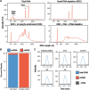
Fig. 3. mRNA purity following three-stage purification pipeline. (A) Bioanalyzer electropherograms displaying the RNA distribution following each stage of our purification pipeline. (B) Average percentage of reads mapping to ncRNA (rRNA, tRNA, snRNA, etc.) and mRNA determined by RNA-seq of two biological replicate total RNA and purified mRNA samples. (C) Representative overlaid extraction ion chromatograms for five RNA modifications that exist solely in ncRNA. These five modifications, in addition to eight additional ncRNA modifications, were detected in our total RNA samples (blue) while not detected in our mRNA samples (red) above our control digestions without RNA added (grey). The LODs for DHU, m3U, mcm5U, t6A, and i6A are 530 amol, 45 amol, 29 amol, 21 amol, and 44 amol, respectively.
Fig. 4
Fig. 4. Enzymatic digestion and LC-MS/MS analysis of S. cerevisiae total RNA and mRNA. (A) RNA is enzymatic digested to ribonucleosides through a two-stage process. RNA is first digested to nucleotide monophosphates by nuclease P1 and then dephosphorylated to ribonucleosides by bacterial alkaline phosphatase. The resulting ribonucleosides are separated using reverse phase chromatography and then quantified using MRM on a triple quadrupole mass spectrometer. (B) S. cerevisiae total RNA and mRNA were analyzed using the LC-MS/MS method developed to quantify 46 modifications in a single analysis. In total RNA, 26 modifications were detected while 13 ribonucleosides were detected in the highly purified mRNA.
Fig. 5
Fig. 5. m1G, m2G, m22G, and m5U are present in S. cerevisiae mRNA. (A) Overlaid extracted ion chromatograms displaying m1G, m2G, m22G, and m5U are detected in our mRNA samples (red) above our digestion control samples without RNA added (grey). (B) m1G, m2G, m22G, and m5U are incorporated into S. cerevisiae mRNA by their corresponding tRNA modifying enzymes (Trm10, trm11, Trm1, and Trm2 respectively). The modification/main base% (e.g., m1G/G%) were normalized to their levels in the average WT mRNA levels. A significant decrease (**p < 0.01) was detected for all cases. The error bars are the standard deviation of the normalized mod/main base%.
Fig. 6
Fig. 6. Methylated guanosine and uridine modifications alter amino acid addition. (A) Watson–Crick base pairing of m1G, m2G and m5U. The added methylation is displayed in red and the hydrogen bond interactions displayed as a dashed orange line. (B) Total peptide formation of translation reactions after 600 seconds using transcribed or single-nucleotide modified mRNAs encoding for either (left panel) Met-Val (GUG) or (right panel) Met-Arg (CGU) dipeptide. Error bars are the standard deviation. (C) Time courses displaying the formation of fMet-Phe dipeptide on an unmodified and singly modified UUC or UUU codons (left panel). Observed rate constants (right panel) were determined from the fit data. The error bars are the standard deviation of the fitted value of kobs.

References

    1. McCown P. J. Ruszkowska A. Kunkler C. N. Breger K. Hulewicz J. P. Wang M. C. Springer N. A. Brown J. A. Naturally occurring modified ribonucleosides. Wiley Interdiscip. Rev.: RNA. 2020;11:e1595. - PMC - PubMed
    1. Ontiveros R. J. Stoute J. Liu K. F. The chemical diversity of RNA modifications. Biochem. J. 2019;476:1227–1245. doi: 10.1042/BCJ20180445. - DOI - PMC - PubMed
    1. Boccaletto P. Machnicka M. A. Purta E. Piątkowski P. Bagiński B. Wirecki T. K. de Crécy-Lagard V. Ross R. Limbach P. A. Kotter A. Helm M. Bujnicki J. M. MODOMICS: a database of RNA modification pathways. 2017 update. Nucleic Acids Res. 2018;46:D303–D307. doi: 10.1093/nar/gkx1030. - DOI - PMC - PubMed
    1. Jonkhout N. Tran J. Smith M. A. Schonrock N. Mattick J. S. Novoa E. M. The RNA modification landscape in human disease. RNA. 2017;23:1754–1769. doi: 10.1261/rna.063503.117. - DOI - PMC - PubMed
    1. Zhang X. Trebak F. Souza L. A. C. Shi J. Zhou T. Kehoe P. G. Chen Q. Feng Earley Y. Small RNA modifications in Alzheimer's disease. Neurobiol. Dis. 2020;145:105058. doi: 10.1016/j.nbd.2020.105058. - DOI - PMC - PubMed

LinkOut - more resources