. 2023 Apr 6;14(18):4845-4856.
doi: 10.1039/d2sc06065e. eCollection 2023 May 10.

Isoamphipathic antibacterial molecules regulating activity and toxicity through positional isomerism

Affiliations

Isoamphipathic antibacterial molecules regulating activity and toxicity through positional isomerism

Swagatam Barman et al. Chem Sci. .

Abstract

Peptidomimetic antimicrobials exhibit a selective interaction with bacterial cells over mammalian cells once they have achieved an optimum amphiphilic balance (hydrophobicity/hydrophilicity) in the molecular architecture. To date, hydrophobicity and cationic charge have been considered the crucial parameters to attain such amphiphilic balance. However, optimization of these properties is not enough to circumvent unwanted toxicity towards mammalian cells. Hence, herein, we report new isoamphipathic antibacterial molecules (IAMs: 1-3) where positional isomerism was introduced as one of the guiding factors for molecular design. This class of molecules displayed good (MIC = 1-8 μg mL-1 or μM) to moderate [MIC = 32-64 μg mL-1 (32.2-64.4 μM)] antibacterial activity against multiple Gram-positive and Gram-negative bacteria. Positional isomerism showed a strong influence on regulating antibacterial activity and toxicity for ortho [IAM-1: MIC = 1-32 μg mL-1 (1-32.2 μM), HC50 = 650 μg mL-1 (654.6 μM)], meta [IAM-2: MIC = 1-16 μg mL-1 (1-16.1 μM), HC50 = 98 μg mL-1 (98.7 μM)] and para [IAM-3: MIC = 1-16 μg mL-1 (1-16.1 μM), HC50 = 160 μg mL-1 (161.1 μM)] isomers. Co-culture studies and investigation of membrane dynamics indicated that ortho isomer, IAM-1 exerted more selective activity towards bacterial over mammalian membranes, compared to meta and para isomers. Furthermore, the mechanism of action of the lead molecule (IAM-1) has been characterized through detailed molecular dynamics simulations. In addition, the lead molecule displayed substantial efficacy against dormant bacteria and mature biofilms, unlike conventional antibiotics. Importantly, IAM-1 exhibited moderate in vivo activity against MRSA wound infection in a murine model with no detectable dermal toxicity. Altogether, the report explored the design and development of isoamphipathic antibacterial molecules to establish the role of positional isomerism in achieving selective and potential antibacterial agents.


Conflict of interest statement

The authors declare no conflicts of interest.

Figures
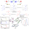
Fig. 1
Fig. 1. (A) Schematic of molecular design of IAMs; (B) synthesis of isoamphipathic antibacterial molecules (IAMs: 1–3); (C) amphiphilicity of IAMs: 1–3 through HPLC retention time measurement; (D) representative structures obtained from the simulations of the three compounds in aqueous solution (pink, IAM-1, green, IAM-2, and light blue, IAM-3) and (E) distance between the centres of mass of the two C6 alkyl chains, obtained from the simulation.
Fig. 2
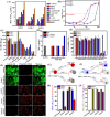
Fig. 2. (A) Antibacterial activity (MIC) and toxicity (HC50) of IAMs: 1–3. The experiment was performed twice in triplicate; (B) hemolysis percentage upon IAMs: 1–3 exposure at variable concentrations. The experiment was performed twice in triplicate; (C) selectivity index of IAMs: 1–3. The SI values were calculated based on the average values of MIC and HC50; (D) antibacterial activity of IAM-1 against MRSA upon human blood plasma and serum pre-incubation at different time intervals (1 h, 2 h and 3 h). The experiment was conducted twice in triplicate; (E) RAW cell viability upon IAMs: 1–3 treatment; (F) fluorescence microscopy of RAW cells upon IAMs: 1–3 treatment at 256 μg mL−1 (257.8 μM); (G) schematic of a MRSA and hRBC co-culture study; (H) co-culture of MRSA and hRBC in the presence and absence of IAM-1, IAM-2 and IAM-3 at 256 μg mL−1 (257.8 μM); (I) schematic of a MRSA and RAW cell co-culture study and (J) co-culture of MRSA and RAW cells in the presence and absence of IAM-1, IAM-2 and IAM-3 at 256 μg mL−1 (257.8 μM). # MRSA: methylene resistant S. aureus, VRSA: vancomycin resistant S. aureus and VRE: vancomycin resistant E. faecium.
Fig. 3
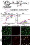
Fig. 3. (A) Schematic of a membrane leakage study using the carboxyfluorescein dye; (B) bacterial model membrane (DPPE : DPPG: 88 : 12) leakage upon treatment with IAMs: 1–3 at 160 μg mL−1 (161.1 μM); (C) mammalian model membrane (DPPC) leakage upon treatment with IAMs: 1–3 at 200 μg mL−1 (201.4 μM). The black arrows indicated the time of compound addition to the vesicles and (D) investigation on the antibacterial mechanism of action with the lead compound, IAM-1: through fluorescence microscopy of MRSA upon IAM-1 treatment for 6 h.
Fig. 4
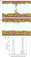
Fig. 4. (A) Structures of the bacterial membrane in the three simulations in which a defect free bilayer was formed. Water molecules and IAM-1 are shown in stick representation and coloured according to atom type (C = grey, O = red, N = blue, and H = white). The phosphorus atoms of the lipids are shown as yellow or orange spheres (for POPE and POPG, respectively). (I) Transmembrane orientation and (II) superficial orientation of IAM-1 in the lipid bilayer. The other parts of the lipid molecules are not shown, for the sake of clarity, so that the light blue area corresponds to the hydrophobic core of the bilayer. (C) Density of selected groups in the equilibrated trajectory (last 20 ns, after the annealing process), showing the different depth of insertion of the analogue. The colour code used in the structural figures is the same as that in Fig. 4B and water is not shown, for the sake of clarity. In the bottom panel the density is shown in black for P atoms, in cyan for water and in red for IAM-1 respectively.
Fig. 5
Fig. 5. Bactericidal kinetics of IAM-1 against planktonic (A) MRSA ATCC 33591 and (B) E. coli MTCC 443; anti-stationary phase activity of IAM-1 (upon 6 h treatment) against stationary cells of (C) MRSA ATCC 33591 and (D) E. coli MTCC 443; anti-persister phase activity of IAM-1 against persister cells of (E) S. aureus MTCC 737 and (F) E. coli MTCC 443. The asterisk (*) corresponds to < 50 CFU. MRSA biofilm disruption upon IAM-1 treatment: (G) crystal violet staining of MRSA ATCC 33591 biofilm; (H) biofilm-embedded MRSA ATCC 33591 cell viability; (I) biofilm-dispersed MRSA cell viability and (J) fluorescence microscopy of MRSA ATCC 33591 biofilm disruption. The live/dead ratio; control: 96% : 4%, vancomycin [64 μg mL−1 (44.2 μM)]: 80% : 20% and IAM-1 [128 μg mL−1 (128.9 μM)]: 15% : 75%. The live/dead ratio was calculated using image J.
Fig. 6
Fig. 6. (A) Histopathology (H and E staining) of untreated and IAM-1 (200 mg kg−1) treated mouse dermal tissue; wound infection with MRSA, (B) experimental plan and (C) experimental outcome. Values are presented as mean ± standard deviation for a group of 4 mice. Statistical analysis was conducted using GraphPad Prism 7 through an unpaired student-t test and P < 0.05 was considered statistically significant.

References

    1. Bush K. Courvalin P. Dantas G. Davies J. Eisenstein B. Huovinen P. Jacoby G. A. Kishony R. Kreiswirth B. Kutter E. et al. . Nat. Rev. Microbiol. 2011;9:894–896. doi: 10.1038/nrmicro2693. - DOI - PMC - PubMed
    1. Boucher H. W. Talbot G. H. Bradley J. S. Edwards J. E. Gilbert D. Rice L. B. Scheld M. Spellberg B. Bartlett J. Clin. Infect. Dis. 2009;48:1–12. doi: 10.1086/595011. - DOI - PubMed
    1. Laxminarayan R. Duse A. Wattal C. Zaidi A. K. M. Wertheim H. F. L. Sumpradit N. Vlieghe E. Hara G. L. Gould I. M. Goossens H. et al. . Lancet Infect. Dis. 2013;13:1057–1098. doi: 10.1016/S1473-3099(13)70318-9. - DOI - PubMed
    1. Davies D. Nat. Rev. Drug Discovery. 2003;2:114–122. doi: 10.1038/nrd1008. - DOI - PubMed
    1. Costerton J. W. Stewart P. S. Greenberg E. P. Science. 1999;284:1318–1322. doi: 10.1126/science.284.5418.1318. - DOI - PubMed