

Nailfold Capillaroscopy: A Promising, Noninvasive Approach to Predict Retinopathy of Prematurity
- PMID: 37182664
- PMCID: PMC12087976
- DOI: 10.1016/j.jpeds.2023.113478
Nailfold Capillaroscopy: A Promising, Noninvasive Approach to Predict Retinopathy of Prematurity
Abstract
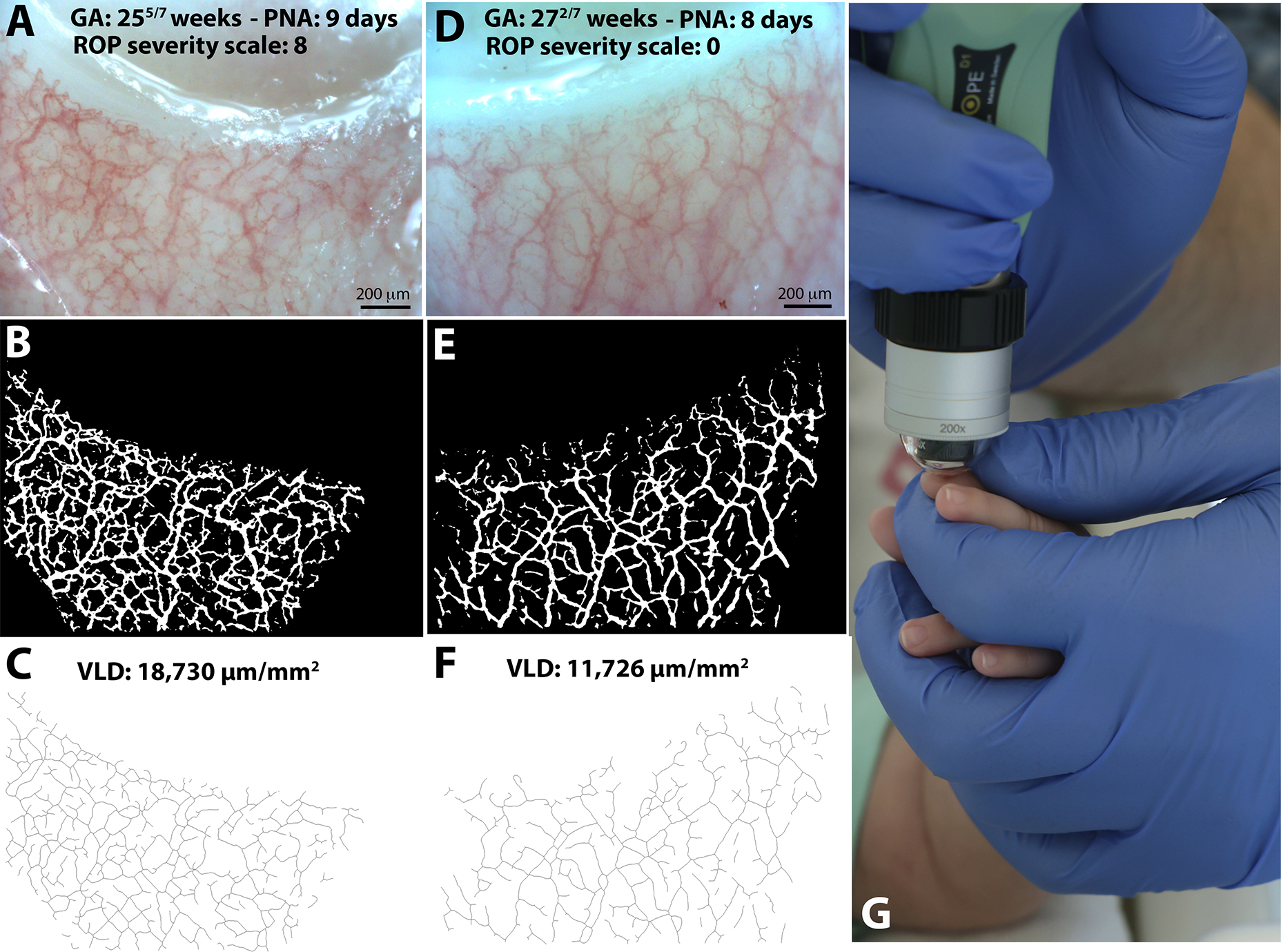
Objective: To test the hypothesis that nailfold capillaroscopy can noninvasively detect dysregulated retinal angiogenesis and predict retinopathy of prematurity (ROP) in infants born premature before its development.
Methods: In a cohort of 32 infants born <33 weeks of gestation, 1386 nailfold capillary network images of the 3 middle fingers of each hand were taken during the first month of life. From these, 25 infants had paired data taken 2 weeks apart during the first month of life. Images were analyzed for metrics of peripheral microvascular density using a machine learning-based segmentation approach and a previously validated microvascular quantification platform (REAVER vascular analysis). Results were correlated with subsequent development of ROP based on a published consensus ROP severity scale.
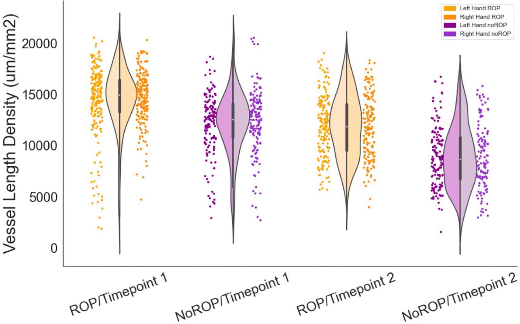
Results: In total, 18 of 32 (56%) (entire cohort) and 13 of 25 (52%) (2-time point subgroup) developed ROP. Peripheral vascular density decreased significantly during the first month of life. In the paired time point analysis, vessel length density, a key metric of peripheral vascular density, was significantly greater at both time points among infants who later developed ROP (15 563 and 11 996 μm/mm2, respectively) compared with infants who did not (12 252 and 8845 μm/mm2, respectively) (P < .001, both time points). A vessel length density cutoff of >15 100 at T1 or at T2 correctly detected 3 of 3 infants requiring ROP therapy. In a mixed-effects linear regression model, peripheral vascular density metrics were significantly correlated with ROP severity.
Conclusions: Nailfold microvascular density assessed during the first month of life is a promising, noninvasive biomarker to identify premature infants at highest risk for ROP before detection on eye exam.
Keywords: microvasculature; neonate.
Copyright © 2023 Elsevier Inc. All rights reserved.
Conflict of interest statement
Declaration of Competing Interest The authors declare no conflicts of interest.
Figures

References
-
- Isaza G, Donaldson L, Chaudhary V. Increased incidence of retinopathy of prematurity and evolving treatment modalities at a Canadian tertiary centre. Can J Ophthalmol. 2019;54(2):269–74. - PubMed
-
- Zin A, Gole GA. Retinopathy of prematurity-incidence today. Clin Perinatol. 2013;40(2):185–200. - PubMed
-
- Fierson WM. Screening Examination of Premature Infants for Retinopathy of Prematurity. Pediatrics. 2018;142(6). - PubMed
Publication types
MeSH terms
Grants and funding
LinkOut - more resources
Full Text Sources
