. 2023 Jul;45(7):e2200251.
doi: 10.1002/bies.202200251. Epub 2023 May 15.

Tender love and disassembly: How a TLDc domain protein breaks the V-ATPase

Affiliations

Tender love and disassembly: How a TLDc domain protein breaks the V-ATPase

Stephan Wilkens et al. Bioessays. 2023 Jul.

Abstract

Vacuolar ATPases (V-ATPases, V1 Vo -ATPases) are rotary motor proton pumps that acidify intracellular compartments, and, when localized to the plasma membrane, the extracellular space. V-ATPase is regulated by a unique process referred to as reversible disassembly, wherein V1 -ATPase disengages from Vo proton channel in response to diverse environmental signals. Whereas the disassembly step of this process is ATP dependent, the (re)assembly step is not, but requires the action of a heterotrimeric chaperone referred to as the RAVE complex. Recently, an alternative pathway of holoenzyme disassembly was discovered that involves binding of Oxidation Resistance 1 (Oxr1p), a poorly characterized protein implicated in oxidative stress response. Unlike conventional reversible disassembly, which depends on enzyme activity, Oxr1p induced dissociation can occur in absence of ATP. Yeast Oxr1p belongs to the family of TLDc domain containing proteins that are conserved from yeast to mammals, and have been implicated in V-ATPase function in a variety of tissues. This brief perspective summarizes what we know about the molecular mechanisms governing both reversible (ATP dependent) and Oxr1p driven (ATP independent) V-ATPase dissociation into autoinhibited V1 and Vo subcomplexes.

Keywords: ADP inhibition; Oxr1p; TLDc domain; V1-ATPase; Vo proton channel; peripheral stalk; protein-protein interaction; reversible disassembly; subunit C; subunit H; vacuolar H+-ATPase.


Conflict of interest statement

Conflict of interest

The authors declare that they have no conflict of interest.

Figures

Figure 1
Figure 1
The eukaryotic vacuolar H+-ATPase and regulation by disengagement of V1-ATPase and Vo proton channel. (A) Schematic of the architecture and mechanism of the yeast V-ATPase. (B) Regulation by reversible disassembly (left side) and Oxr1p mediated disassembly (right side).
Figure 2
Figure 2
Structure of the yeast vacuolar ATPase in lipid nanodisc. (A) Left panel: CryoEM structure of lipid nanodisc yeast reconstituted V-ATPase in rotary state 1[32] (PDBID 7FDA). Middle panel: V-ATPase central rotor subcomplex. Two of the c subunits are not shown to allow a view inside the c-ring cylinder. Right panel: V-ATPase stator subcomplex. (B) Cross section of states 1–3 at the level of the phosphate binding loops (P-loops) of the V1 A subunits as indicated with the dashed line in (A), left panel. The blue arrowheads point to the open catalytic sites, with the P-loops highlighted in blue. The closed and half-closed catalytic sites are indicated by black arrowheads. (C) Cross section of the V-ATPase Vo at the level indicated by the dashed line in (A), left panel. For details, see text.
Figure 3
Figure 3
Structure of autoinhibited yeast V1-ATPase. Upper panel: CryoEM structure of autoinhibited V1 in state 2 (PDBID 7TMM),[27] highlighting the interaction between HCT and the N-termini of EG2. Lower panel: Region of the cryoEM structure of nanodisc bound holo V-ATPase in state 2 (PDBID 7FDB),[32] highlighting the binding site of HCT on aNT.
Figure 4
Figure 4
Structure of autoinhibited yeast Vo proton channel. (A) CryoEM structure of autoinhibited Vo in lipid nanodisc (PDBID 6M0R)[75] as seen parallel to the membrane (left panel) and from the cytoplasm towards the bilayer (right panel). The interaction of aNT and d is highlighted by the dashed pink circles. (B) CryoEM structure of yeast holo V-ATPase in state 3 (PDBID 7FDC)[32] seen as in the panels in (A). The conformational change of aNT from a peripheral location in V1Vo to its binding site on d in free Vo is highlighted by the pink arrow in the view from the cytoplasm towards the membrane.
Figure 5
Figure 5
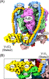
Structure of yeast V1 in complex with Oxr1p and the C subunit and comparison to mEAK7 bound human V-ATPase. (A) Two views of the cryoEM structure of V1(C)Oxr1 (PDBID 7FDE),[32] highlighting Oxr1p’s binding site formed by EG2, subunit C, and the B subunit of the open catalytic site. V1(C)Oxr1 is halted in rotary state 1. (B) CryoEM structure of human V-ATPase in complex with mEAK7 (PDBID 7UNF).[38] Left panel, same view as in left panel 5A. Right panel, like yeast Oxr1p, mEAK7 binds the N-termini of a EG heterodimer and the C-terminal domain of a B subunit with the open catalytic site conformation. However, note that mEAK7 binds to peripheral stalk EG3 rather than EG2 as Oxr1p. (C) Zoomed in views of the Oxr1p binding site (upper panel) and comparison to the same site in state 1 V1Vo without Oxr1p bound (PDBID 7FDA). (D) Zoomed in view of the mEAK7 binding site (upper panel), and comparison to the same site in state 2 V1Vo (PDBID 6WM3).

References

    1. Collins MP, Forgac M. 2020. Regulation and function of V-ATPases in physiology and disease. Biochim Biophys Acta Biomembr 1862: 183341. - PMC - PubMed
    1. Eaton AF, Merkulova M, Brown D. 2021. The H(+)-ATPase (V-ATPase): from proton pump to signaling complex in health and disease. Am J Physiol Cell Physiol 320: C392–C414. - PMC - PubMed
    1. Marshansky V, Futai M. 2008. The V-type H+-ATPase in vesicular trafficking: targeting, regulation and function. Curr Opin Cell Biol 20: 415–26. - PMC - PubMed
    1. Maxson ME, Grinstein S. 2014. The vacuolar-type H(+)-ATPase at a glance - more than a proton pump. J Cell Sci 127: 4987–93. - PubMed
    1. Santos-Pereira C, Rodrigues LR, Côrte-Real M. 2021. Emerging insights on the role of V-ATPase in human diseases: Therapeutic challenges and opportunities. Med Res Rev. - PubMed

Publication types

MeSH terms

Substances