Review
. 2023 Jun;17(2):299-312.
doi: 10.1007/s12105-023-01548-8. Epub 2023 May 15.

Proceedings of the 2023 North American Society of Head and Neck Pathology Companion Meeting, New Orleans, LA, March 12, 2023: Navigating New Developments in High Grade Sinonasal Neuroendocrine and Neuroectodermal Neoplasms

Affiliations
Review

Proceedings of the 2023 North American Society of Head and Neck Pathology Companion Meeting, New Orleans, LA, March 12, 2023: Navigating New Developments in High Grade Sinonasal Neuroendocrine and Neuroectodermal Neoplasms

Lisa M Rooper. Head Neck Pathol. 2023 Jun.

Abstract

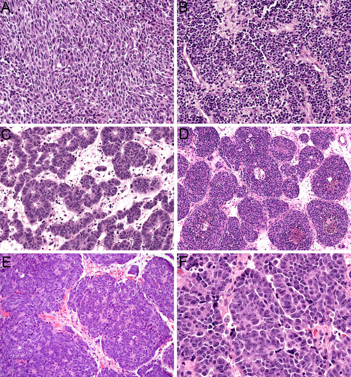
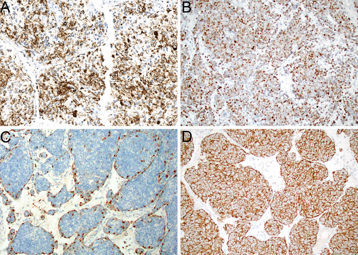
Although the definitions of sinonasal neuroendocrine and neuroectodermal neoplasms did not change substantially in the 5th edition WHO Classification of Head and Neck Tumours, the diagnosis of olfactory neuroblastoma (ONB), small cell neuroendocrine carcinoma, and large cell neuroendocrine carcinoma remains quite challenging in practice. Ambiguities surrounding the amount of keratin expression allowable in ONB and the amount of neuroendocrine differentiation seen in sinonasal undifferentiated carcinoma (SNUC) lead to significant diagnostic discrepancies at the high grade end of this tumor spectrum. Furthermore, a group of problematic neuroepithelial tumors that show overlapping features of ONB and neuroendocrine carcinoma have never been recognized in formal classification schemes. Since publication of the 5th edition WHO, two new tumor entities have been proposed that help resolve these problems. Olfactory carcinoma is defined by high grade keratin-positive neuroectodermal cells with frequent intermixed glands and shows recurrent Wnt pathway, ARID1A, and RUNX1 alterations. IDH2-mutant sinonasal carcinoma is a molecularly-defined category that encompasses tumors with undifferentiated (SNUC), large cell neuroendocrine, and neuroepithelial phenotypes. This review will provide a practical overview of these emerging entities and their application to diagnostic challenges in the post-WHO sinonasal neuroendocrine and neuroectodermal tumor classification.

Keywords: IDH2-mutant sinonasal carcinoma; Immunohistochemistry; Molecular diagnostics; Nasal neoplasms; Neuroendocrine carcinoma; Olfactory carcinoma; Olfactory neuroblastoma; Sinonasal undifferentiated carcinoma.


Conflict of interest statement

The author certifies that she has no affiliations with or involvement in any organization or entity with any financial interest or non-financial interest in the subject matter or materials discussed in this manuscript.

Figures

Fig. 1
Fig. 1
ONB is composed of lobules and nests of neuroectodermal cells that are surrounded by hypervascular stroma (A, × 10). The tumor cells have syncytial borders and monotonous round to oval nuclei with speckled chromatin (B, × 40). Low grade ONB tends to have abundant neurofibrillary stroma with pseudorosette formation (C, × 20). High grade ONB shows increased mitotic activity and necrosis (D, × 20)
Fig. 2
Fig. 2
Sinonasal small cell neuroendocrine carcinoma displays sheets of markedly atypical epithelial cells with scant cytoplasm (A, × 20). The nuclei are hyperchromatic with frequent molding, mitotic figures, and apoptotic bodies (B, × 40). Large cell neuroendocrine carcinoma tends to show nested architecture (C, × 10). The tumor cells have abundant cytoplasm and large nuclei with vesicular chromatin and prominent nucleoli that demonstrate peripheral palisading (D, × 40)
Fig. 3
Fig. 3
Olfactory carcinoma is predominantly composed of sheets and lobules of neuroectodermal cells (A, × 4). These cells have syncytial cytoplasm that ranges from scant to abundant (B, × 40). The nuclei are hyperchromatic and angulated with prominent nuclear molding and cell-cell wrapping (C, × 40). Most cases display overt neural differentiation with variable amounts of neurofibrillary matrix (D, × 40) including occasional expansile neurofibrillary zones with ganglion cells (E, × 10). Many cases also show well-formed true rosettes (F, × 20)
Fig. 4
Fig. 4
Olfactory carcinoma also displays a well-developed glandular component that can include either complex, expansile proliferations of back-to-back acini (A, × 20) or simple ducts and tubules that are closely intermixed with surrounding neuroectodermal cells (B, × 20). Many of the glands show well-developed cilia formation (C, × 40). The glandular cells have abundant eosinophilic cytoplasm and round to oval nuclei that lack the atypia of surrounding neuroectodermal cells (D, × 40). It can be difficult to differentiate glands from rosettes in some cases (E, × 20). Rare cases show squamous morules intermixed with the glands (F, × 20)
Fig. 5
Fig. 5
Olfactory carcinoma generally shows strong positivity for low molecular weight keratin Cam 5.2 (A, × 20) with variable expression of pankeratin AE1/AE3 that is stronger in glands than neuroectodermal cells (B, × 20). The neuroectodermal cells also display variable expression of neuroendocrine markers including INSM1 (C, × 20) and synaptophysin (D, × 20). A subset of cases show peripheral p40 expression in the neuroectodermal cells (E, × 20) and S100-protein-positive sustentacular cells (F, × 20)
Fig. 6
Fig. 6
IDH2-mutant sinonasal carcinoma can show variable architecture, including sheet-like growth in undifferentiated cases (A, × 20). Tumors with neuroendocrine differentiation frequently display cells arranged in lobules and nests (B, × 20) or cords and trabeculae (C, × 20). Rare tumors with neuroepithelial differentiation are composed of compact nests (D, ×20) and demonstrate well-developed rosette formation and neurofibrillary matrix (E, × 20). Across histologic phenotypes, prominent macronucleoli are consistently seen (F, × 40)
Fig. 7
Fig. 7
IDH2-mutant sinonasal carcinomas are consistently positive for keratin (A, × 20). Tumors with neuroendocrine and neuroepithelial differentiation show expression of neuroendocrine markers such as INSM1 (B, × 20). Neuroepithelial tumors also frequently have an S100-protein-positive sustentacular network (C, × 20). Mutant IDH1/IDH2 immunohistochemistry demonstrates strong cytoplasmic positivity in tumor cells (D, × 20)

References

    1. Thompson LDR, Bishop JA. Update from the 5th edition of the world health organization classification of head and neck tumors: nasal cavity, paranasal sinuses and skull Base. Head Neck Pathol. 2022;16:1–18. doi: 10.1007/s12105-021-01406-5. - DOI - PMC - PubMed
    1. Bell D, Classe M, Perez-Ordonez B et al (2022) Olfactory neuroblastoma. In: WHO Classification of Tumours Editorial Board, ed. WHO Classification of Head and Neck Tumours. Lyon, France: International Agency for Research on Cancer
    1. Mete O, Wenig BM. Update from the 5th edition of the world health organization classification of head and neck tumors: overview of the 2022 WHO classification of head and neck neuroendocrine neoplasms. Head Neck Pathol. 2022;16:123–142. doi: 10.1007/s12105-022-01435-8. - DOI - PMC - PubMed
    1. Rooper LM, Classe M, Nose V et al (2022) Small cell neuroendocrine carcinoma. In: WHO Classification of Tumours Editorial Board, ed. WHO Classification of Head and Neck Tumours. Lyon, France: International Agency for Research on Cancer
    1. Rooper LM, Classe M, Nose V et al (2022) Large cell neuroendocrine carcinoma. In: WHO Classification of Tumours Editorial Board, ed. WHO Classification of Head and Neck Tumours. Lyon, France: International Agency for Research on Cancer

MeSH terms

Supplementary concepts